

Diagnostic devices and apparatus for the controlled movement of reagents without membranes
- Summary
- Abstract
- Description
- Claims
- Application Information
AI Technical Summary
Benefits of technology
Problems solved by technology
Method used
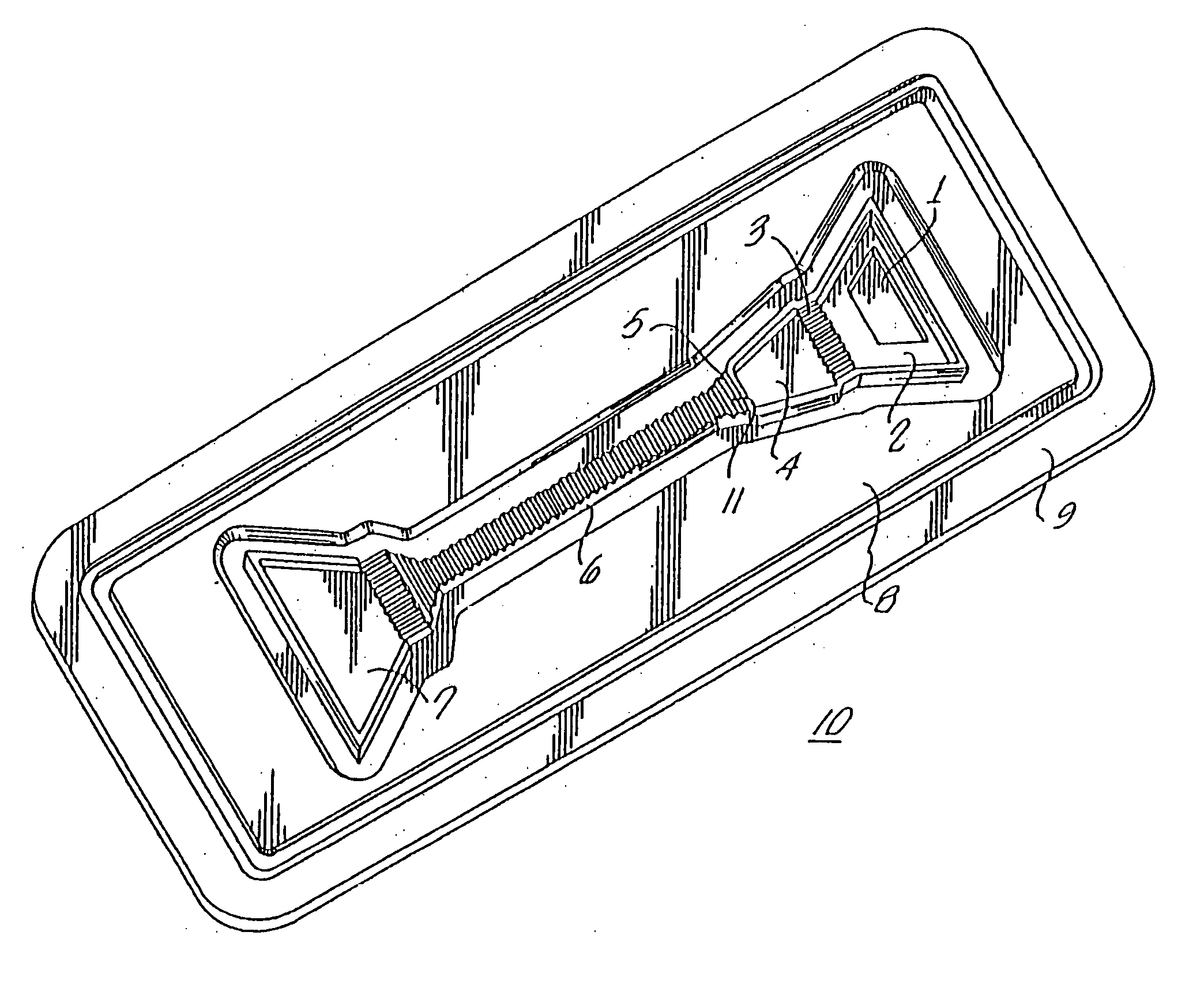
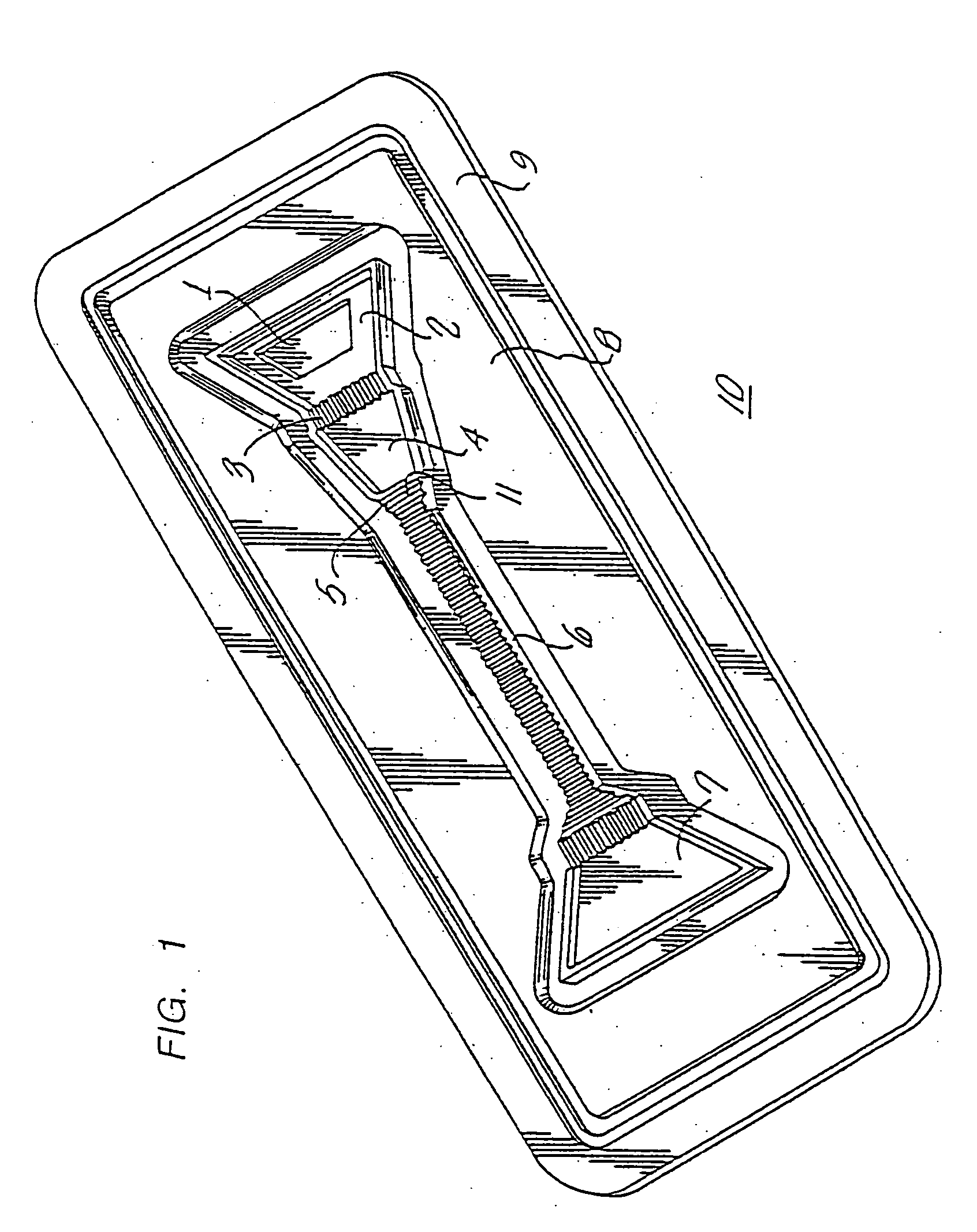
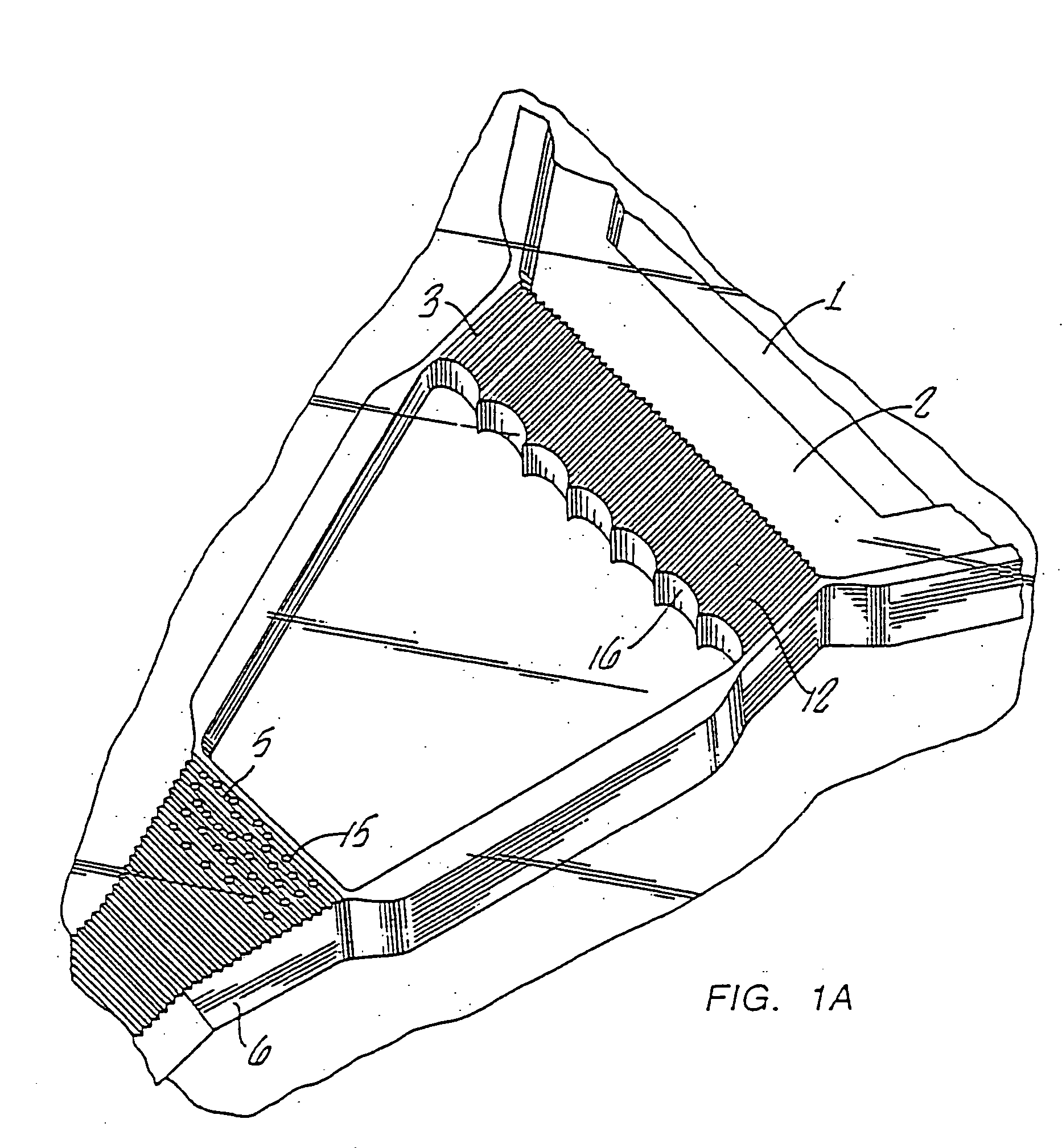
Image

Examples
example 1
Preparation of Anti-βhCG Antibody-Colloidal Gold Conjugate
[0126] Colloidal gold with an average diameter of 45 nm was prepared according to the method of Frens, Nature, Physical Sciences, 241, 20 (1973). The colloidal gold conjugate was prepared by first adding 5.6 ml of 0.1 M potassium phosphate, pH 7.58, dropwise with rapid stirring to 50 ml of colloidal gold. Anti B-subunit monoclonal antibody to hCG (Applied Biotech, San Diego, Calif.; 1 ml of 4.79 mg / ml in phosphate buffered saline, 0.02% sodium azide, pH 7) was added in a bolus to the colloidal gold with rapid stirring. After complete mixing the stirring was stopped and the solution was incubated at room temperature for 1 h. Polyethylene glycol (average molecular weight=20,000) was added (0.58 ml) as a 1% solution to the colloidal gold solution and the solution was mixed. The colloidal gold solution was subjected to centrifugation at 27,000 g and 5 C for 20 min. The supernatant was removed and each pellet was washed twice by...
example 2
Preparation of Anti-hCG Antibody Latex
[0127] Surfactant-free polystyrene particles (Interfacial Dynamics Corp., Portland, Oreg.; 0.106 ml of 9.4% solids, 0.4 μm) was added while vortexing to anti α-subunit hCG monoclonal antibody (Applied Biotech, San Diego, Calif.; 0.89 ml of 6.3 mg / ml in 0.1 M 2-(N-morpholino) ethane sulfonic acid, (MES), pH 5.5) and the suspension was incubated at room temperature for 15 min. The suspension was subjected to centrifugation to pellet the latex particles. The pellet was washed three times by centrifugation and resuspension of the pellet with 10 mM MES, 0.1 mg / ml trehalose, pH 5.5. The final pellet was resuspended in the wash buffer at a solids concentration of 1%.
example 3
Preparation of Goat Anti-Mouse Latex
[0128] Surfactant-free polystyrene particles (Interfacial Dynamics Corp., Portland, Oreg.; 0.11 ml of 9.4% solids, 0.6 μm) were added while vortexing to goat IgG antibody against mouse IgG (Jackson ImmunoResearch Laboratories, Inc.; 0.89 ml of 0.34 mg / ml in 0.1 M MES, pH 5) and the suspension was incubated at 45° C. for 2 h. The suspension was subjected to centrifugation to pellet the latex particles. The pellet was washed three times by centrifugation and resuspension of the pellet with 10 mM MES, 0.2 mg / ml trehalose, pH 5.5. The final pellet was resuspended in the wash buffer at a solids concentration of 1%.
PUM
| Property | Measurement | Unit |
|---|---|---|
| Volume | aaaaa | aaaaa |
| Electrochemical properties | aaaaa | aaaaa |
Abstract
Description
Claims
Application Information