Device for sensing contaminants

a technology of contaminants and sensors, applied in the field of devices, can solve the problems of rapid methods, possibly as little as 30 minutes or less, and achieve the effects of simple and easy to interpret, easy to use, and no loss of effectiveness

Inactive Publication Date: 2005-06-09
EASTMAN KODAK CO
View PDF9 Cites 26 Cited by
  • Summary
  • Abstract
  • Description
  • Claims
  • Application Information

AI Technical Summary

Benefits of technology

[0009] This technology is simple and easy to interpret and the test strip can be used by personnel with very minimal laboratory training. The test is flexible and can be taught in a protocol without losing effectiveness. The test strips are disposable or storable for later reading by more trained personnel. They can be used on any material suspected of contamination, and can be easily used at ports of entry, in production agriculture, and in natural resource environments. In fact, the technology is designed to be so simple that it could be used any place that food is processed, prepared, served, or consumed (kitchens, mess kits, food cartons, etc.).
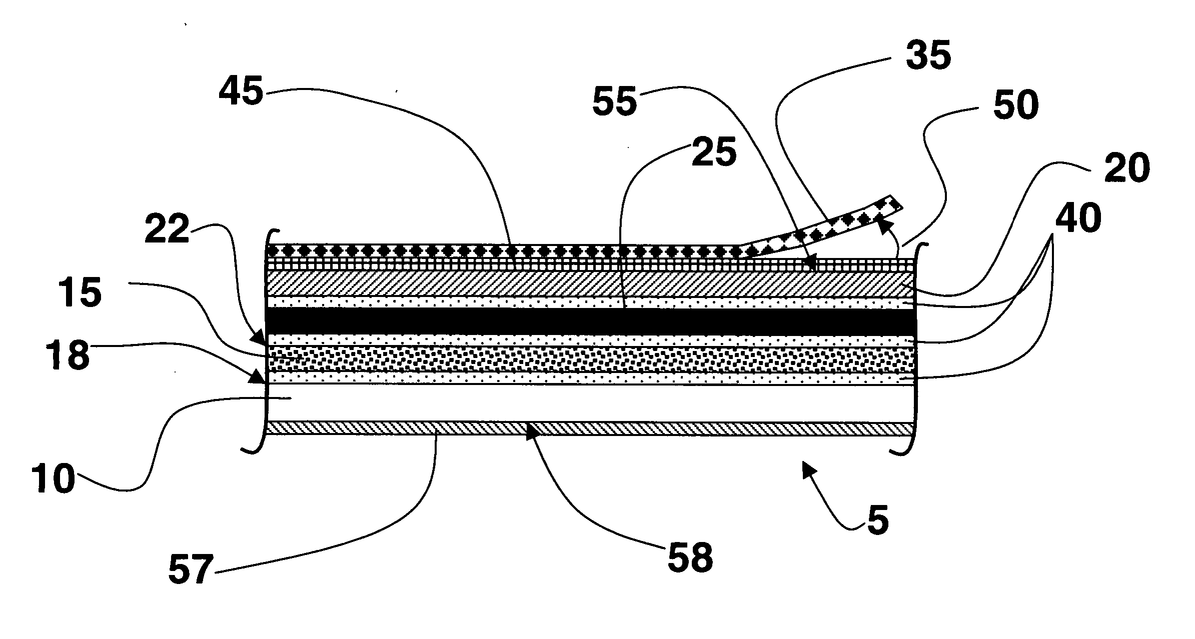
[0010] The test strip can be used with a simple swab of suspected material. A positive result will be indicated by the development of a visual indicator, the amount roughly proportional to the amount of target contaminant. The indicator can be visually detected, or further quantified by use of a hand-held thermal processor and densitometer reader, equipment easy to use by non-technical personnel. The method is rapid, giving results in possibly as little as 30 minutes or less. Due to the unique silver halide based amplification technology, the test strip will be able to detect very low levels of a contaminant, without preamplification. In one embodiment the appearance of a color will provide a signal of the presence of a contaminant. The amount of color is proportional to the amount of contaminant present and can be more carefully measured to determine the extent of the suspected contaminant. The test strip may use any number of detection methods.
[0011] The test strip can be designed with multiple coatings so that areas of the test strip are selective to different suspect pathogens. The coating process is well known and is very reproducible and consistent. Both the upper layers and the silver halide layers can be coated to known thickness, with known silver content and silver grain size. In this way, sensor elements can be fabricated that provide the same response for the same amount of suspect material. The ability to formulate a multiple test strip has a distinct advantage in cost and efficiency in usage.
[0012] Additionally the test strip of the invention could improve sample preparation. Foods could be tested without extensive handling or preparation.

Problems solved by technology

The method is rapid, giving results in possibly as little as 30 minutes or less.

Method used


Image

  • Device for sensing contaminants
  • Device for sensing contaminants
  • Device for sensing contaminants

Examples


example 1

[0210] In this example, no blocking layer was used. All steps occurred in a dark room with safe lights. Standard wet chemical development, including a fix and wash, were used. We used Kodak Polymax II RC paper. A strip of this paper was cut to about 2.5 cm×15 cm. The bottom 1 to 1.5 cm of this film was suspended in a solution consisting of D85 developer. D85 developer is a black-and-white photographic developer containing primarily the LIFCS hydroquinone in a boric acid buffer. The strip was incubated for 5 minutes at 37-40° C. This was done at 3 concentrations of hydroquinone: 0.2 M, 0.02 M, and 0.002 Molar.

[0211] The strip was rinsed for 5 seconds and then the bottom 2.5 cm of this film was developed in D76 for 1 minute. The bottom 5 cm region was fixed. The change in size of development and fixing regions allowed a clear comparison of the exposed (to the LIFCS hydroquinone) and unexposed regions of the strip. The density in the different regions was not quantified, but showed co...

example 2

[0214] This is a prophetic example. In this example, no blocking layer is used. All steps occur in a dark room with safe lights. Standard wet chemical development, including a fix and wash, are used. The substrate for the film is PET, prepared with a gel sub, and then coated with 1.6×10³ mg / m2 silver as a silver chloride cubic emulsion and 3.2×10³ mg / m2 gel. A sample layer of gelatin incorporating hydroquinone at 1000 mg / m2 is coated on top. A strip of this film is cut to about 2.5 cm×7.5 cm.

[0215] A 1 mm spot of anti-E. coli antibody in phosphate buffered saline (PBS) is placed on the film and allowed to dry for 1 minute. At a concentration of approximately 100 microgram / ml and a volume per spot of 20 nl, the coverage is estimated at 0.05 mg / m2 of antibody.

[0216] This 1 mm spot is exposed to E. coli in a solution at approximately 1×10⁴ cfu / ml, and incubated at 37° C. for 10 minutes. Concurrently, on the same strip for the same 10 minutes, but in a different location, a 1 mm spot ...

example 3

[0219] This is a prophetic example. In this example, no blocking layer is used. All steps occur in a dark room with safe lights. Standard wet chemical development, including a fix and wash, are used. The substrate for the film is PET, prepared with a gel sub, and then coated with 1.6×10³ mg / m2 silver as a silver chloride cubic emulsion and 3.2×10³ mg / m2 gel. A sample layer of gelatin incorporating polystyrene beads (2. A strip of this film is cut to about 2.5 cm×7.5 cm. An E. coli solution of approximately 1×10⁴ cfU / ml is spotted on the film (0.01 ml). A PBS solution is spotted on the film (0.01 ml). The films are incubated at 37° C. for 20 minutes, and then developed for 1 minute in Kodak D76. The film strip is fixed for 30 seconds, and then washed for 30 seconds. The developed film shows a black spot at approximately the location of the exposure to E. coli, and very little black elsewhere, including the spot exposed to only PBS. The ratio of the density of the E. coli spot to the ...


PUM

PropertyMeasurementUnit
thicknessesaaaaaaaaaa
thicknessaaaaaaaaaa
thicknessaaaaaaaaaa

Abstract

This invention relates to a test strip comprising a support having thereon a non-sampling area and a sampling area wherein said sampling area further comprises a sampling layer which can react with a target species to form or release a signal compound which is capable of effecting a reaction with silver halide to form a latent image, and a signal amplification layer comprising silver halide. It further relates to a kit containing the test strip and a method of using the test strip.

Description

FIELD OF THE INVENTION [0001] This invention relates to a test strip, particularly a test kit, for detecting contaminants in the environment and more specifically in food and water and a method for processing and viewing the sensor. The test strip uses silver halide amplification technology. BACKGROUND OF THE INVENTION [0002] Easy and effective methods for detecting contaminants, especially of food and water have long been sought. Antibody technology comprises the largest group of rapid methods; a large number of immunology-based rapid assays have been successfully used for detection of toxins, cells and viruses. Many forms of immunology-based rapid assays have been investigated and developed, including immunofiltration (IMF), micro array immunoassay (MAI), enzyme-linked immunofiltration (ELIFA), chemiluminescent immunoassay (CLIA), immunomagnetic separation (IMS), immunoliposome sandwich assay (ILSA), immunochromatography and improved and standard applications of sandwich ELISA. Ma...

Claims


Application Information

Patent Timeline
Patent Type & Authority Applications(United States)
IPC IPC(8): C12Q1/00G01N31/22G01N33/48G01N33/52G01N33/58
CPCG01N33/525G01N31/22
Inventor PATTON, DAVID L.WIEN, RICHARD W.SWITALSKI, STEVEN C.WILLIAMS, KEVIN W.
Owner EASTMAN KODAK CO
Why Eureka
Try Eureka