Method for detecting microorganisms

a microorganism and detection method technology, applied in the field of detection methods, can solve the problems of unintended effect of breeding resistant strains of bacteria, many symptoms only evident, and severe, even life-threatening infections

Inactive Publication Date: 2005-06-30
WOUNDCHEK LAB US
View PDF57 Cites 46 Cited by
  • Summary
  • Abstract
  • Description
  • Claims
  • Application Information

AI Technical Summary

Benefits of technology

[0012] In still another aspect, the present invention features a kit for detecting a wound infection, comprising a biosensor for detecting the presence or absence of a microorganism in a sample, and one or more reagents for detecting the presence of the microorganism that is the causative agent of the wound infection. For example, the reagent can be used to detect an enzyme secreted by the microorganism. In particular, the reagent can be used to detect the modification of the substrate of the biosensor.
[0013] In yet another aspect, the present invention features a polypeptide comprising or consisting of the sequence of SEQ ID NO: 1, 2, 3, 4, 5, 9, or 10. These polypeptides are useful for the identification and / or detection of the presence of wound-specific enzymes as described herein in a sample. In one embodiment, the polypeptide is detectably labeled.
[0014] In another aspect, the present invention features a nucleic acid comprising or consisting of the sequence of SEQ ID NO: 6, 7, or 8. This nucleic acid sequences are useful for the identification and / or detection of the presence of wound-specific enzymes as described herein in a sample. In one embodiment, the nucleic acid is detectably labeled.

Problems solved by technology

Infection of wounds is a major source of healthcare expenditure in the United States.
Staphylococci are usually carried as harmless commensals, however given a breach in the epidermis, they can cause severe, even life threatening infection.
While generally effective, this strategy has the unintended effect of breeding resistant strains of bacteria.
Unfortunately, many symptoms are only evident after the infection is already established.
Furthermore, after a patient is discharged from the hospital they become responsible for monitoring their own healthcare, and the symptoms of infection may not be evident to the unskilled patient.

Method used


Image

  • Method for detecting microorganisms
  • Method for detecting microorganisms
  • Method for detecting microorganisms

Examples


example 1

Identification of Wound-Specific Proteins

[0090] The TIGR comprehensive microbial resource multi genomic analysis tools located at the following Internet site: http: / / www.tigr.org / tigr-scripts / CMR2 / select_genomes.spl?showref=true&reforg=0&cutoff=60&logic=AND&showheader=true, as available on Jun. 18, 2001 were used to analyze the complete genome sequences of the following common wound pathogen species: Staphylococcus aureus (S. aureus), Staphylococcus epidermidis, Streptococcus pyogenes, Pseudomonas aeruginosa, Enterococcus faecalis, and Escherichia coli. Specifically, each gene in the S. aureus genome was compared for homologs in each of the other above specified genomes. Any S. aureus gene that did not have a homolog (with at least 45% identity at the amino acid level) to all 5 pathogens, was discarded. The remaining pool contained only those genes that are common to these six major wound pathogens. These searches were conducted using the default settings as parameters.

[0091] To i...

example 2

Preparation of Bacteria for Detection of the Absence or Presence of Bacteria in a Sample

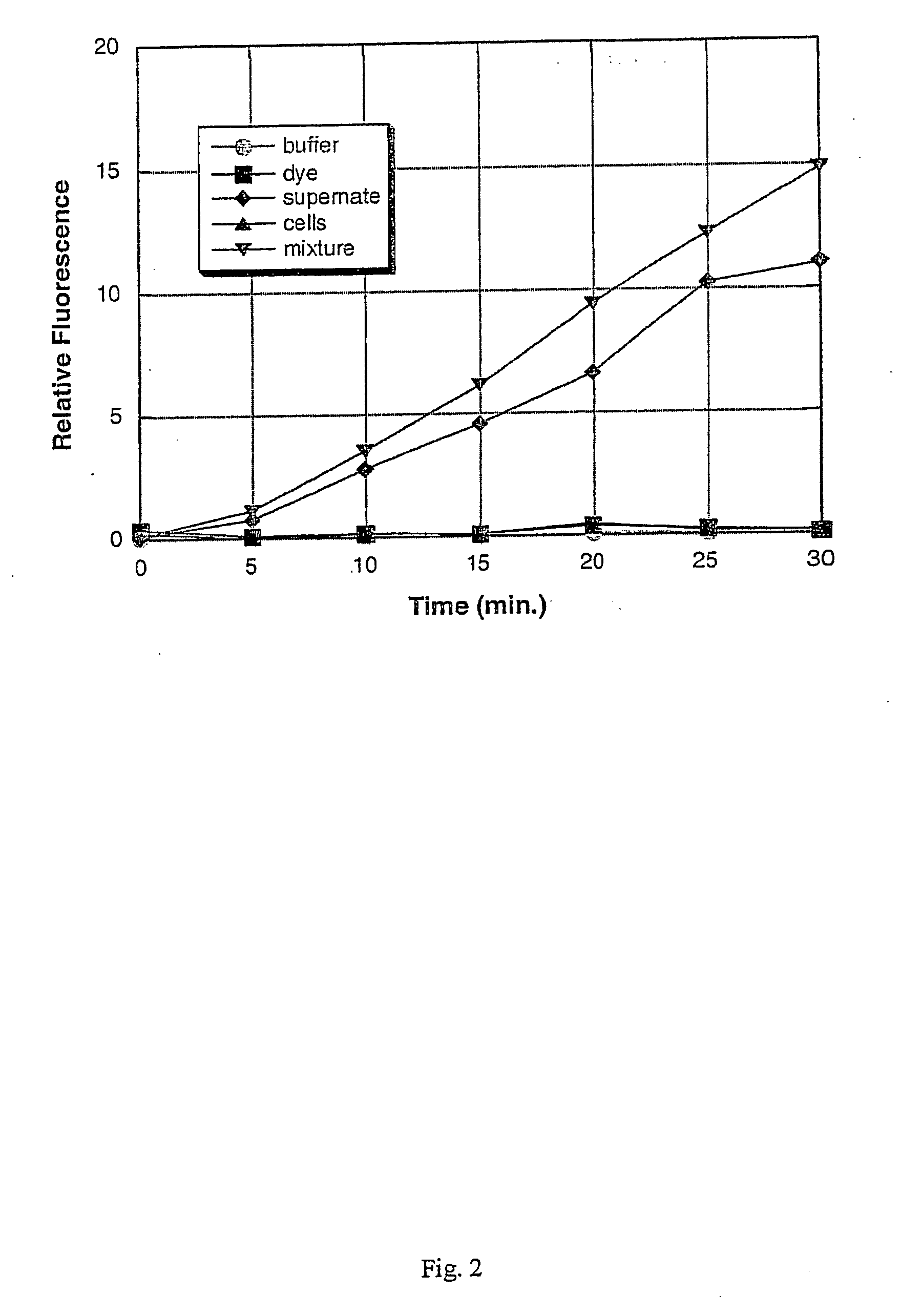
[0102] A culture of each of the following bacterial species was grown to saturation in Brain Heart Infusion (BHI) broth at 37° C. with vigorous shaking (−200 rpm), using methods that are standard in the art: Staphylococcus aureus; Staphylococcus epidermidis; Serratia marcescens; Streptococcus salivarius; Escherichia coli; Pseudomonas aeruginosa; and Enterococcus faecalis. After overnight growth (to saturation), a 1 mL sample of each culture was obtained, and the cells were removed from the culture supernatant by centrifugation at 12,000×g for 5 minutes. The remaining culture supernatants were stored on ice until required (less than one hour). The bacteria were assayed for the presence or absence of specific enzymes as described below. Alternatively, the bacterial cells are not separated from the culture supernatant, but rather, the assay is carried out on a sample containing the cells still in s...

example 3

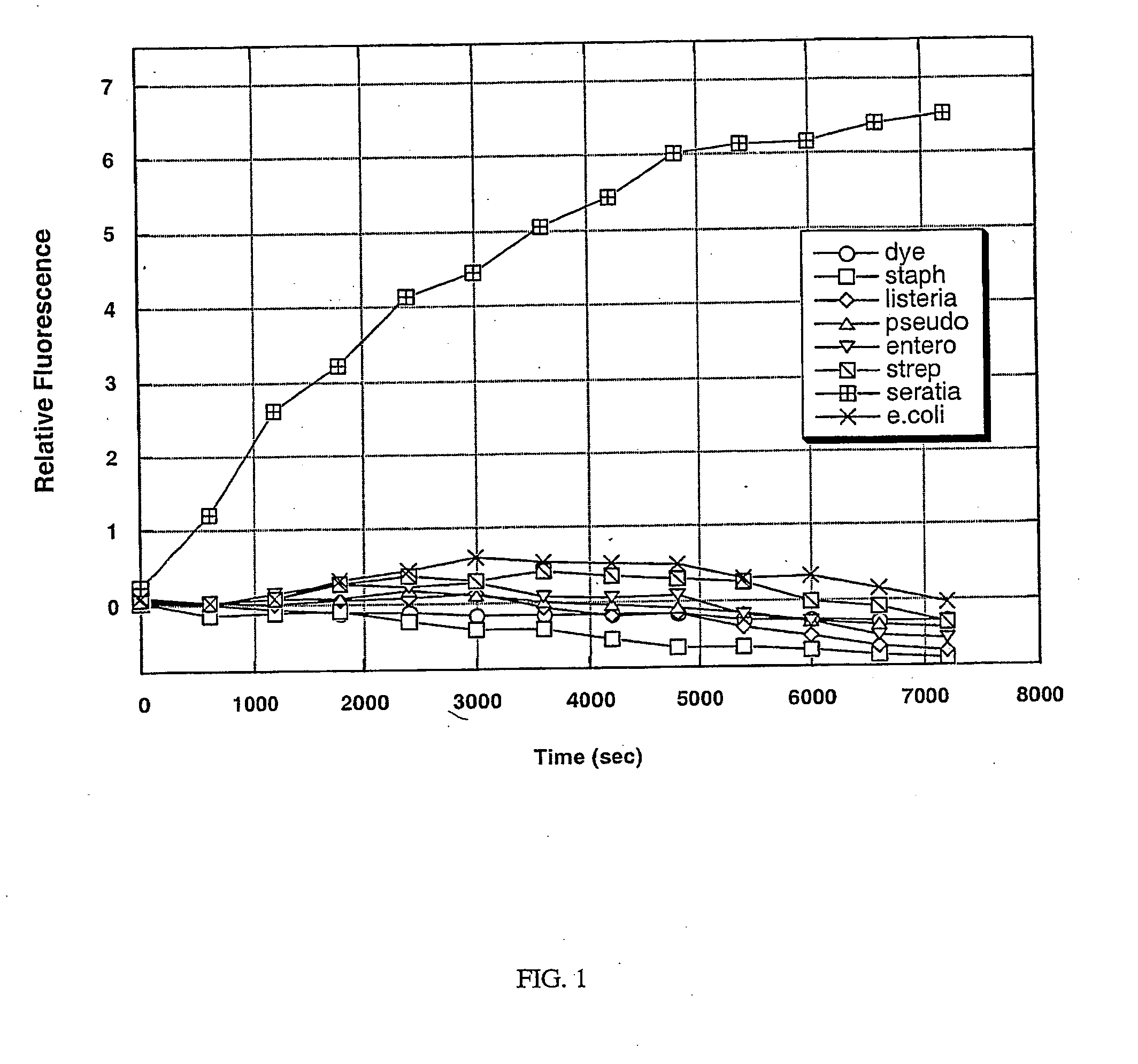
Detection of Serratia marcescens Using a Protease Assay

[0103] A protease is an enzyme that is responsible for the degradation of proteins by hydrolysis of peptide bonds. A protease can be either general or specific in its target sequence, depending on its purpose. Pathogenic bacteria secrete some proteases that are specific in nature and target a select protein or peptide for the purpose of either attack of other cells or as a defense mechanism. The target of a specific protease is identified by the amino acid sequence of the protein adjacent to the cleavage site.

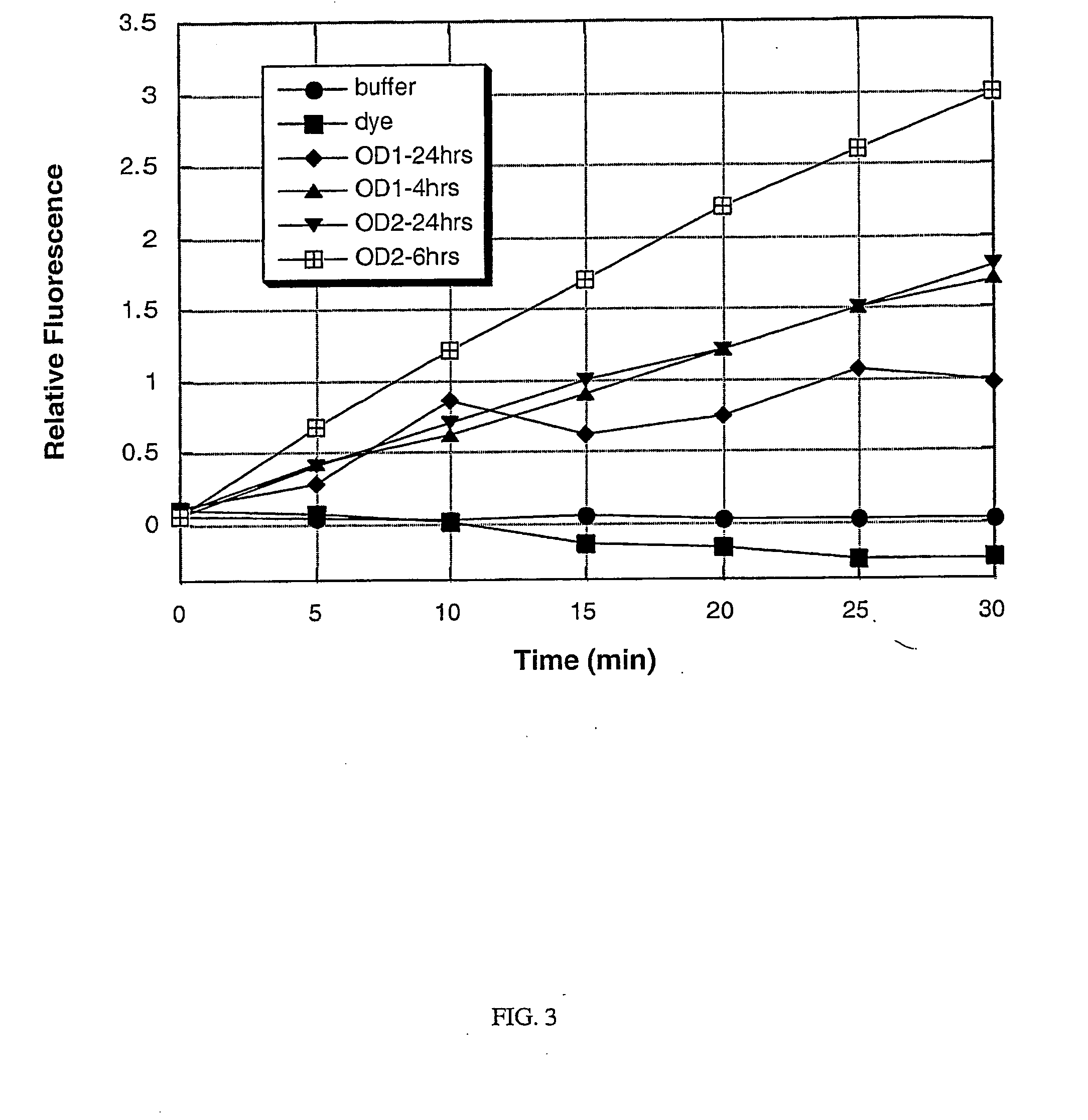
[0104] A Serratia marcescens specific protease was identified based on a homology search using the sequence of a known sspB protease found in the Staphylococcus species. This protease has homology to the cysteine protease precursor (sspB) protein of Staphylococcus aureus. The corresponding Serratia protease has not been previously characterized. To test for the presence of the specific protease in a bacterial culture, a s...


PUM

PropertyMeasurementUnit
ODaaaaaaaaaa
ODaaaaaaaaaa
pHaaaaaaaaaa

Abstract

Described herein are methods of detecting a wound infection and for detecting the presence or absence of microorganisms, for example, wound pathogens in a sample, by contacting a sample with an enzyme produced and/or secreted by the bacteria, and detecting modification or the absence of modification of the substrate, as an indicator of the presence or absence of the enzyme in the sample. The present invention also features a biosensor for detecting the presence or absence of bacteria in a sample.

Description

RELATED APPLICATIONS [0001] This application relates to U.S. Provisional Application No. 60 / 383,847 filed May 28, 2002 and 60 / 354,001 filed Jan. 31, 2002. The entire teachings of the above applications are incorporated herein by reference.BACKGROUND OF THE INVENTION [0002] Infection of wounds is a major source of healthcare expenditure in the United States. Approximately 5% of all surgical wounds become infected with microorganisms, and that figure is considerably higher (10-20%) for patients undergoing abdominal surgery. Bacterial species, such as Staphylococci are the most frequently isolated organisms from infected wounds. This is probably because humans are the natural reservoir for staphylococci in the environment, with up to 50% of the population colonized at any given time. Colonization rates are significantly higher in the hospital setting, both among healthcare workers, and among patients. Moreover, the colonizing organisms in the hospital environment are likely to be resis...

Claims


Application Information

Patent Timeline
Patent Type & Authority Applications(United States)
IPC IPC(8): C12M1/34C12Q1/04C12Q1/25C12Q1/34C12Q1/37C12Q1/54C12R1/445G01N33/554G01N33/569
CPCC12Q1/04C12Q1/37C12Q1/54C12Q1/44C12Q1/14C12Q1/34C12Q1/10
Inventor SANDERS, MITCHELLLOWE, ADRIANHAMILTON, MAUREENCOLPAS, GERARD
Owner WOUNDCHEK LAB US
Why Eureka
Try Eureka