Bodily lumen closure apparatus and method

a body lumen and tube technology, applied in the field of medical devices, can solve the problems of increased risk of post-operative infection, patient pain, and substantial blood loss, and achieve the effects of facilitating the rolling of the sheet, preventing blood leakage, and superior remodeling properties

Active Publication Date: 2005-07-21
OREGON HEALTH & SCI UNIV +2
View PDF83 Cites 143 Cited by
  • Summary
  • Abstract
  • Description
  • Claims
  • Application Information

AI Technical Summary

Benefits of technology

[0014] In another aspect of the invention, the distal end of the hemostatic member includes a plurality of slits, such as two slits dividing the hemostatic member lengthwise into quarters and which extend for about 25-30% of its length. Slitting the distal portion of hemostatic member allows the distal end to expand outward to facilitate the sealing process.
[0016] In yet another aspect of the invention, the hemostatic member delivery apparatus can be adapted to introduce the hemostatic member into an aneurysm to prevent leakage around a stent graft. In one embodiment, the stent graft includes an open section through which an outer delivery catheter could be introduced that would provide a means to deliver the, hemostatic members to the aneurysm after the stent graft had been placed. Afterward, another section of the stent graft would be introduced through the original stent graft and positioned over the open section. A second option would be to include a sleeve or other type of valve in the graft material through which the delivery system could be introduced. The valve would then close to prevent leakage of blood. One example of a hemostatic closure member delivery system for treatment of an aneurysm would comprise a series of hemostatic members placed adjacently over a wire guide and loaded into an outer sheath member, such as an introducer sheath or delivery catheter, typically with the assistance of a pusher member. A second method involves loading one or more closure members into a loading cartridge which is inserted into the delivery catheter, whereby a pusher member urges them into the catheter's passageway for final deployment. In one method of deployment, the pusher member individually deploys the hemostatic member or members loaded in the catheter. This procedure is repeated with additional closure members until the aneurysm is filled. Alternatively, the closure member(s) can be expelled from the delivery catheter by infusing saline or another fluid via a syringe through a side port in the delivery system. This method advantageously pre-hydrates the closure member prior to it reaching the deployment site in the body.
[0017] In another aspect of the invention, the hemostatic member and delivery system is adapted for delivery into an aneurysm, such as an abdominal aortic aneurysm, such that the delivery catheter is positioned outside of the graft prosthesis, between the graft and the vessel wall. The graft prosthesis is then deployed, leaving the catheter tip inside the excluded aneurysm. This placement method takes advantage of the fact that the technique is already well known for placement of contrast media infusion catheters in this manner. Conveniently, the same catheter for infusion of contrast can be used for the delivery of the hemostatic members. Another advantage is that the graft prosthesis need not be modified to provide temporary access into the aneurysm so that the catheter, which would likely be the case if the hemostatic members are td be delivered from the inside of the graft prosthesis.
[0018] In yet another aspect of the invention, the closure member construct comprises a plurality of layers of the second sheet material, such as a single-layer SIS or another biomaterial, preferably lyophilized, that are configured to readily separate in the presence of bodily fluid. The separating layers create a large amount of surface area to absorb fluid and fill a body lumen or cavity when deployed singularly or as a multiplicity of closure members. SIS or other ECM sheet material typically has superior remodeling properties over sponge material, which is subject to additional processing steps, such as cross-linking and comminution that may degrade the matrix structure and growth factors therein and thus, represents a construct that may be more advantageous in clinical situation where rapid swelling is less important than the need to stimulate remodeling by native tissue within a body lumen, such as when attempting the exclusion of an aneurysm.
[0019] In a first embodiment, the layered sheet closure member is formed by rolling one of more sheets of SIS to form a tightly rolled configuration that optionally, but not necessarily, includes a functional pathway or lumen extending therethrough. In a second embodiment, the closure member is created from a stack of individual sheets that are either cut into a tubular construct or a squared / rectangular shaped construct that are preferably unattached to one another or partially laminated such that they readily separate when exposed to fluid. In a third embodiment, one or more sheets of material are rolled around a somewhat rigid or semi-rigid core member that in one preferred embodiment, comprises two narrow strips of SIS that are tightly intertwined or braided into an elongate member that is air-dried. The elongate twisted member acts as a mandril that facilitates the rolling of the sheet to form the construct and is absorbed or replaced with adjacent tissue along with the sheet layers following deployment in the body. To facilitate rapid separation of layers of material in each of the above embodiments, a closure member may include one or more longitudinal splits extending the length of the construct. The closure member can be formed by rolling or stacking hydrated ECM sheets, then lyophilizing them, which may result in some degree of lamination between layers of material. Alternatively, the sheet or sheets may be lyophilized first, then rolled or stack to produce a looser construct than may facilitate entry of fluid between the layers of material. A less dense ECM material, such as stomach mucosa, can also represent an alternative larger-cell material that when lyophilized has superior swelling properties similar to sponge material.

Problems solved by technology

Open surgical procedures which require incisions through skin, tissue, and organs have a traumatic effect on the body and can lead to substantial blood loss.
In addition, such procedures expose tissue and organs to the outside environment which creates an increased risk of post-operative infection.
After open surgical procedures, patients are generally in pain, require substantial recovery time, and are susceptible to post-operative complications.
As a result, open surgical procedures are generally higher in cost and have a higher degree of risk.
One of the important benefits of minimally invasive intravascular procedures is less patient blood loss; however, particularly in procedures in which the femoral artery is accessed, achieving quick and effective hemostasis at the puncture site still can be problematic.
More recently, the increased use of heparin and larger sized introducer sheaths have presented additional challenges.
Even after hemostasis, the site remains vulnerable to further bleeding, especially if the patient is moved.
In addition, the complexity of the device has led to reports of failures such as in the ability to form a proper knot and other problems.
Another known complication is when the device is deployed such that the needles penetrate completely through the opposite wall of the target vessel, which can inadvertently lead to the vessel being closed off, a potentially serious event for the patient.
Re-puncture at the site can typically occur at that time at the site, but may be problematic if the anchor device has not been reabsorbed.
Additionally both closure devices, being made of bovine collagen, can cause the formation of fibrotic tissue in some patients, which in severe cases, has been known to be sufficient to restrict blood flow within the vessel.
Even suturing devices are intended for closing puncture sites in the small to moderate range, although some physicians have reportedly been able to perform an additional series of steps to suture larger arterial puncture sites, adding to the time and complexity of the procedure.
Both of these procedures, however, are invasive, are not generally performed in the doctor's office, and can be expensive.
The fallopian tube occlusion device of Nikolchev et al. is complicated requiring the metallic coil to be pre-formed into a flower shape which must have a diameter larger than the interior of the fallopian tube, or the device will not lodge in the fallopian tube.

Method used


Image

  • Bodily lumen closure apparatus and method
  • Bodily lumen closure apparatus and method
  • Bodily lumen closure apparatus and method

Examples


Embodiment Construction

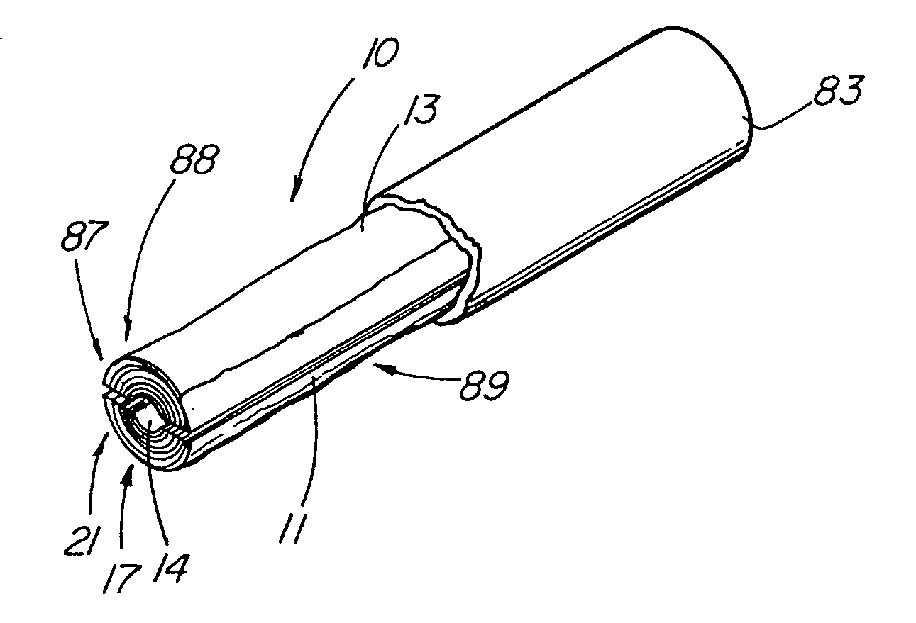
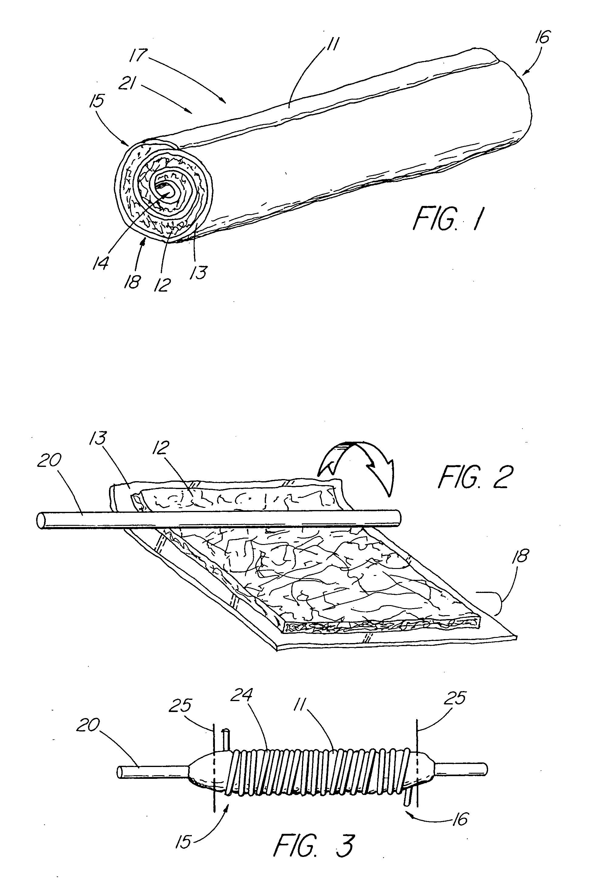
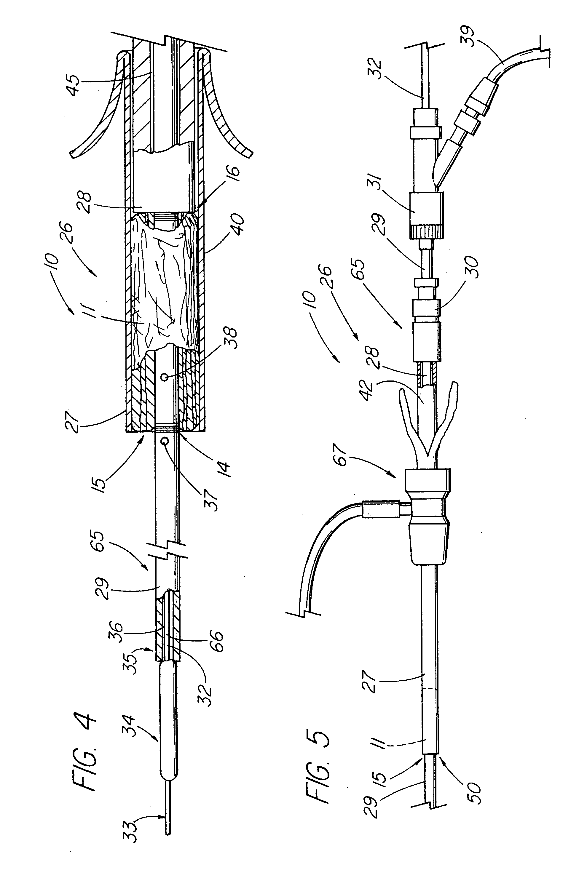
[0046] In one embodiment of the present invention, depicted in FIGS. 1-16, the closure member is a hemostatic member 11 which is delivered to a treatment site within the body of a patient to provide an external hemostatic seal or intravascular occlusion to prevent blood flow, such as from a blood vessel 48 punctured during a procedure using an introducer sheath 27 to gain access of a patient's artery or vein, or to fill an aneurysm 58, especially where a stent graft 57 has been placed. The hemostatic member 11 comprises a construct that is able to absorb blood and swell in diameter, yet has sufficient structural integrity in its expanded state to exert a gentle expansile force that provides a more effective seal for achieving hemostasis than collagenous foam alone, particularly in larger puncture channels (above 8 Fr). The illustrative hemostatic member 11, depicted in FIG. 1, includes a rolled configuration 17 comprising a layer 18 of two materials formed by rolling together a firs...


PUM


Abstract

An absorbable and expandable closure member used to occlude or exclude a body lumen or cavity, such as a blood vessel, fallopian tube, duct, aneurysmal sac, etc., comprising a closure member comprising one of more sheets of a biomaterial that are rolled, stacked, or folded to form a multilayer construct of a generally cylindrical configuration for deployment through a delivery system, either as a singularly or part of a multiplicity of closure members. The biomaterial is derived from a source material, such as small intestinal submucosa or another remodelable material (e.g., an extracellular matrix) having properties for stimulating ingrowth of adjacent tissue into the biomaterial deployed within the bodily lumen. The closure member is deployed to the bodily lumen from a delivery sheath, cartridge, and / or over a inner guiding member, such as a wire guide or catheter.

Description

CROSS-REFERENCE TO RELATED APPLICATIONS [0001] This application claims priority of provisional application Ser. No. 60 / 470,611, filed May 15, 2003, and is a continuation-in-part of co-pending U.S. patent application Ser. No. 10 / 206,480, filed Jul. 26, 2002, which claims priority to provisional application Ser. No. 60 / 307,893, filed Jul. 26, 2001.TECHNICAL FIELD [0002] This invention relates to medical devices, more particularly to vessel closure members, delivery apparatuses, and methods of inserting the closure members. BACKGROUND OF THE INVENTION [0003] Open surgical procedures which require incisions through skin, tissue, and organs have a traumatic effect on the body and can lead to substantial blood loss. In addition, such procedures expose tissue and organs to the outside environment which creates an increased risk of post-operative infection. After open surgical procedures, patients are generally in pain, require substantial recovery time, and are susceptible to post-operativ...

Claims


Application Information

Patent Timeline
Patent Type & Authority Applications(United States)
IPC IPC(8): A61B17/00A61B17/12A61F2/06A61F6/22A61F6/24
CPCA61B17/00234A61F2230/005A61B17/12022A61B17/12109A61B17/12113A61B17/12118A61B17/1219A61B2017/00004A61B2017/00637A61B2017/00641A61B2017/00654A61B2017/00898A61F2/07A61F6/225A61F6/24A61F2002/061A61F2002/065A61F2002/077A61F2/89A61B17/0057
Inventor PAVCNIK, DUSANKAUFMAN, JOHNFETTE, CLAY D.
Owner OREGON HEALTH & SCI UNIV
Why Eureka
Try Eureka