Method for detecting artifacts in data

Active Publication Date: 2005-07-28
ABBOTT DIABETES CARE INC
View PDF19 Cites 96 Cited by
  • Summary
  • Abstract
  • Description
  • Claims
  • Application Information

AI Technical Summary

Benefits of technology

[0021] The invention provides a safeguard against inclusion of erroneous data resulting from an inadvertent movement of a body part with respect to a measuring probe in a non-invasive optical measurement. Inclusion of erroneous data in the subsequent calculation of the concentration of a me

Problems solved by technology

Failure to maintain proper contact between the probe and the surface of the tissue can result in artificial perturbations (artifacts) in some or all of the recorded data.
An artifact can result in an erroneous calculation of the concentration of the analyte because the artifact distorts the actual trend of the intensity of the radiation emitted over a period of time.
Contact between the apparatus and the tissue can be compromised by improper application of the probe to the surface of the tissue, by accidental movement of the probe or the tissue during the measurement, and perhaps by any substances on the surface of the tissue that interfere with the collection of radiation emitted.
Most of these artifacts were assumed to result from human error by the patient or by the clinician and cannot be totally prevented.
It is believed that a primary cause of data artifacts is movement of the forearm during a meas

Method used


Image

  • Method for detecting artifacts in data
  • Method for detecting artifacts in data
  • Method for detecting artifacts in data

Examples


example 1

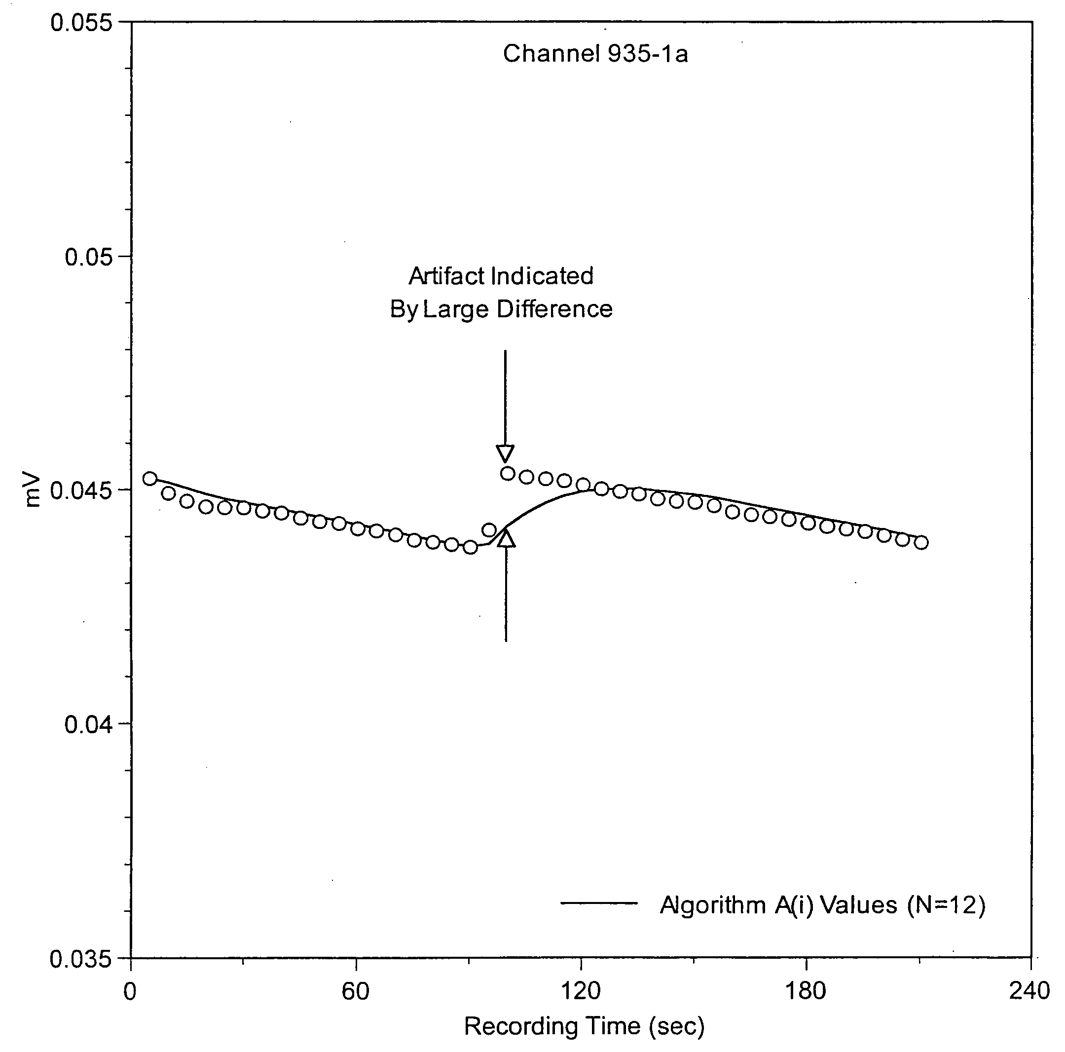
[0081]FIG. 8A depicts measurements of normalized optical data recorded on patient no. 11, test no. 17, in the first clinical trial from four (4) of the 32 channels. The values of A(i) closely followed the data beginning with the sixth (6th) point. The data were determined to be completely free of artifacts. FIG. 8B depicts the corresponding values of D(i), which represent the absolute difference between each value of A(i) and each value of the normalized data. The values of D(i) remained well below the threshold of 0.02, thereby confirming the absence of artifacts in these measurements.

example 2

[0082]FIG. 9A depicts measurements of normalized optical data recorded on patient no. 10, test no. 9, in the first clinical trial from four (4) of the 32 channels. The values of A(i) failed to closely follow the data at various locations between the sixth (6th) point and the end of the test. Data from all four (4) channels were determined to have artifacts at one or more locations. FIG. 9B depicts the corresponding algorithm values of D(i), all of which intermittently exceeded the threshold of 0.02, thereby confirming the presence of artifacts.

example 3

[0083]FIG. 10 summarizes the number of tests in the first clinical trial that contained data artifacts detected by the method of this invention. A total of 37 out of 400 tests were identified as having artifacts in some or all of the measurements taken in the 32 optical channels. Patient no. 19 experienced the highest incidence of artifacts, i.e., 6 artifacts out of 20 tests.


PUM


Abstract

A method for identifying artifacts occurring during a measurement of the concentration of an analyte in a biological sample by means of an apparatus that employs temperature-controlled optical probes, introduces electromagnetic radiation into tissue, and collects and detects radiation emitted at a distance from the point at which the electromagnetic radiation is introduced. The values of intensity of radiation emitted at different wavelengths, at different distances between the light introduction site(s) and the light collection site(s), and at different temperatures are collected and used in the method to generate a relationship between these values and the concentration of an analyte in the tissue or the disease state of a patient. The method involves the use of an algorithm that identifies artifacts in the data resulting from motion of the patient and allows the rejection of data sets that contain these artifacts. The algorithm identifies sudden changes in the magnitude and direction in a sequence of collected signals.

Description

BACKGROUND OF THE INVENTION [0001] 1. Field of the Invention [0002] This invention relates to a method for detecting artifacts in data, more particularly, data observed during physiological testing. [0003] 2. Discussion of the Art [0004] U.S. Pat. No. 6,526,298 discloses an apparatus and method for measuring the concentration of an analyte (such as glucose) within a biological tissue. The method involves the steps of controlling the temperature of the tissue, introducing electromagnetic radiation into the tissue, and measuring the intensity of the electromagnetic radiation emitted from the surface of the tissue. The period of contact between the apparatus and the tissue lasts several minutes. Optical data from a plurality of channels of the apparatus are recorded at regular time intervals during each measurement. See FIGS. 1A, 1B, 1C, and 1D, which show various trends of data. The optical data, which are converted to voltage levels by a photodetector, are used to determine the conce...

Claims


Application Information

Patent Timeline
IPC IPC(8): A61B5/00G01N21/35G01N21/47G01N21/49G01N33/487
CPCA61B5/14532A61B5/14535G01N21/49A61B5/7207A61B5/1455
Inventor LOWERY, MICHAEL G.SHAIN, ERIC B.KHALIL, OMAR S.
Owner ABBOTT DIABETES CARE INC
Why Eureka
Try Eureka