
Medical devices having MRI-enhancing encapsulated fluids

Inactive Publication Date: 2005-08-04
PHANTOMS BY DESIGN
View PDF18 Cites 24 Cited by
  • Summary
  • Abstract
  • Description
  • Claims
  • Application Information

AI Technical Summary

Benefits of technology

[0019] In view of the foregoing, it is an object of the present invention to provide medical devices, including interventional devices, that comprise materials that enhance visibility under MRI, which are biocompatible, and that are sufficiently robust to withstand the manufacturing processes typically encountered in device construction.
[0020] It is another object of this invention to provide materials that may be incorporated into medical devices, including interventional devices, with enhanced visibility under MRI, but which do not substantially impact the mechanical properties of the devices.
[0021] It also is an object of the present invention to provide materials that may be incorporated into medical devices, such as interventional devices and contrast agents, with enhanced visibility under MRI, wherein the materials comprise fluid encapsulated in thin, environmentally robust shells capable of withstanding manufacturing processes employed with medical grade polymers and environmental conditions encountered in the human body.
[0022] It is a further object of this invention to provide a stable, biocompatible contrast additive for use with medical grade polymers that allows medical devices, such as catheters, to be directly visualized under clinical MRI conditions.
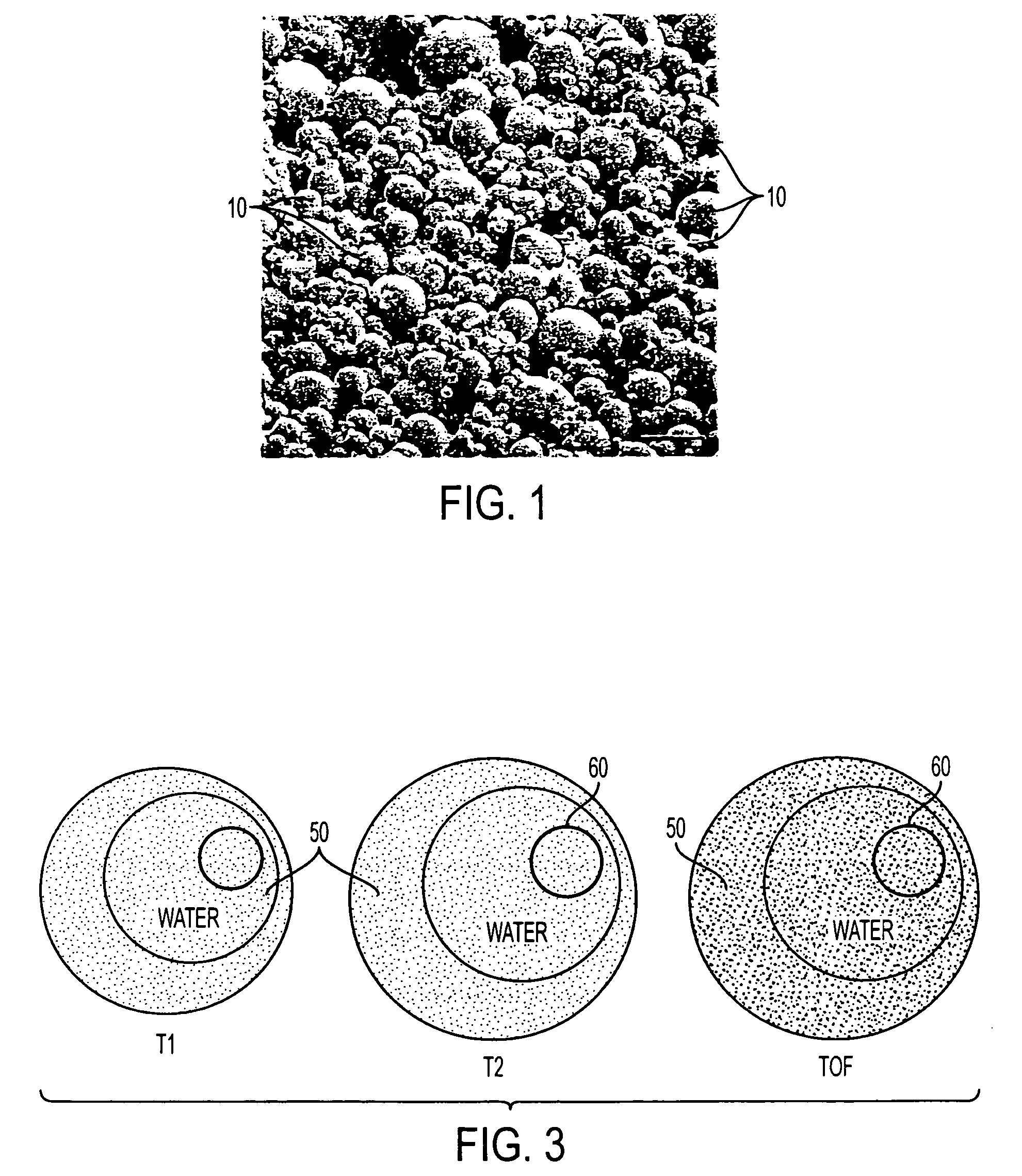
[0024] In one embodiment of the present invention, the microcapsules comprise thin, environmentally robust shells formed by coacervation that contain lipids or oils, such as mineral oil, cod liver oil, terpene or polyunsaturated fatty acids. The fluids contained within the microcapsules provide a source of free protons to enhance visibility under MRI, while the shells of the microcapsules physically isolate the fluid from the polymer matrix in which the microcapsules are incorporated. According to some embodiments, the device body may further comprise relaxation modifying agents, such as iron oxide particles or anchored gadolinium based T1 reducing agents, that limit fluid mobility.
[0025] Further in accordance with the principles of the present invention, neither the shells nor cores of the microcapsules have air inclusions that could reduce the population of available protons and / or produce susceptibility artifacts in the MR images. In addition, the shells of the microcapsules preferably comprise materials that provide stability, and the ability to withstand prolonged exposure to organic solvents, high temperatures and high pressures.

Problems solved by technology

Cardiovascular disease is currently one of the world's biggest health care issues, and it is the leading cause of death in many countries including the United States.
Despite the promise offered by MRI, there exist technical challenges to successful widespread adoption of this technique.
For example, MRI is a relatively slow imaging technique, and diagnostic procedures suffer from image artifacts caused by tissue motion occurring during the extended periods required for image formation.
Moreover, most interventional devices are not compatible with MRI, either because the devices incorporate metal components, or are manufactured from polymers that are not visible in magnetic resonance images.
While it would be desirable to incorporate fluids into the polymer matrix of interventional devices to enhance visibility, the presence of fluids may either inhibit polymerization of the polymer matrix of the interventional device, or lead to materials having poor mechanical properties.
A drawback of such devices, however, is that the mechanical properties of the catheters are expected to vary from catheters manufactured from more conventional materials, thus hindering adoption of MRI techniques.
While such contrast agents of the foregoing patents may be safe and effective for introduction into a patient's circulatory system, it is expected that such contrast agents could not withstand the manufacturing processes needed to incorporate into interventional devices to enhance the visibility of such devices under MRI.
However, these nanoparticles remain in the patient's blood pool for extended periods of time.
However, most existing microparticle contrast agents are not directly visible in MRI and suffer from poor signal strength and tissue contrast.
Additionally, most microparticle contrast agents are not compatible with commonly-used medical grade polymers and solvents.

Method used


Image

  • Medical devices having MRI-enhancing encapsulated fluids
  • Medical devices having MRI-enhancing encapsulated fluids
  • Medical devices having MRI-enhancing encapsulated fluids

Examples


Embodiment Construction

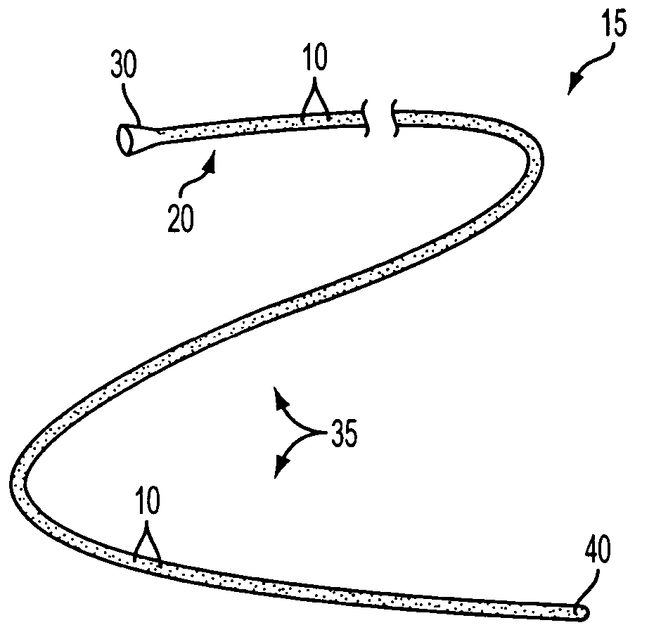
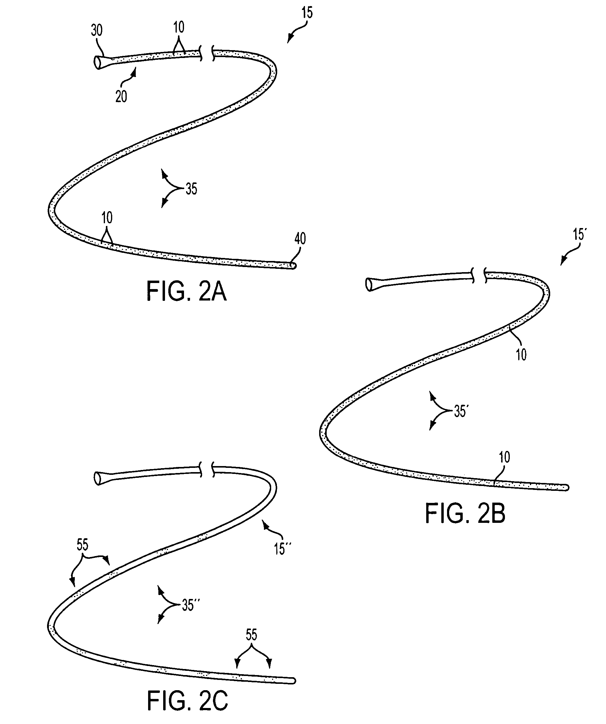
[0030] The present invention is directed to medical devices having enhanced visibility under MRI, wherein microcapsules containing MRI contrast additives are dispersed in at least a portion of the device comprising medical grade polymer matrices. The microcapsules allow the construction of MRI compliant medical devices that exhibit clinically relevant MRI visibility while retaining favorable mechanical and manufacturing properties. The present invention may be especially advantageous for construction of diagnostic and therapeutic interventional devices, such as catheters. The contrast additive of the present invention is compatible with materials and manufacturing methods currently used to make medical devices, is patient-safe, and produces clinically acceptable visibility independent of viewing angle or pulse sequence.

[0031] While the present invention is illustratively described in the context of catheters suitable for use in MRI-guided diagnostic or treatment procedures, it will...


PUM


Abstract

A medical device is described that comprises a polymeric component and a microcapsule additive including a fluid that enhances direct visibility during magnetic resonance imaging of at least a predetermined portion of the polymeric component.

Description

FIELD OF THE INVENTION [0001] The present invention relates to medical devices and, more particularly, to interventional medical devices that include microencapsulated fluids to enhance MRI visibility. BACKGROUND OF THE INVENTION [0002] Cardiovascular disease is currently one of the world's biggest health care issues, and it is the leading cause of death in many countries including the United States. Despite a variety of clinical presentations, the majority of cardiovascular diseases are related to atherosclerosis. Emerging research has identified pathogenic attributes associated with disease progression. Some of these attributes include subtle vessel wall features linked to unstable plaque as well as microvascular dysfunction linked to angina and ischemia. [0003] New techniques offer the promise of early detection with minimally invasive treatment prior to acute clinical syndromes, arterial occlusions and infarction. To maximize clinical utility, diagnosis, risk stratification, it ...

Claims


Application Information

Patent Timeline
IPC IPC(8): G01R33/28
CPCG01R33/286
Inventor BURKE, THOMAS M.
Owner PHANTOMS BY DESIGN
Learn More