Method and apparatus for registration, verification, and referencing of internal organs

a technology of internal organs and methods, applied in the field of methods and apparatus for registration, verification, and referencing of internal organs, can solve the problems of reducing accuracy, electromagnetic tracking devices are subject to interference from ferromagnetic materials and conductors, and optical tracking devices suffer from line-of-site constraints

Inactive Publication Date: 2005-08-18
PHILIPS ELECTRONICS LTD
View PDF100 Cites 459 Cited by
  • Summary
  • Abstract
  • Description
  • Claims
  • Application Information

AI Technical Summary

Benefits of technology

[0058] In some embodiments, the integrated system may include a referencing device. In some embodiments data may be sent and received between the referencing device and computer element. The referencing device may, inter alia, aid in providing image data, location data, position data, coordinate data, and / or motion data regarding an anatomical region of the patient. The referencing device may otherwise enable dynamic referencing of an anatomical region of a patient, (including soft tissues and / or deformable bodies).

Problems solved by technology

However, these optical tracking devices suffer from line-of-site constraints, among other things.
One disadvantage, however, is that electromagnetic tracking devices are subject to interference from ferromagnetic materials and conductors.
This interference may degrade accuracy when such ferromagnetic materials or conductors are placed in the proximity of position indicating elements or EM tracking devices.
There are many difficulties and problems in image guided surgery and the prior techniques.
Paired-point registration is less-attractive when the anatomical object is either inaccessible, non-rigid, or both.
Registration and referencing of non-rigid and / or moving organs such as the liver, gall bladder, stomach, pancreas, kidney, lung, colon, heart, prostate gland, etc. is a difficult task.
Furthermore, it is difficult to attach any kind of dynamic reference to a soft moving object.
Techniques using ultrasound are complicated by different sound velocities and attenuation from different tissues.
However, problems still exist since multiple dynamic references must be temporarily fixed to a deformable anatomical object.
Another limitation of current dynamic referencing techniques stems from the use of reference sensors.
In the event that they are attached some distance from the site of intervention / procedure, or attached to a non-rigid object, the motion of the dynamic reference may not accurately track the motion at the site of intervention / procedure.
Another limitation of current image guided surgery techniques may include the difficulty of verifying that a registration has been performed correctly.
This technique suffers from the same issues as registration itself in soft tissue, e.g., deformation of soft tissue during verification, access to the tissue, paucity of verification landmarks, and other problems.
A further limitation of current approaches is the amount of fluoroscopy that must be used to correctly position the therapy device in the event that image guided surgery is not used.
These land other problems exist.

Method used


Image

  • Method and apparatus for registration, verification, and referencing of internal organs
  • Method and apparatus for registration, verification, and referencing of internal organs
  • Method and apparatus for registration, verification, and referencing of internal organs

Examples


Embodiment Construction

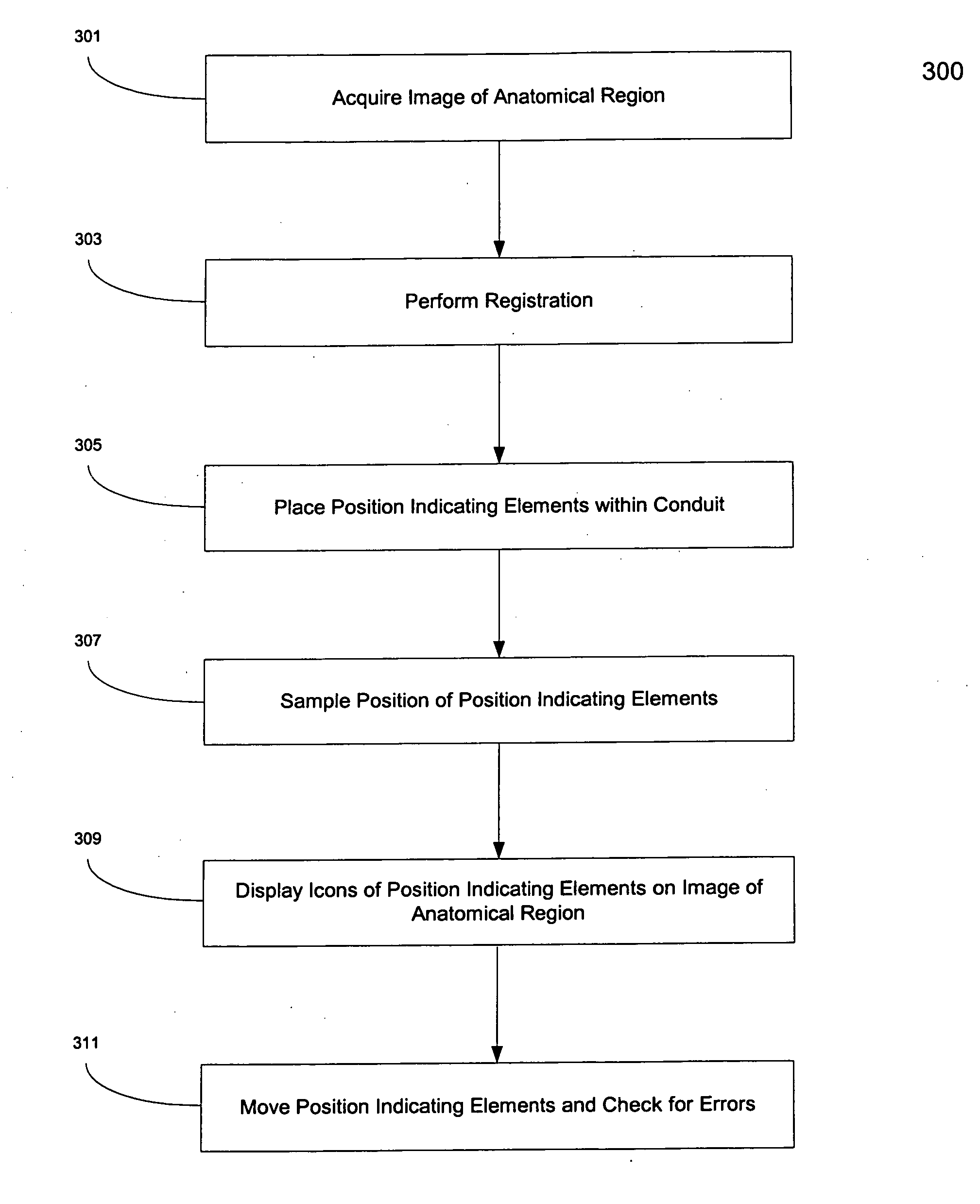
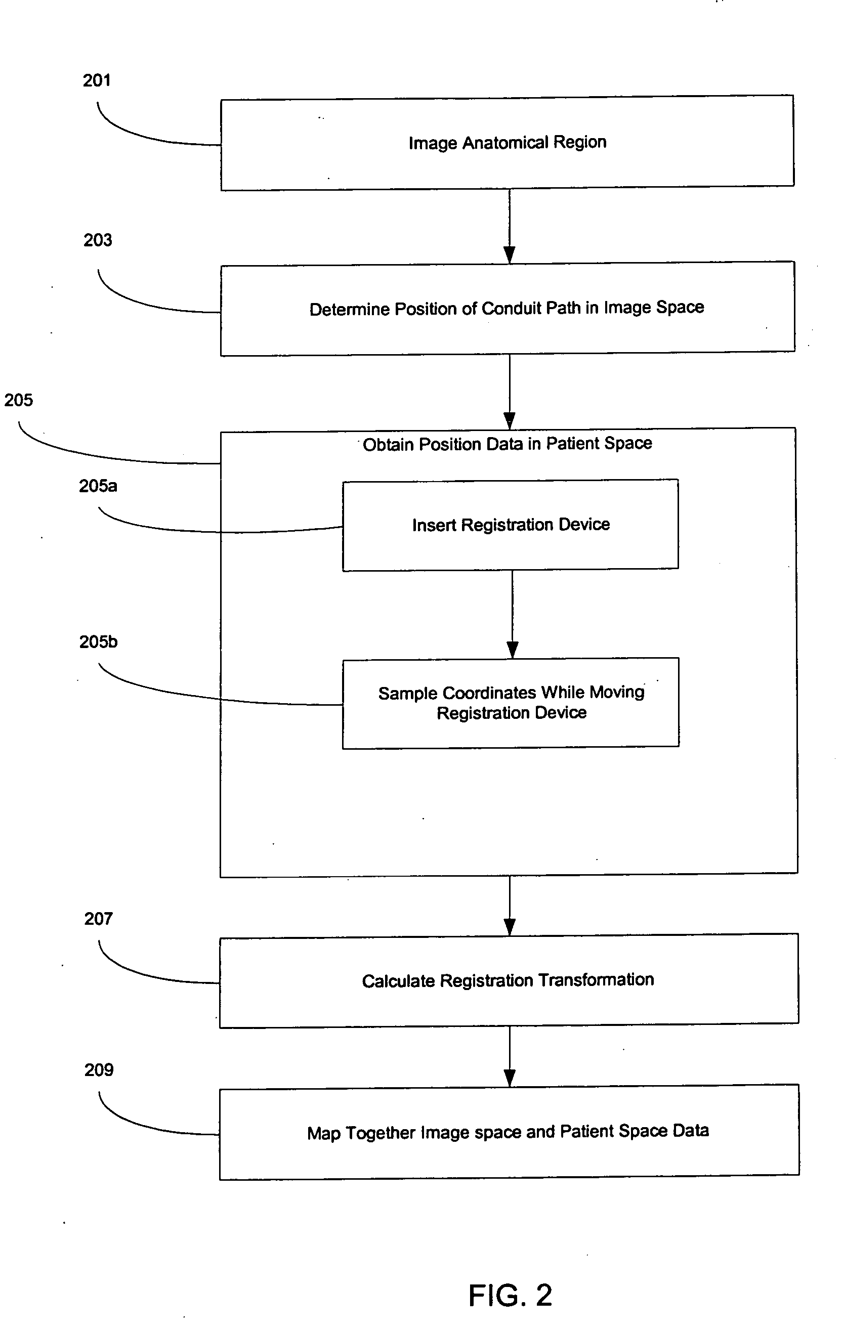
[0081] The invention provides systems and methods for registration of an anatomical region of a patient, verification of the registration of the anatomical region, and dynamic referencing of the anatomical region, wherein the anatomical region may include soft tissue and / or deformable bodies.

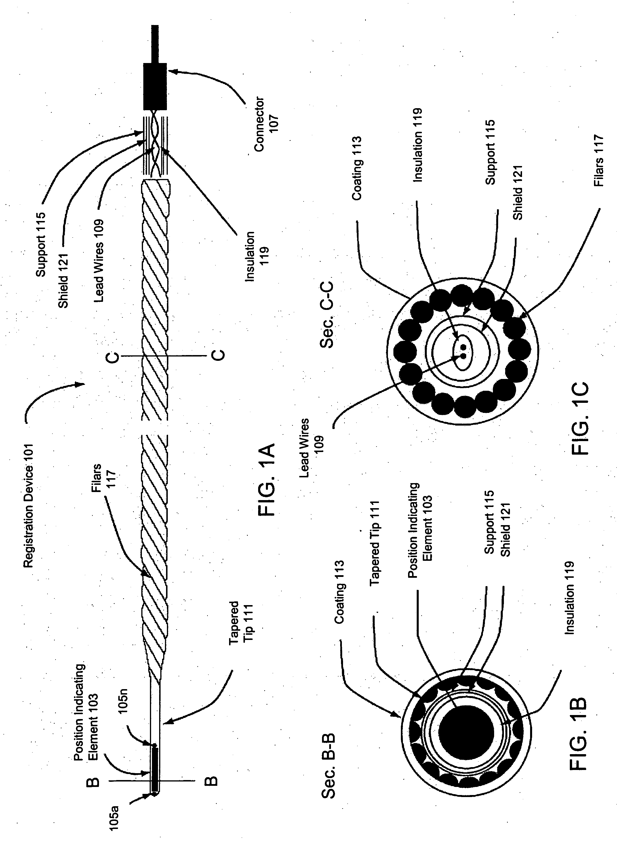
[0082] In one embodiment, the invention may use a conduit within an anatomical region of a patient to, inter alia, aid in providing image information and position information from within the anatomical region. This conduit may supply sufficient coordinate information regarding the anatomical region to be used for registration of the anatomical region. For example, a coronary artery surrounding the heart may provide sufficient topographical coordinate information regarding the heart to be used as a conduit for registration by a method of the invention.

[0083] In one embodiment, a conduit as used herein may include a naturally existing conduit within the anatomical region such as, for example, an...


PUM


Abstract

Systems and methods for registering, verifying, dynamically referencing, and navigating an anatomical region of interest of a patient are provided. In one embodiment, the anatomical region of interest is imaged using an imaging device such as, for example, an x-ray device. A tracked registration device may then be removably inserted in a conduit within the anatomical region and the position of the registration device may be sampled by a tracking device as the registration device is moved within the anatomical region through the catheter. The sampled position data is registered to the image data to register the path of the conduit to the anatomical region of interest. The same or a similar device may be used to dynamically reference the movements affecting the anatomical region and modify the registration in real time. The registration may also be verified.

Description

RELATED APPLICATIONS [0001] This application claims priority to U.S. Provisional Patent Application Ser. No. 60 / 544,344, filed Feb. 17, 2004; U.S. Provisional Patent Application Ser. No. 60 / 605,139 filed Aug. 30, 2004; U.S. Provisional Patent Application Ser. No. 60 / 626,422, filed Nov. 10, 2004; and to U.S. Provisional Patent Application Ser. No 60 / 626,488, filed Nov. 10, 2004, each of which are incorporated herein by reference in their entirety.FIELD OF THE INVENTION [0002] This invention relates to methods and devices for registering an anatomical region with images of the anatomical region, verifying registration of an anatomical region, and dynamically referencing the anatomical region. BACKGROUND OF THE INVENTION [0003] Image Guided Surgery (IGS), also known as “frameless stereotaxy” has been used for many years to precisely locate and position therapeutic or medical measurement devices in the human body. Proper localization including position and orientation of these devices i...

Claims


Application Information

Patent Timeline
Patent Type & Authority Applications(United States)
IPC IPC(8): A61B5/03A61B5/042A61B5/05A61B5/06A61B6/12A61B8/08A61B19/00
CPCA61B5/03A61M25/0105A61B5/061A61B5/418A61B5/7285A61B6/12A61B6/5247A61B6/541A61B8/0833A61B8/5238A61B19/5244A61B2019/5236A61B2019/5238A61B2019/5251A61B2019/5255A61B2019/5289A61B2562/222A61B5/042A61B8/4245A61B8/4263A61B2034/2055A61B34/20A61B2090/364A61B2090/376A61B2090/374A61B2034/2051A61B5/283A61B2034/107A61B2034/2061A61B2090/3925A61B2090/3954A61B2090/3966A61B5/02A61B5/031A61B5/062A61B5/066A61B5/08A61B5/12A61B5/20A61B5/42A61B5/43A61B5/7221A61B6/032A61B6/037A61B8/0841A61B8/12A61B8/481A61B8/5261A61M25/0108A61M25/0127
Inventor GLOSSOP, NEIL DAVID
Owner PHILIPS ELECTRONICS LTD
Why Eureka
Try Eureka