Method for inhibiting fibrogenesis by a mixture of natural peptides from porcine liver extract
a technology of natural peptides and liver extract, which is applied in the field of liver fibrogenesis, can solve the problems of ineffective methods for reversing treatment, inability to be used, and inability to achieve the effects of reducing the pathologic changes of fibrogenesis, inhibiting the proliferation of fibroblasts, and good safety profil
- Summary
- Abstract
- Description
- Claims
- Application Information
AI Technical Summary
Benefits of technology
Problems solved by technology
Method used
Image
Examples
example 1
Inhibition of Rat Liver Fibrogenesis through Fibroscutum
Material and Methods
Animals and Experimental Design
[0066] All experiments were carried out using accepted ethical guidelines. Male Wistar rats (Beijing Medical University) weighing 200-250 g were used in this study. The animals had free access to food and drinking water. 40% CCl4 (Sigma) was prepared by mixing CCl4 with olive oil. Liver damage and fibrosis was induced through giving 40% CCL4 at 3 ml / kg body weight by subcutaneous injection, two times a week for four weeks. Control animals for CCL4 received only olive oil.
[0067] Fibroscutum was intraperitoneally administered daily at the dose of 1.25 ug / kg / d body weight.
[0068] Thus, 3 groups including each 7 animals could be distinguished. Group I: olive oil alone; group II: 40% CCL4; group III: 40% CCL4 plus Fibroscutum. Fibroscutum treatment was started at the third week and lasted for three weeks. In another series of experiments, animals were similarly treated by olive ...
example 2
Inhibition of Fibrogenesis by Fibroscutum Through Suppressing Activation of HSC Cells
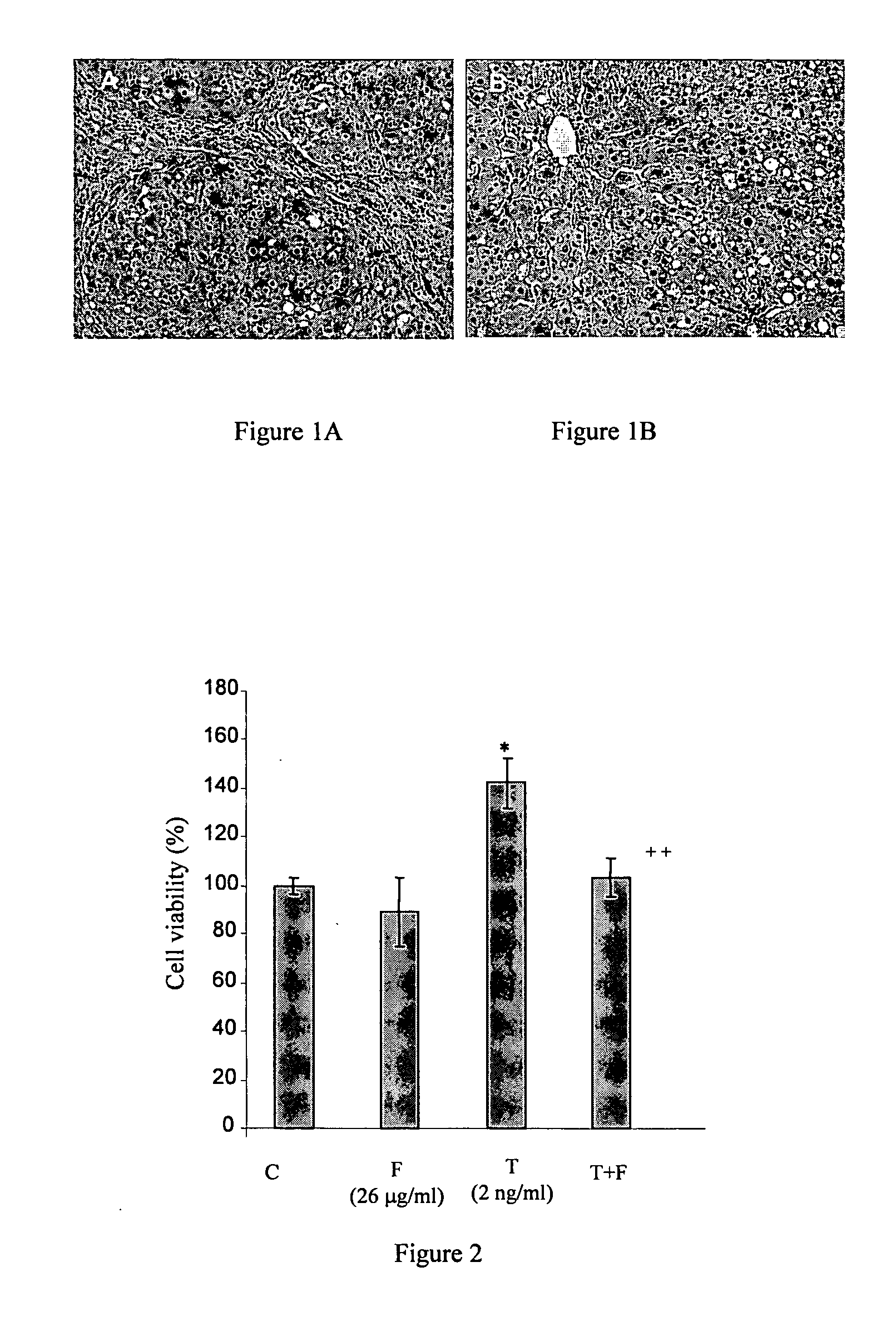
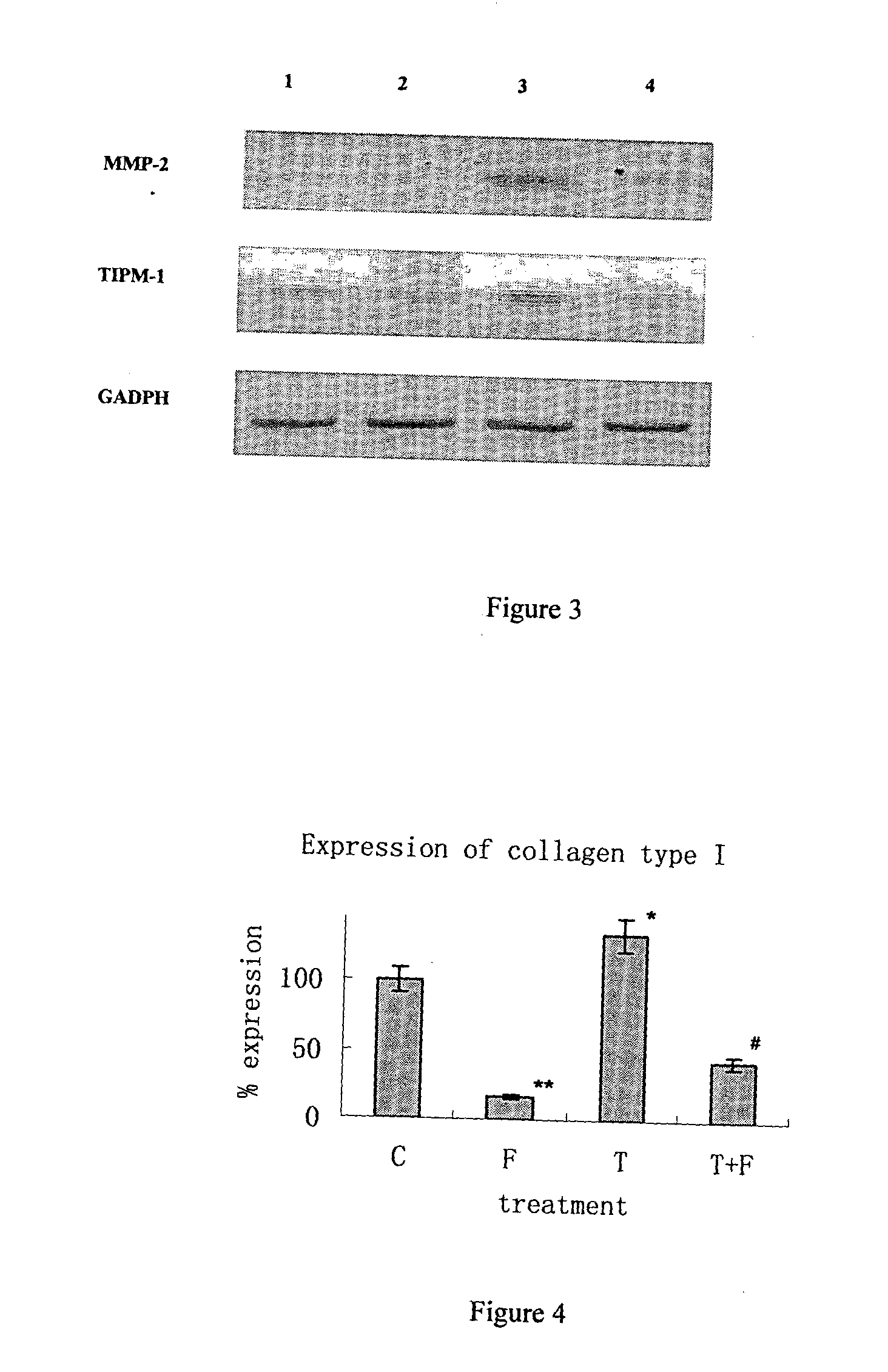
[0083] To elucidate the action of Fibroscutum in fibrogenesis, the effect of Fibroscutum on activation of HSC cells stimulated by TGF-β 1 was measured. HSC activation has been examined and is an essential process of liver fibrogenesis. It presents as proliferation of HSC and fibroblast, and remodeling of extra cellular matrix, including, degradation of collagen IV in basement membrane and deposition of an excess of extracellular matrix components, such as collagen I and collagen III, and change of expression of MMPs and TIMPs in mRNA level. TGF-β 1 is one of the most important activation factors in the process.
Material and Methods
Cells
[0084] An established Human HSC cell line was used, named LX-2. These cells have been extensively characterized and exhibit many similarities with primary cultures of HSC.
[0085] LX-2 cells (2000 / well) were seeded in 96-well microplate for ...
example 3
The Effect of Fibroscutum on Proliferation of NIH 3T3 Cells Stimulated by TGF.beta.1
[0092] Proliferation of fibroblasts is also involved in fibrogenesis. The effect of Fibroscutum on fibroblast proliferation was therefore examined.
Cells
[0093] NIH 3T3 fibroblasts were bought from ATCC.
Proliferation
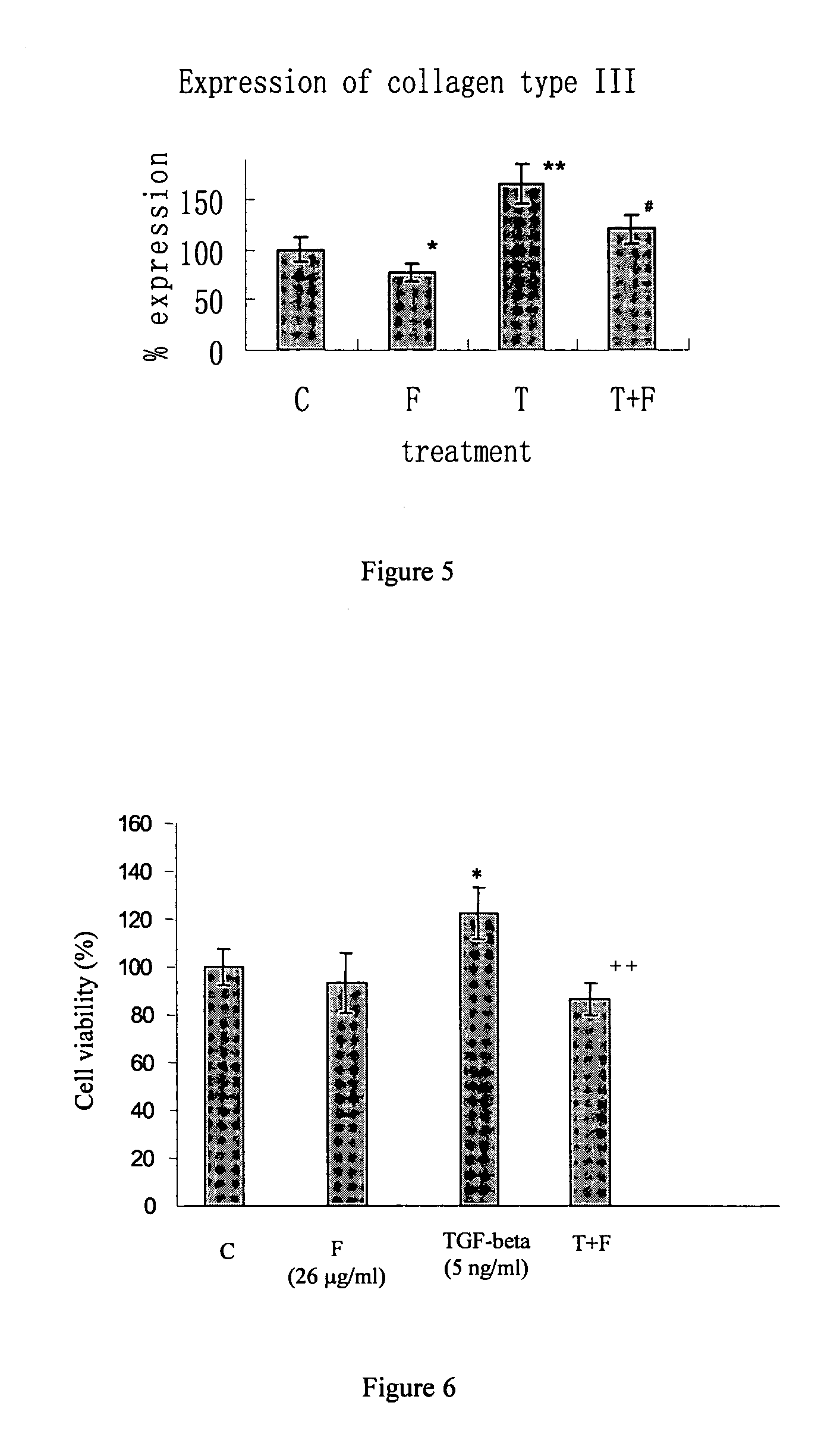
[0094] NIH 3T3 fibroblasts (2000 / well) were seeded in 96-well microplate for 24 hours. The culture medium was replaced with DMEM supplemented with 0.5% FBS and the cells were starved for 48 hours. After starvation, the medium was replaced with 2% serum supplemented DMEM medium. The reagents were then added to treat cells; and NIH 3T3 cells were exposed to Fibroscutum, either alone, or in the presence of 5 ng / ml TGF.beta.1 for 48 hours. Cell number was evaluated through the measurement of the reduction of the dye 3-(4,5-dimethylthiazol-2yl)-2,5 diphenyltetrazolium (MTT).
Results
[0095]FIG. 6 shows that TGF-β 1 increased the proliferation of NIH-3T3 fibroblast cells. However, the mitogen...
PUM
 Abstract
Description
Claims
Application Information
 - R&D
- Intellectual Property
- Life Sciences
- Materials
- Tech Scout
- Unparalleled Data Quality
- Higher Quality Content
- 60% Fewer Hallucinations
Browse by: Latest US Patents, China's latest patents, Technical Efficacy Thesaurus, Application Domain, Technology Topic, Popular Technical Reports.
© 2025 PatSnap. All rights reserved.Legal|Privacy policy|Modern Slavery Act Transparency Statement|Sitemap|About US| Contact US: help@patsnap.com


