
Method for reducing electronic artifacts in ultrasound imaging

Inactive Publication Date: 2005-10-27
ETHICON ENDO SURGERY INC
View PDF99 Cites 10 Cited by
  • Summary
  • Abstract
  • Description
  • Claims
  • Application Information

AI Technical Summary

Benefits of technology

[0007] The present invention provides a method for the reduction of artifacts in ultrasound imaging of anatomical tissue. The method begins with receiving at least two calibration signals of imaging ultrasound waves that have been reflected from different regions

Problems solved by technology

Ultrasound images frequently include electronic artifacts that appear as non-random noise in the image and that may obscure significant image information.
These artifacts are especially endemic to devices used for both imaging and medical treatment of tissue because of the conflicting design constraints of high power required for medical treatment and wide bandwidth required for imaging.
Other artifacts or undesirable signals include image degradation due to acoustic reflection caused by an intervening structure between the transducer and the tissue, such as a coupling balloon or even an air bubble.

Method used


Image

  • Method for reducing electronic artifacts in ultrasound imaging
  • Method for reducing electronic artifacts in ultrasound imaging
  • Method for reducing electronic artifacts in ultrasound imaging

Examples


Embodiment Construction

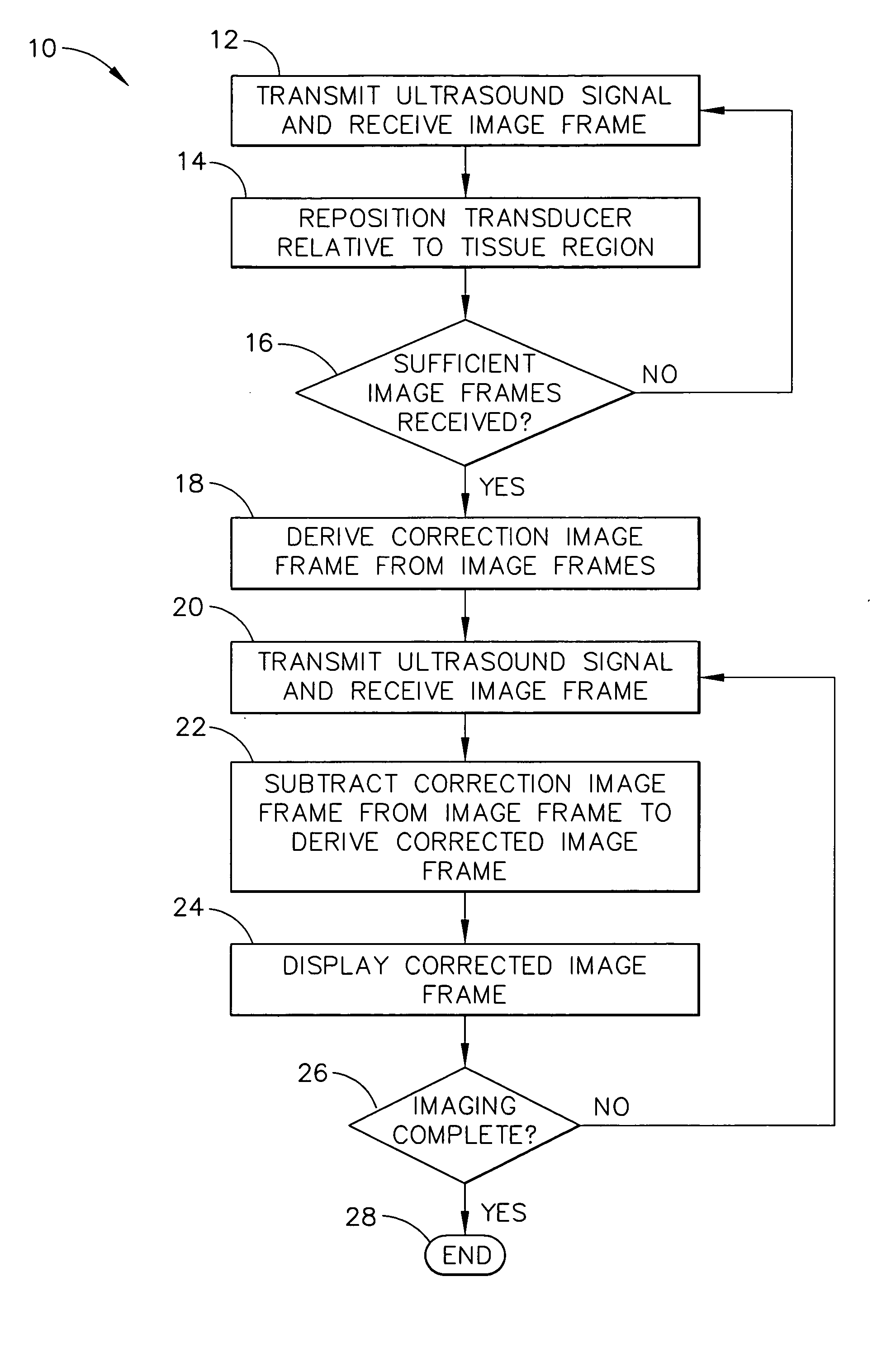
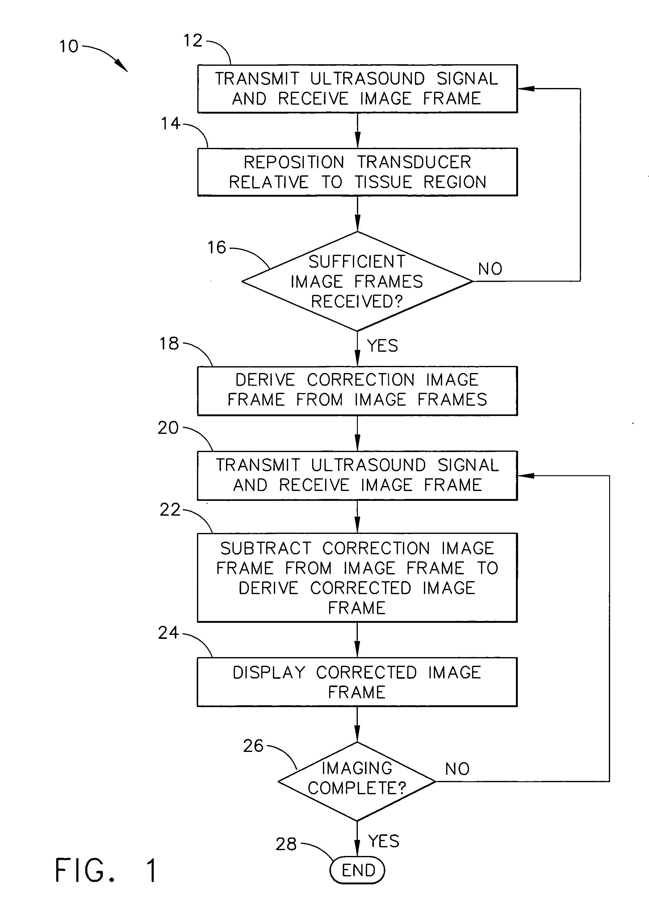
[0013] Referring now to the Figures, in which like numerals indicate like elements, FIG. 1 discloses an overview of an ultrasound imaging method with artifact reduction 10 according to an embodiment of the present invention. The method 10 begins at step 12 by transmitting a low-intensity ultrasound signal and receiving reflected echo signals to form an image frame. It is understood that the terminology “image” includes, without limitation, creating an image in a visual form and displayed, for example, on a monitor, screen or display, and creating image data in electronic form that, for example, can be used by a computer without first being displayed in visual form. One possible embodiment of an image frame consists of a two dimensional array, where each element in the array corresponds to a location in the anatomical tissue and has a value corresponding to the RF signal reflected from that location. After the image frame is received in step 12, either the transducer or the tissue is...


PUM


Abstract

A method for reducing electronic artifacts in ultrasound images of anatomical tissue. At least two calibration signals are received from imaging ultrasound waves that have been reflected from different regions in anatomical tissue. A correction signal is derived from the calibration signals. The correction signal is subtracted from a signal to derive a corrected signal. An image generated from the corrected signal is then displayed. The correction signal may be derived using weighted averaging and may be updated upon receipt of additional signals. The correction signal may be initialized to zero periodically, in response to a change in the system, or at the direction of the operator.

Description

FIELD OF THE INVENTION [0001] The present invention relates generally to ultrasound, and more particularly, to a method for reducing electronic artifacts in pulse-echo ultrasound imaging. BACKGROUND OF THE INVENTION [0002] Ultrasound medical systems and methods include ultrasound imaging of anatomical tissue to identify tissue for medical treatment. Ultrasound may also be used to medically treat and destroy unwanted tissue by heating the tissue. Imaging is done using low-intensity ultrasound waves, while medical treatment is performed with high-intensity ultrasound waves. [0003] Ultrasound waves may be emitted and received by a transducer assembly. The transducer assembly may include a single transducer element, or an array of elements acting together, to image the anatomical tissue and to ultrasonically ablate identified tissue. Transducer elements may employ a concave shape or an acoustic lens to focus or otherwise direct ultrasound energy. Transducer arrays may include planar, co...

Claims


Application Information

Patent Timeline
IPC IPC(8): A61B8/00A61B8/12A61B8/14G01S7/52
CPCA61B8/14G01S7/5205G01S7/52046A61B8/58
Inventor MAST, T. DOUGLASFAIDI, WASEEMMAKIN, INDER RAJ S.BARTHE, PETER G.SLAYTON, MICHAEL H.KOUKLEV, VADIM V.
Owner ETHICON ENDO SURGERY INC
Learn More