
Closed automated system for tissue based therapy

Inactive Publication Date: 2005-11-03
MEDGENICS INC
View PDF16 Cites 11 Cited by
  • Summary
  • Abstract
  • Description
  • Claims
  • Application Information

AI Technical Summary

Benefits of technology

[0010] The term “micro-organ” as used herein refers to a tissue structure derived from an explant that has been prepared in a manner conducive for cell viability and function, while maintaining at least some in vivo interactions. Micro-organs are comprised of two or more adjacent layers of tissue, retain the micro-architecture of the organ or organs from which they were derived, and enable passive diffusion of adequate nutrients and gases to its cells and diffusion of cellular waste out of said cells so as to minimize cellular toxicity and concomitant death due to insufficient nutrition and accumulation of waste.
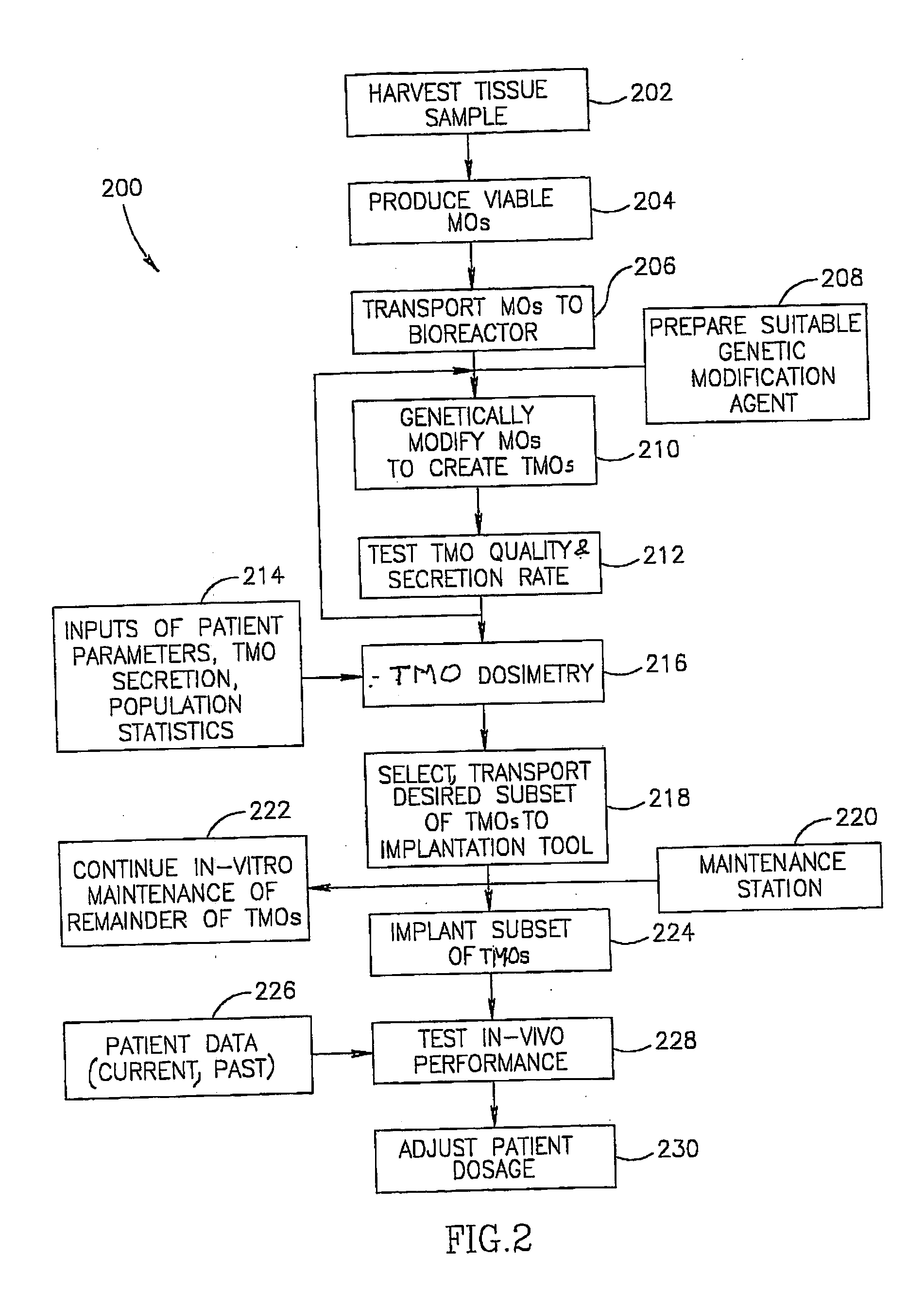
[0018] In an exemplary embodiment of the invention, for example when skin is the tissue being harvested, the tissue sample is harvested by lifting the surface of the tissue and cutting a section of the skin to a specified depth. The section is thick enough to include all of the desired layers of the skin. Optionally, the desired layers include the entire epidermis and at least some portion of the underlying dermis (up to and including the fill thickness of the skin) and corresponds in thickness from 0.3 to 3 mm depending on the location of the skin from which the sample is taken. When a skin structure is used that includes both epidermis and some dermis (including all the cellular layers, matrix and stromal architecture of the dermis which compose it), and processing it into micro-organs, the viability of the harvested tissue can be maintained for long periods both in vitro and in vivo, following implantation. As used herein, the verbs “cut” and “slice” are used to denote separation of one portion of tissue from another using a sharp blade or blade-like object.
[0034] In an embodiment of the invention, the modules have matching ports so that there can be an easy transfer of micro-organ / TMO between the modules and so that the modules can cooperate to carry out the processes.
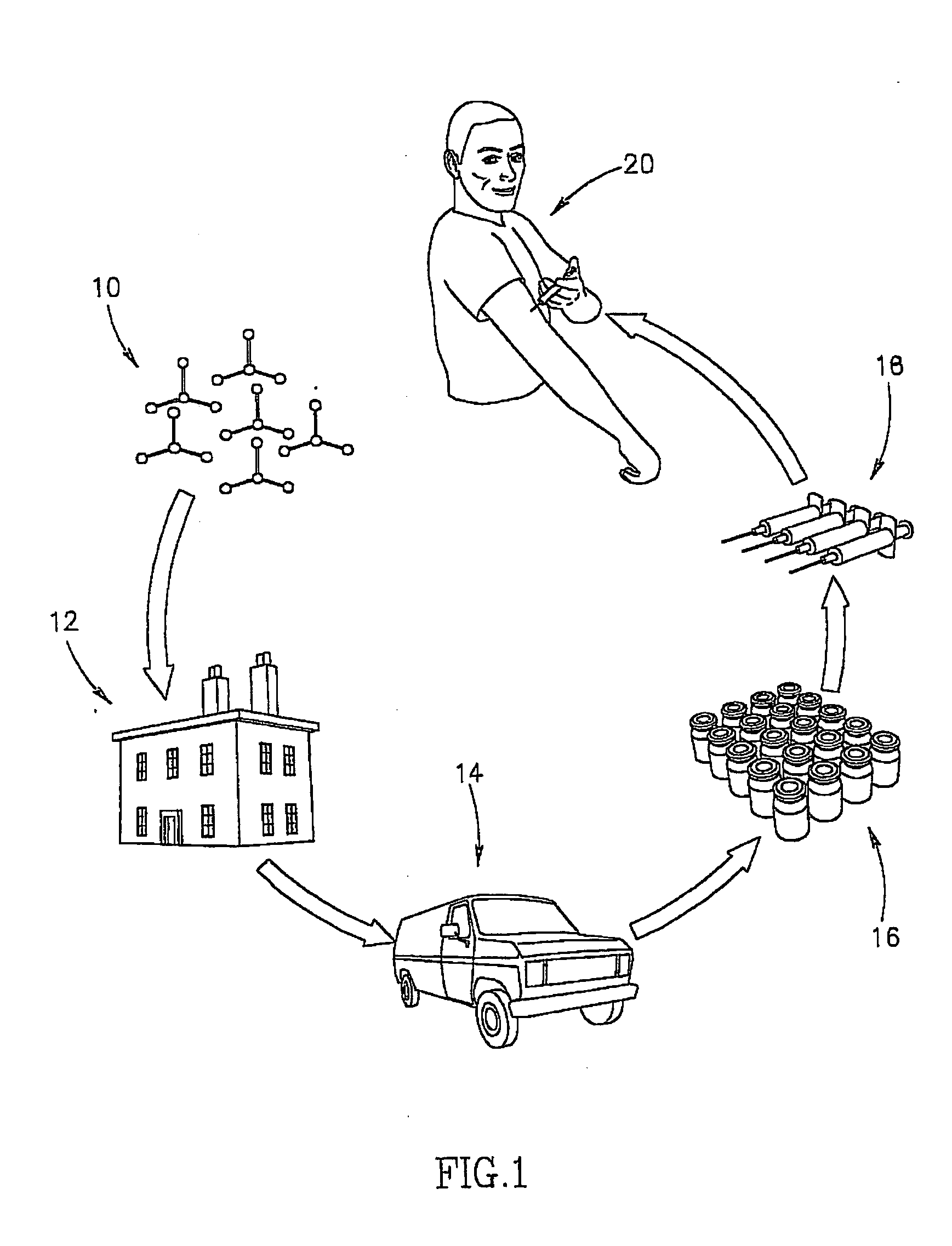
[0037] It is a characteristic of some embodiments of the invention that a large amount of micro-organ is processed into a TMO together. This allows for easier and more exact determination of the secretion of the TMO and for determination of dosage.

Problems solved by technology

In each of these cases the method of delivery is limited by the body processes that the agent is subjected to, by the requirement for frequent administration, and limitations on the size of molecules that can be utilized.

Method used


Image

  • Closed automated system for tissue based therapy
  • Closed automated system for tissue based therapy
  • Closed automated system for tissue based therapy

Examples


example 1

Human Skin TMOs, Expressing Mouse Interferon Alpha (mIFNα?, Implanted in SCID Mice.

[0269] Human skin micro-organs were prepared from fresh skin tissue samples, obtained from tummy-tuck surgery procedure. A section of 1.4-1.5 mm skin thickness (depth) was removed and cleaned using hypochloride solution (10% Milton solution). A cleaned skin sample was sectioned, using a tissue chopper (TC-2 chopper, Sorval, Du-pont instruments) into 450 micrometer sections (width) under sterile conditions. The resulting micro-organs were placed, one per well, in a 48-well micro-plate containing 400 μl per well of DMEM (Biological Industries - Beit Haemek) in the absence of serum, under 5% CO2- at 37° C. for 24 hours. Thereafter, each well underwent a transduction procedure in order to generate a therapeutic micro-organ (TMO) using an adeno viral vector (1×10⁹ IP / ml) carrying the gene for mouse interferon alpha (Adeno-mIFNα. Thereafter, the TMOs were again maintained in 400 μl per well of DMEM. The m...

example 2

Human Skin TMOs, Expressing Mouse Interferon Alpha (mIFNα)?, Show High Reproducibility From Patient to Patient in Protein Output

[0271] TMOs were prepared and transduced with Ad5 / CMV-mIFNα vector using a standard (but non-optimized) protocol, as describe above, including an adeno viral titer of 1×10⁹ IP / ml.

[0272] Transduction was performed 24 hours post micro-organ preparation. Medium was assayed for in-vitro mIFNα secretion on day 6 following transduction by using a specific ELISA kit (Cat. # CK2010-1, Cell Science Inc.). FIG. 15 shows that the degree of variability between skin samples from different patients, processed at different times, is remarkably small. This low variation between human patients indicates that sufficiently comparable levels of protein secretion can be obtained from a standard sized skin sample taken from patients in practical use for dosing and filtrating the amount of TMOs to be implanted in order to achieve the desired therapeutic effect.

example 3

Human Skin Linear TMOs, Expressing Human Erythropoietin (hEPO), Implanted in SCID Mice, Including Re Implantation.

[0273] Linear (20 mm long and 0.4 micrometer wide) human skin micro-organs were prepared from fresh skin tissue samples obtained from a tummy-tuck surgery procedure. Tissue samples of 0.85-1.1 mm split skin thickness (depth) were removed and cleaned using DMEM containing glutamine and Pen.-Strep in Petri dishes (90 mm).

[0274] In order to generate the linear micro-organs, the above tissue samples were cut by a press device using a blade structure as described above, into the desired dimensions: 20 mm×400 micrometers. The resulting linear micro-organs were placed, one per well, in a 24-well micro-plate containing 500 μl per well of DMEM (Biological Industries—Beit Haemek) in the absence of serum under 5% CO2 at 37° C. for 24 hours. Each well underwent a transduction procedure in order to generate a therapeutic micro-organ (TMO) using an adeno viral vector (1×10¹⁰IP / ml) ...


PUM


Abstract

A micro-organ structure comprising at least two micro-organ portions formed from a tissue, in which said at least two micro-organs are linked one to the other by means of a junction formed from said tissue of which the micro-organs were formed therefrom.

Description

RELATED APPLICATIONS [0001] This application claims the benefit under 35 U.S.C. 119(e) of U.S. Provisional Application 60 / 330,959 filed Nov. 5, 2001 and U.S. provisional Applications 60 / 393,746 and 60 / 393,745 filed Jul. 8, 2002, the disclosures of all of which are incorporated herein by reference.FIELD OF THE INVENTION [0002] The invention relates to the field of tissue based micro-organs such as therapeutic tissue based micro-organs. BACKGROUND OF THE INVENTION [0003] Various methods for delivering therapeutic agents are known. For example, therapeutic agents can be delivered orally, transdermally, by inhalation, by injection and by depot with slow release. In each of these cases the method of delivery is limited by the body processes that the agent is subjected to, by the requirement for frequent administration, and limitations on the size of molecules that can be utilized. For some of the methods, the amount of therapeutic agent varies between administrations. [0004] This documen...

Claims


Application Information

Patent Timeline
IPC IPC(8): C12M3/00C12N5/08
CPCC12M21/08C12M35/04C12M23/44
Inventor PEARLMAN, ANDREW L.BELLOMO, STEPHEN F.GARFINKEL, LEONARD I.PIVA, GUILLERMO ALBERTOSHAVITT, MENACHEM D.ROSENBERG, LIORLIPPIN, ITZHAKBUKHMAN, MORDECHAYALMON, EINATSHER, NIV
Owner MEDGENICS INC
Learn More