Methods for rapid screening of mad cow disease and other transmissible spongiform encephalopathies

a spongiform encephalopathy and rapid screening technology, applied in the field of rapid screening of mad cow disease and other transmissible spongiform encephalopathies, can solve the problems of not being suited for in vivo testing, not optimal for rapid screening of large numbers of animals, and taking hours to compl

Inactive Publication Date: 2006-01-19
U S GOVERNMENT REPRESENTED BY THE DEPT OF VETERANS AFFAIRS +1
View PDF2 Cites 4 Cited by
  • Summary
  • Abstract
  • Description
  • Claims
  • Application Information

AI Technical Summary

Benefits of technology

[0012] The present invention is useful in the diagnosis of any diseases which alter neuropathology (e.g. the pathology of the nervous system). In particular, the present invention is useful in the diagnosis of any diseases which alter vacuoles or, alternatively, form plaques in a tissue. For example, the present invention teaches the diagnosis of transmissible spongiform encephalopathies (TSE) such as, but not limited to, bovine spongiform encephalopathy (BSE or Mad Cow disease), scrapie in sheep, and chronic wasting disease (CWD) of deer. In an alternative embodiment, the subject invention is used to identify human patients with Creutzfield-Jakob disease (CJD) In a further embodiment, the present invention provides a method of distinguishing sporadic from variant and / or familial forms of the disease. It is contemplated that the methods described herein are further useful for the diagnosis of Gerstmann-Streussler-Sheinker Disease (GSS), fatal familial insomnia (FFI), hereditary Icelandic syndrome, senility and multiple myeloma, for example.
[0013] Method of Diagnosis in Dead Animal
[0014] Included are methods of diagnosis of a dead animal. Tissue of slaughtered animals is provided. The tissue may be any body tissue known to be vulnerable to the pathological effect of the disease, such as, for example, neural tissue, including, but not limited to brain and spinal cord tissues. Tissue deep in the brain is also contemplated. For example, the tissue is accessed by the use of a probe. More specifically, a needle type probe may be inserted directly through thin regions of the skull. Alternatively, the probe may be inserted through the roof of the orbit below the eye brow to sample the frontal cortex.
[0015] The tissue is imaged. For example, a radial scan is performed to image the brain. The probe may be advanced to sample a volume of tissue. The data may be analyzed by the operator in real time. Alternatively, the data may be stored for off-line processing. A skilled artisan is aware of methods well known in the art for processing such data regardless of whether the processing is performed at the time of data acquisition. It is contemplated that software may be developed to automatically identify, measure, and count the number of vacuole per volume of tissue sampled. For example, the index of refraction of the vacuole may also be determined based on the amplitude of reflected light using methods well known in the art. These data will be analyzed using statistical criteria that define the likelihood of TSE in specific brain regions, the animal, and stage of the disease.
[0017] Various imaging techniques are useful in the methods of the present invention. Exemplary techniques are described in International Application Number PCT / US2003 / 028352, which is hereby incorporated by reference herein. In an exemplary embodiment, imaging is performed using a needle-type probe. Other, non-limiting, examples of imaging techniques are contemplated and include, for example, contact but non-penetrating imaging, and non-contact imaging.
[0019] In the non-contact imaging, a ‘stand-back’ scanning method, which does not require contact with the affected tissue, may also be used. Non-contact imaging provides the least risks for contamination and spread of contagious tissue. In this method, the pathology needs to be close to the surface of the tissue. The tissue may or may not be sliced in preparation.

Problems solved by technology

These tests require biopsy of tissue and typically take hours to complete.
These tests are not optimal for rapid screening of large numbers of animals.
Furthermore, they are not well suited for in vivo testing.

Method used


Image

  • Methods for rapid screening of mad cow disease and other transmissible spongiform encephalopathies
  • Methods for rapid screening of mad cow disease and other transmissible spongiform encephalopathies

Examples


example 1

CJD, Scrapie, and BSE Diagnosis Using Catheter Based OCT Probe to Visualize Vacuolar Appearance

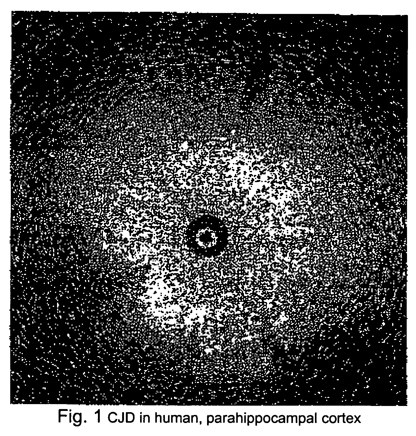
[0027] As illustrated in FIG. 1, brain tissue from a patient who died of CJD was imaged using a catheter based OCT probe manufactured by LightLab Imaging (of Westford, Mass.). Large numbers of vacuoles of different diameters were observed. The high degree of back scattering by the vacuoles suggests that they are not simple vacuoles filled with CSF-like fluid. Vacuoles having the observed OCT appearance shown in FIG. 1 have not been observed in human brain stored in the same manner.

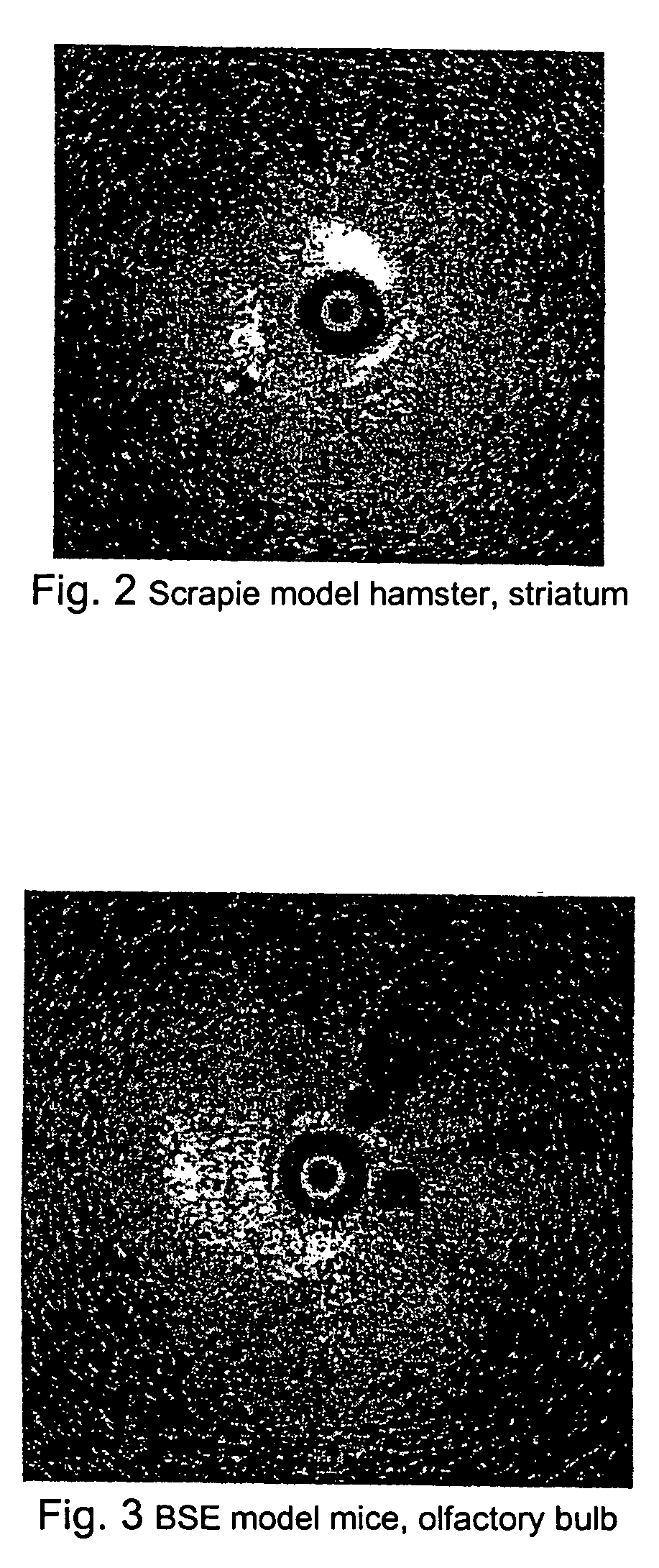
[0028] As illustrated in FIG. 2, a hamster infected with scrapie was sacrificed shortly before OCT imaging. Highly reflective vacuoles similar to that observe in CJD brain were observed in the striatum and possibly in the cortex.

[0029] As illustrated in FIG. 3, OCT was performed in a mouse brain infected with BSE. Large vacuoles were identified in the olfactory bulb.

example 2

Methods for Screening Tissue of Slaughtered Animals (Imaging Using a Needle-Type Probe)

[0030] A catheter-based OCT probe packaged within a rigid cannula (needle-type probe) is inserted into an exposed tissue (i.e. brain, spinal cord, etc) of a slaughtered animal. The approach is used when tissue deep in the brain is desired for sampling and / or testing. A needle type probe may also be inserted directly through thin regions of the skull (i.e. through the roof of orbit below the eye brow to sample the frontal cortex). A radial scan may be performed to image the brain as illustrated in the proceeding figures. The probe will be advanced to sample a volume of tissue. The data may be interpreted by the operator in real time or may be stored for off-line processing. Software may be developed to automatically identify, measure, and count the number of vacuole per volume of tissue sampled. The index of refraction of the vacuole may also be determined based on the amplitude or reflected light...

example 3

Methods for Screening Tissue of a Slaughtered Animal (Contact but Non-Penetrating Imaging)

[0031] A clear disposable window may be placed against the tissue to separate the OCT probe from the brain. These probes may or may not need to be catheter based. Catheter-based probes may have a linear scanning movement, similar to the ‘push-pull’ design of LightLab Imaging and probes currently designed for GI endoscopy and dermatology. Non-catheter-based probes may use designs similar to those used for OCT opthalmoscope and OCT microscope. This method is best suited for pathology that is located at a relative short distance from the surface of the tissue. Most spongiform lesions in the cortex are within the detection distance from the surface of the cortex. It is also possible to cut the sample so that pathology anywhere within the brain may be detected. In such case, the tissue would need to be handled but still would not need to be extensively prepared as for conventional histology.


PUM

PropertyMeasurementUnit
optical coherence tomographyaaaaaaaaaa
OCT imagingaaaaaaaaaa
volumeaaaaaaaaaa

Abstract

Methods for diagnosing altered neuropathology in an animal are disclosed, wherein said methods comprise imaging brain, spinal cord, or other neural tissue of the animal, analyzing the appearance of the tissue, and determining whether the appearance of the tissue is altered relative to corresponding unaltered tissue. Also disclosed are methods for diagnosing spongiform encephalopathies in an animal, wherein said methods comprise imaging brain, spinal cord, or other neural tissues of the animal, analyzing the appearance of vacuoles in the tissue, and determining whether the appearance of the vacuoles in the tissue is altered relative to corresponding spongiform encephalopathy-free tissue. Also disclosed are automated methods for diagnosing altered neuropathy and spongiform encephalopathies.

Description

[0001] This work was supported by NINDS Grant No. NS44627, and therefore the government may have certain rights to the invention.FIELD OF THE INVENTION [0002] The present invention relates to methods of diagnosing diseases involving altered neuropathology. Included are methods for rapid screening of mad cow disease and other transmissible spongiform encephalopathies. These methods utilize visualization techniques such as optical coherence tomography (OCT). BACKGROUND OF THE INVENTION [0003] Mad Cow disease (also know as BSE, bovine spongiform encephalopathy) has had an enormous negative impact on the economies Great Britain, Canada, and now the US. The definitive means for documenting transmissible spongiform encephalopathies (TSE) such as Creutzfield-Jakob disease (CJD) in humans, bovine spongiform encephalopathy (BSE or Mad Cow disease), scrapie in sheep, and chronic wasting disease (CWD) in deer and elk is to transmit disease to another animal. But practical diagnosis is generall...

Claims


Application Information

Patent Timeline
Patent Type & Authority Applications(United States)
IPC IPC(8): G01N33/53A61B5/00G01N33/68
CPCA61B5/0064A61B5/0066G01N2800/2828G01N33/6896A61B5/6852
Inventor TANG, CHA MINROHWER, ROBERT G.
Owner U S GOVERNMENT REPRESENTED BY THE DEPT OF VETERANS AFFAIRS
Why Eureka
Try Eureka