
Method of coating implantable medical devices

Inactive Publication Date: 2006-01-26
ABBOTT CARDIOVASCULAR
View PDF99 Cites 6 Cited by
  • Summary
  • Abstract
  • Description
  • Claims
  • Application Information

AI Technical Summary

Benefits of technology

[0013] In accordance with another aspect of the invention, a method for coating an implantable medical device is provided, comprising forming a polymer layer containing a drug on the device, applying a polymer in a liquid state and free from any solvents to the polymer layer to form a topcoat layer, wherein the during the application of the topcoat layer the migration of the drug from the polymer layer is prevented or significantly minimized.

Problems solved by technology

A problem associated with the above procedure includes formation of intimal flaps or torn arterial linings which can collapse and occlude the conduit after the balloon is deflated.
Vasospasms and recoil of the vessel wall also threaten vessel closure.
Although stents work well mechanically, the chronic issues of restenosis and, to a lesser extent, thrombosis remain.
These events are adversely affected by the mechanical aspects of the stent such as the degree of injury and disturbance in hemodynamics caused by the stent.
A challenge is maintaining the necessary concentration of a therapeutic substance at the lesion site for the necessary period of time.
This can be done via brute force methods using oral or intravenous administration but the issues of systemic toxicity and side effects arise.
Being made of metal, plain stents are not useful for therapeutic substance delivery.
However, when a topcoat composition is applied to the polymer-drug layer, the solvent may extract the drug out from the polymer-drug layer, therefore reducing the effectiveness of the topcoat layer.

Method used


Image

  • Method of coating implantable medical devices
  • Method of coating implantable medical devices
  • Method of coating implantable medical devices

Examples


Embodiment Construction

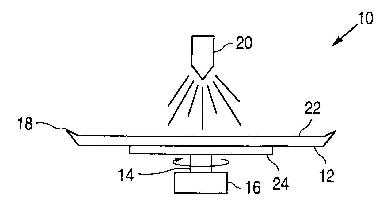
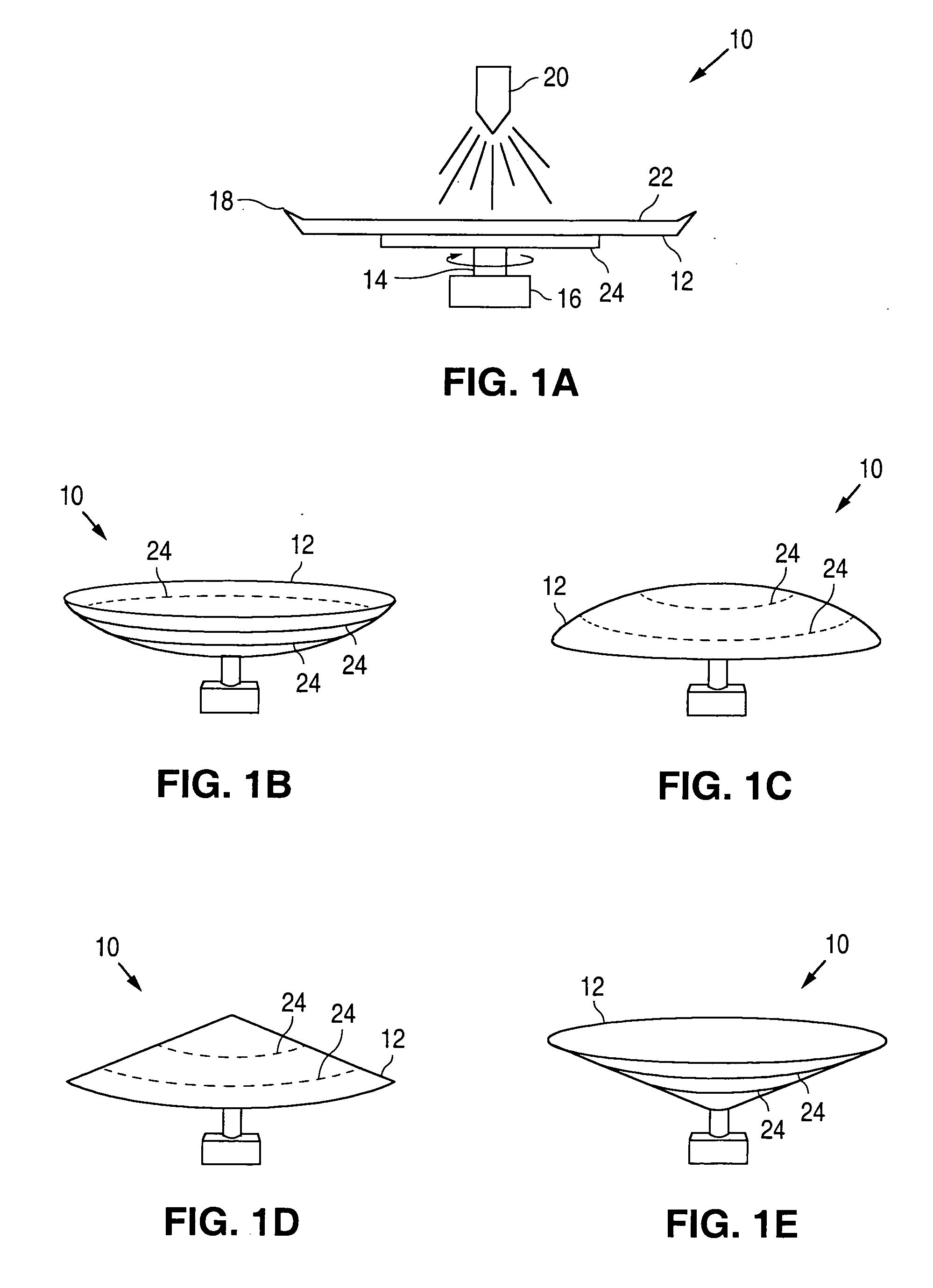
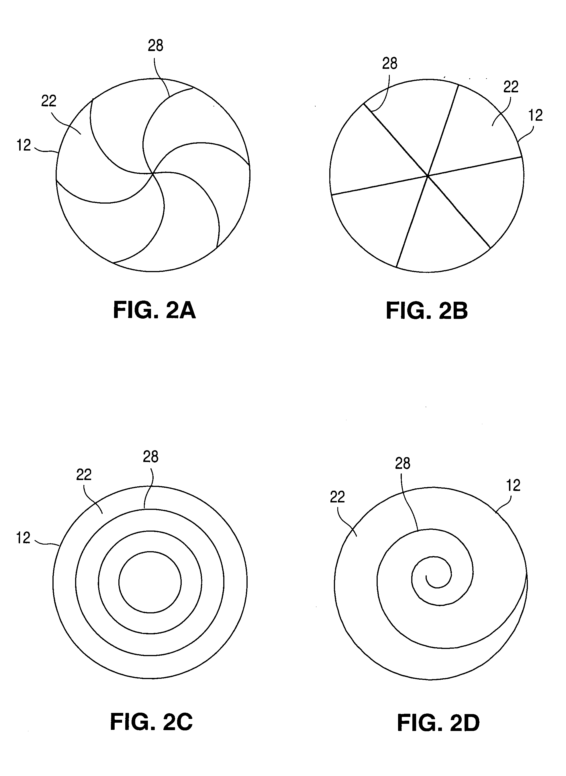
[0018]FIGS. 1A-1E illustrate embodiments of an apparatus 10 for coating medical devices, such as stents. Apparatus 10 can include a disk member 12 mounted on a shaft 14. The shaft 14, in turn, can be connected to a motor 16 for rotating the disk member 12 in a clockwise or counterclockwise direction. The disk member 12 can be flat (FIG. 1A), concave (FIG. 1B), convex (FIG. 1C) or conical (FIGS. 1D and 1E) in shape. The disk member 12 can optionally include a lip 18 disposed about the periphery thereof. The lip 18 can extend in an upwardly direction, towards a nozzle 20.

[0019] The disk member 12 can be made from any suitable material or can be coated with the desired material so as to minimize the ability of the composition to adhere to a surface 22 of the disk member 12 on which the composition is applied via the nozzle 20. One suitable non-stick surface 22 can be TEFLON. A temperature adjustor 24 can also be provided for adjusting the temperature of the composition during the coat...


PUM

PropertyMeasurementUnit
Percent by volumeaaaaaaaaaa

Abstract

A method of coating a medical device, such as a stent is provided.

Description

CROSS REFERENCE [0001] This application is a divisional of application Ser. No. 10 / 772,858 filed on Feb. 4, 2002, which is a continuation-in-part of application Ser. No. 10 / 040,574 filed on Dec. 28, 2001.BACKGROUND OF THE INVENTION [0002] 1. Field of the Invention [0003] This invention relates to a method for coating implantable medical devices, such as stents. [0004] 2. Description of the Background [0005] Percutaneous transluminal coronary angioplasty (PTCA) is a procedure for treating heart disease. A catheter assembly having a balloon portion is introduced percutaneously into the cardiovascular system of a patient via the brachial or femoral artery. The catheter assembly is advanced through the coronary vasculature until the balloon portion is positioned across the occlusive lesion. Once in position across the lesion, the balloon is inflated to a predetermined size to radially press against the atherosclerotic plaque of the lesion for remodeling of the vessel wall. The balloon i...

Claims


Application Information

Patent Timeline
IPC IPC(8): A61L33/00B05D3/00B05B3/10B05B13/02
CPCA61F2/82B05B3/001Y10S118/16B05B13/0228B05B3/1014
Inventor HOSSAINY, SYED F.A.
Owner ABBOTT CARDIOVASCULAR
Learn More