T-statistic method for suppressing artifacts in blood vessel ultrasonic imaging

Inactive Publication Date: 2006-02-09
THE BOARD OF TRUSTEES OF THE LELAND STANFORD JUNIOR UNIV
View PDF9 Cites 12 Cited by
  • Summary
  • Abstract
  • Description
  • Claims
  • Application Information

AI Technical Summary

Benefits of technology

[0011] In one aspect, the present invention provides a computationally efficient and effective technique for suppressing the time varying blood scatter signal and improving contrast in intravascular ultrasound imaging. By imaging the instantaneous t-statistic of repeated radiofrequency echoes, the lumen to blood vessel contrast is significantly improved as compared with averaging the radiofrequency of the repeated echoes. The technique is simple and fast to implement. Moreover, the improvement in contrast ratio can make feasible the use of forward-directed ultrasound beams. Because drop out is particularly severe at oblique angles between the blood vessel wall and the ultrasound beam, conventional intravascular ultrasound transducers direct pulses radially within the lumen rather than forward along the length of the vessel. With the significant improvement in contrast ratio at oblique angles provided by the technique of the present invention, however, forward-directed ultrasound beams become practical.
[0012] In one embodiment, a method for generating an enhanced ultrasound image from ultrasound echo amplitudes is provided. A temporal sequence of n image frames containing data samples representing the ultrasound echo amplitudes at image points in the frame are stored in a computer-readable memory and processed to produce an enhanced image. Portions of the enhanced image representing time-varying ultrasound echo amplitudes are suppressed to provide increased contrast between moving blood and the relatively still vessel wall. An image generated from the enhanced image is then displayed. The processing of the image frames includes calculating a point-wise t-statistic value for each image point. The t-statistic value for each image point may be calculated, for example, by computing a mean value of data samples for the image point in the n image frames, computing a standard deviation of data samples for the image point in the n image frames, and computing the ratio of the mean value to the standard deviation. This calculation is done point-wise, i.e., using sample data for individual points independent of data for other points in the image. Consequently, the calculation is simple and efficient. Moreover, the t-statistic method provides large contrast enhancement using only a few image frames, e.g., less than ten. Even with four or fewer frames significant enhancement is obtained, making the technique very fast to implement.

Problems solved by technology

At these high frequencies, however, the backscatter from blood increases, resulting in significant decreases in contrast ratio between the blood vessel wall and the lumen of the blood vessel.
The contrast, however, is less than perfect.
It provides only the images of the blood lumen and is therefore of limited use.
However, this technique is not applicable for low blood velocity and suffers from reduced lateral resolution without gray-scale.
This approach, however, was limited by low spatial resolution, poor sensitivity to vessel wall motion, and the requirement of high frame rate.
While the blood suppression was significantly improved, a significant trade-off requiring reduction of both frame-rate and angular resolution resulted.
Unfortunately, this technique reduces the spatial resolution of the image.
Moreover, the technique requires complex signal processing circuitry.

Method used


Image

  • T-statistic method for suppressing artifacts in blood vessel ultrasonic imaging
  • T-statistic method for suppressing artifacts in blood vessel ultrasonic imaging
  • T-statistic method for suppressing artifacts in blood vessel ultrasonic imaging

Examples


Embodiment Construction

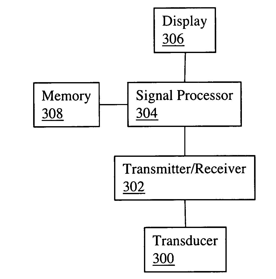
[0023] Embodiments of the present invention may be implemented using various types of intravascular ultrasound systems, suitably modified to process signals as will be described in more detail later. A schematic diagram of a generic ultrasound system is shown in FIG. 3. An ultrasound transducer 300 is connected to a transmitter / receiver 302. A signal processor 304 connected to transmitter / receiver 302 processes the signals, stores them in connected memory 308, and produces a digital image for viewing on connected display 306. Transducer 300 is conventionally attached to the end of a catheter which may be inserted into a blood vessel. Various types of transducer 300 may be used, including sideways-directed, forward-directed, and a combination of both. Signal processor 304 may be a programmable digital signal processor (DSP) or other processor built into an ultrasound imaging device, or it may be software running on a conventional desktop computer. Ultrasound systems may manifest the ...


PUM


Abstract

A technique for enhancing the image quality in intravascular ultrasound imaging increases contrast between blood and vessel wall processes image data using a point-wise t-statistic technique. Data from an ultrasound transducer is digitized and stored in a memory buffer [500]. For each point in the image, a t-statistic value is derived from signal amplitude values for the same point at a sequence of previous frames [502]. An image is then generated and displayed using the t-statistic values for the intensity of each point [504]. The improvement in contrast ratio as compared to averaging techniques is most significant at highly oblique angles when contrast ratio is particularly poor in the unprocessed signal.

Description

CROSS-REFERENCE TO RELATED APPLICATIONS [0001] This application claims priority from U.S. provisional patent application No. 60 / 592,848 filed Jul. 30, 2004, which is incorporated herein by reference.FIELD OF THE INVENTION [0002] This invention relates generally to methods and devices for ultrasound imaging. More specifically, it relates to signal processing techniques for enhancing the quality of images generated using very high frequency intravascular ultrasound. BACKGROUND OF THE INVENTION [0003] Intravascular ultrasound is a medical imaging technique used in the study of blood vessels in vivo. A long and thin catheter is used to guide an ultrasound transducer through the interior of the blood vessel while computerized ultrasound equipment processes the ultrasound echoes and generates an image. Detailed information on the subject of intravascular ultrasonography is contained in U.S. Pat. No. 4,794,931 and U.S. Pat. No. 5,000,185, which are incorporated herein by reference. [0004] ...

Claims


Application Information

Patent Timeline
IPC IPC(8): A61B8/00
CPCA61B8/12G06T7/0012G06T2207/30101G06T5/50G06T2207/10132G06T5/008G06T5/94
Inventor LIANG, DAVID H.YANG, PHILLIPKOOLWAL, ADITYAPARK, BYONG-HO
Owner THE BOARD OF TRUSTEES OF THE LELAND STANFORD JUNIOR UNIV
Why Eureka
Try Eureka