
Endoluminal occlusion-irrigation catheter with aspiration capabilities and methods of use

Inactive Publication Date: 2006-02-23
BOSTON SCI SCIMED INC
View PDF91 Cites 8 Cited by
  • Summary
  • Abstract
  • Description
  • Claims
  • Application Information

AI Technical Summary

Benefits of technology

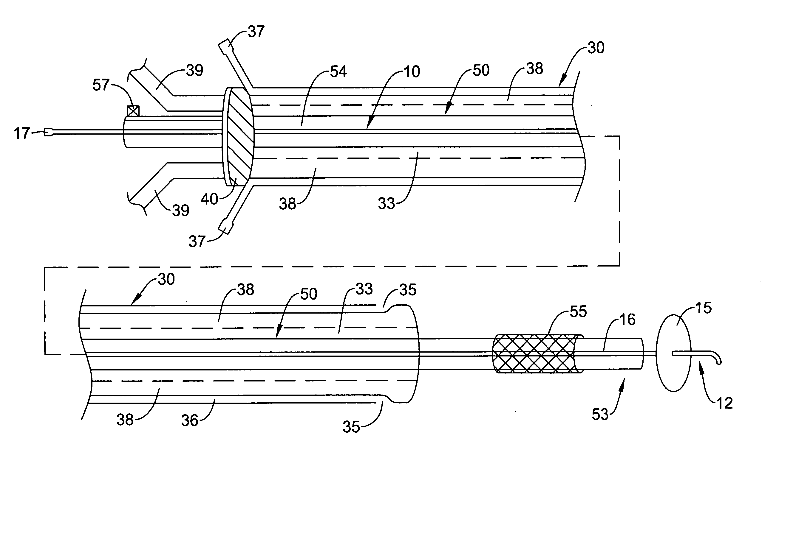
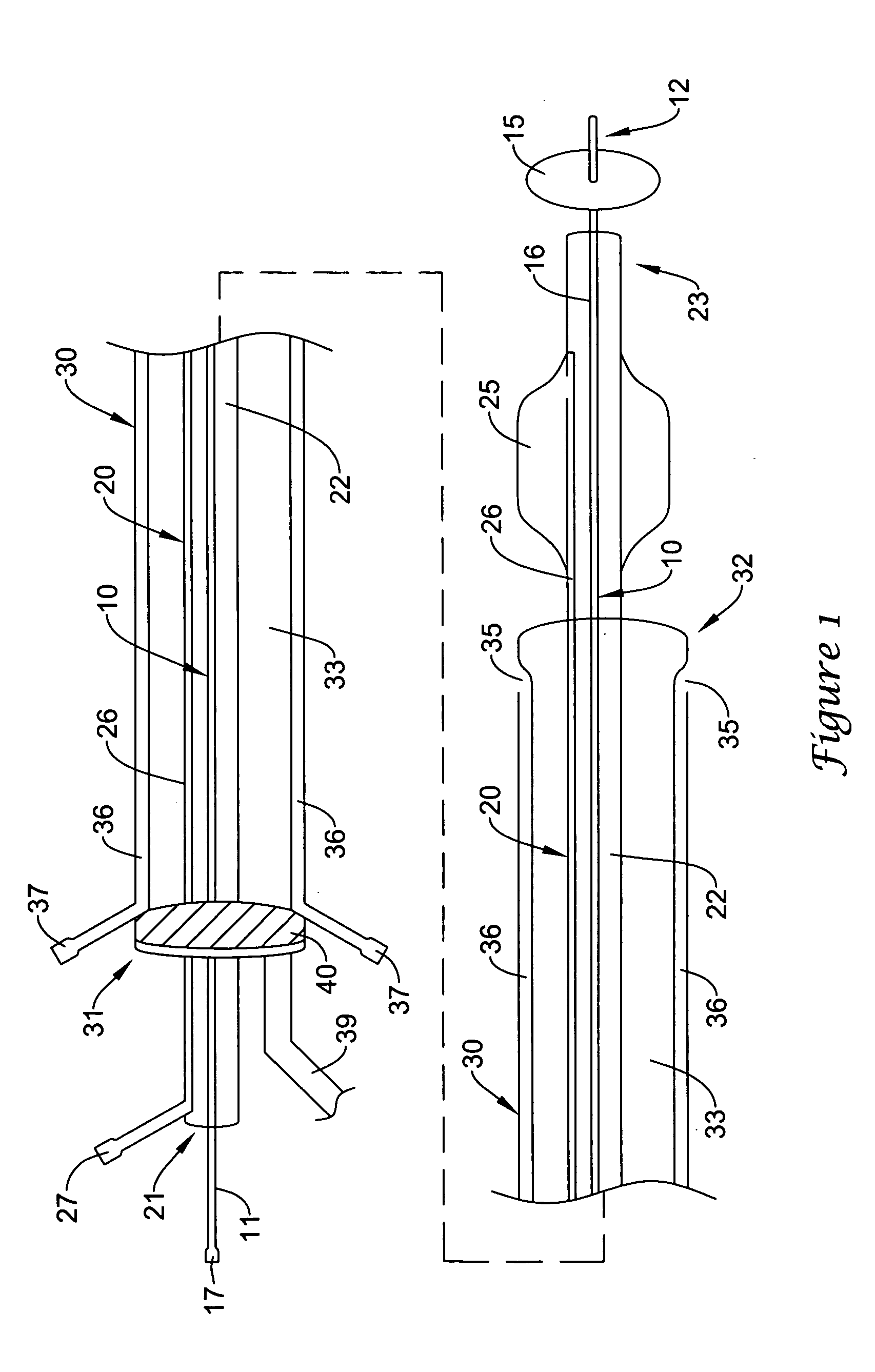
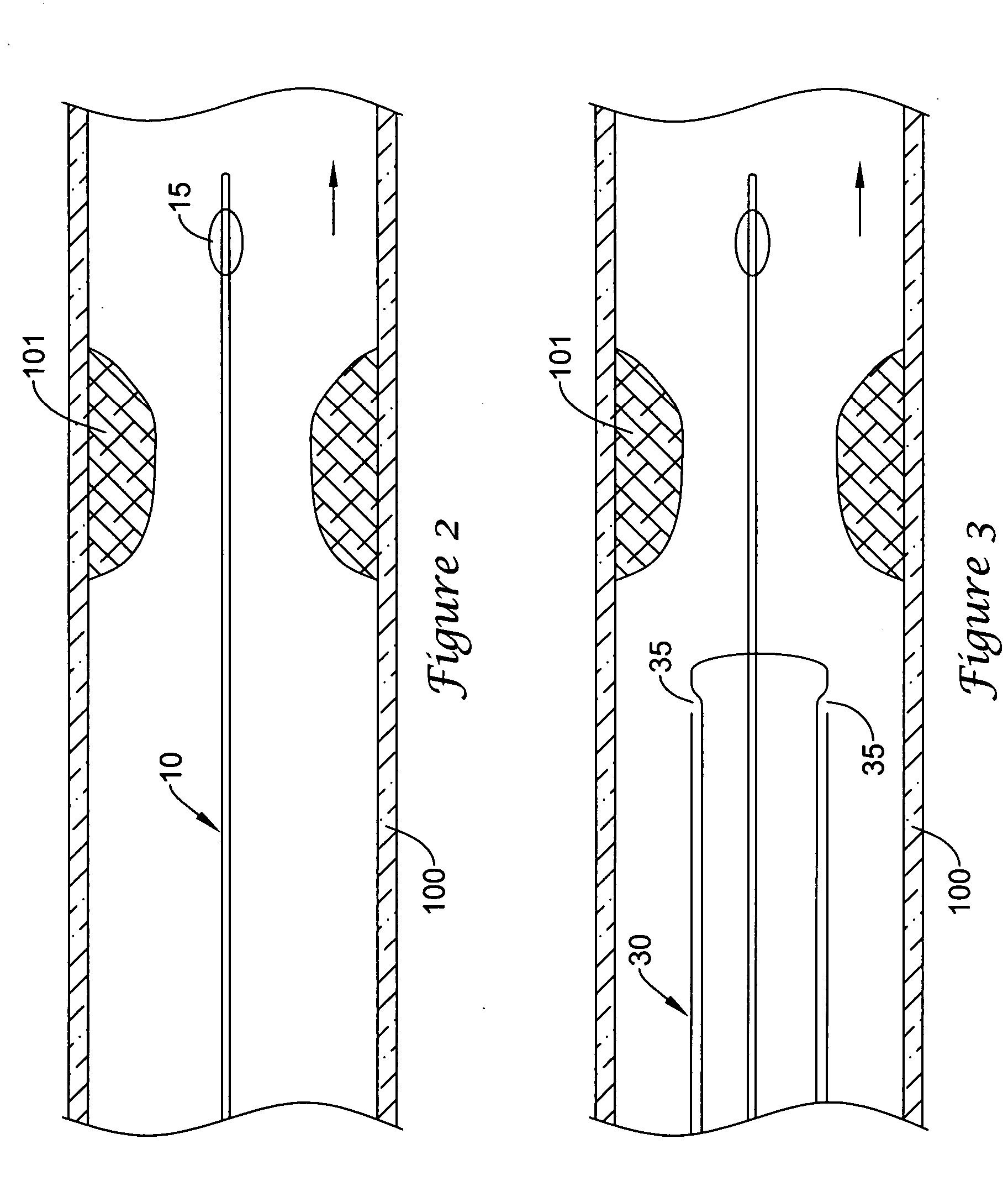
[0012] In a first method of treating an endovascular lesion using the catheter system described above, the guidewire, which has the expandable occluder placed in a collapsed state, is inserted in the lumen of the aspiration catheter. The aspiration catheter carrying the guidewire is then inserted into a vessel, and the guidewire is advanced to position the occluder distal to a region of interest. An endovascular device, e.g., an angioplasty catheter, is inserted over the proximal end of the guidewire, and advanced to position a dilatation member within the region of interest. The aspiration catheter is advanced over the guidewire and positioned proximal the dilatation member. The occluder on the guidewire is expanded to occlude the vascular lumen distal to the lesion. The dilatation member is expanded to treat the vascular lesion and collapsed after luminal patency is achieved. Fluid, such as saline or Ringer's lactate solution, is infused through the infusion lumen and ports to irrigate the treated lumen. Fluid, blood, and embolic debris are removed through the aspiration lumen under suction. The occluder on the guidewire remains expanded during irrigation and aspiration. In certain embodiments, infusion and aspiration of fluid can create a venturi effect in the vascular lumen between the expanded occluder and the aspiration catheter to facilitate removal of loose emboli and embolic material partially attached to the vascular wall. The surgeon can tell that removal of embolic material is complete after the aspirated fluid turns from red to clear and is free of any debris. In this way, the catheter system is capable of complete removal of emboli.
[0014] It will be understood that there are several advantages in using the catheter systems and methods disclosed herein for treating a vascular lesion. For example, the devices (1) can be inserted in arteries or veins of various diameter, (2) provide near-total capture of embolic material, thereby dramatically reducing the risk of distal embolization, (3) accommodate a variety of endovascular instruments, and (4) provide treatment of vascular lesions and emboli protection utilizing one catheter system, thereby obviating the need for device exchange.

Problems solved by technology

Treatments of vascular stenosis or lesions during endovascular procedures, such as atherectomy, balloon angioplasty with or without stent placement, or ablation therapy, are associated with increased risk of distal embolization.
Tissue debris, calcium, atheromatous plaque, and / or thrombi generated during the procedure often become lodged downstream in a small vessel of vital organs, causing tissue ischemia or infarction.
However, disadvantages associated with the blood filters are that (1) dislodgment of embolic material can occur during insertion and retrieval of the filter device, and (2) blood filters cannot easily be used in small vessels, (e.g., a saphenous vein graft measuring 3 or 4 mm).
The major disadvantage associated with the device is that some of the embolic material will not be removed by irrigation and suction, thereby leaving the patient at risk for embolic complication.

Method used


Image

  • Endoluminal occlusion-irrigation catheter with aspiration capabilities and methods of use
  • Endoluminal occlusion-irrigation catheter with aspiration capabilities and methods of use
  • Endoluminal occlusion-irrigation catheter with aspiration capabilities and methods of use

Examples


Embodiment Construction

[0035] Referring now to the drawings, an embodiment of the catheter system for treating a vascular lesion is depicted in FIG. 1. The system generally comprises guidewire 10, angioplasty catheter 20, and aspiration catheter 30. Guidewire 10 has proximal end 11 and distal end 12. An expandable occluder, shown as balloon 15 which communicates with inflation lumen 16 and proximal port 17, is mounted on distal end 12. Balloon 15 can be expanded by infusing air, gas, or saline through proximal port 17. Guidewire 10 is inserted through lumen 22 of angioplasty catheter 20. Lumen 22 communicates with proximal end 21 and distal end 23. Dilatation member, shown as expandable balloon 25 which communicates with inflation lumen 26 and inflation port 27, is mounted on distal end 23 of the catheter. Dilatation balloon 25 can be expanded by infusing air, gas, or saline through proximal port 27. Angioplasty catheter 20 is inserted through lumen 33 of aspiration catheter 30. Lumen 33 communicates with...


PUM


Abstract

A catheter system comprising a guidewire, an endovascular catheter, and an aspiration catheter. The guidewire has an expandable occluder mounted on a distal end. The guidewire and the endovascular catheter are insertable into a lumen of the aspiration catheter. The aspiration catheter also includes infusion and aspiration lumen(s) and port(s). Methods of using the catheter system for treating a vascular lesion and removing embolic material during the procedure are also disclosed.

Description

[0001] This is a continuation application of U.S. application Ser. No. 09 / 470,026 filed Dec. 22, 1999.FIELD OF THE INVENTION [0002] The present invention generally relates to medical devices for protecting a patient from distal embolization during interventional procedures, such as angioplasty or stent placement for treatment of vascular stenosis. More particularly, the devices comprise a catheter having irrigation and aspiration capabilities, and a guidewire carrying a distal expandable occluder. An endovascular instrument, such as an angioplasty catheter, is insertable into a lumen of the aspiration catheter. BACKGROUND OF THE INVENTION [0003] Treatments of vascular stenosis or lesions during endovascular procedures, such as atherectomy, balloon angioplasty with or without stent placement, or ablation therapy, are associated with increased risk of distal embolization. Tissue debris, calcium, atheromatous plaque, and / or thrombi generated during the procedure often become lodged dow...

Claims


Application Information

Patent Timeline
IPC IPC(8): A61F2/06A61M25/00A61B17/22A61M1/00A61M25/10
CPCA61B17/22A61B2017/22067A61B2217/005A61M2206/16A61M25/007A61M25/1011A61B2217/007
Inventor JANG, YUE-TEHTSUGITA, ROSS S.ADDIS, BRUCE S.MAAHS, TRACY D.CHANG, JEAN C.
Owner BOSTON SCI SCIMED INC
Learn More