
Methods in the diagnosis of pulmonary embolism

Inactive Publication Date: 2006-03-16
AEROCRINE AB
View PDF1 Cites 2 Cited by
  • Summary
  • Abstract
  • Description
  • Claims
  • Application Information

AI Technical Summary

Benefits of technology

[0031] The present invention is further directed to a method wherein the level of NO is measured in at least two samples of expire

Problems solved by technology

Pulmonary embolism is a common disorder accompanied by a significant morbidity and mortality.
Both pulmonary gas embolism and thromboembolism are known to cause severe haemodynamic and gas exchange abnormalities, including acute respiratory distress, pulmonary infarct and pleuritis.
The clinical diagnosis of pulmonary embolism is difficult and unreliable.
Inhaled nitric oxide (NO) has been tried experimentally, but consensus has not been reached if such treatment is efficacious or not.
Deem et al. also discuss the limitations of the model, venous gas embolisation using a continuous infusion of small bubbles, and states that it may be dissimilar to cases of air embolisation in the clinical setting, and that extrapolation of the data to clinical management is difficult.
In the light of the inconclusive evidence presented in the prior art, it remains a problem to identify alternative markers, with the aim to find a reliable marker for non-invasive, rapid and sensitive diagnosis of pulmonary embolism, in particular pulmonary thromboembolism.

Method used


Image

  • Methods in the diagnosis of pulmonary embolism
  • Methods in the diagnosis of pulmonary embolism
  • Methods in the diagnosis of pulmonary embolism

Examples


Embodiment Construction

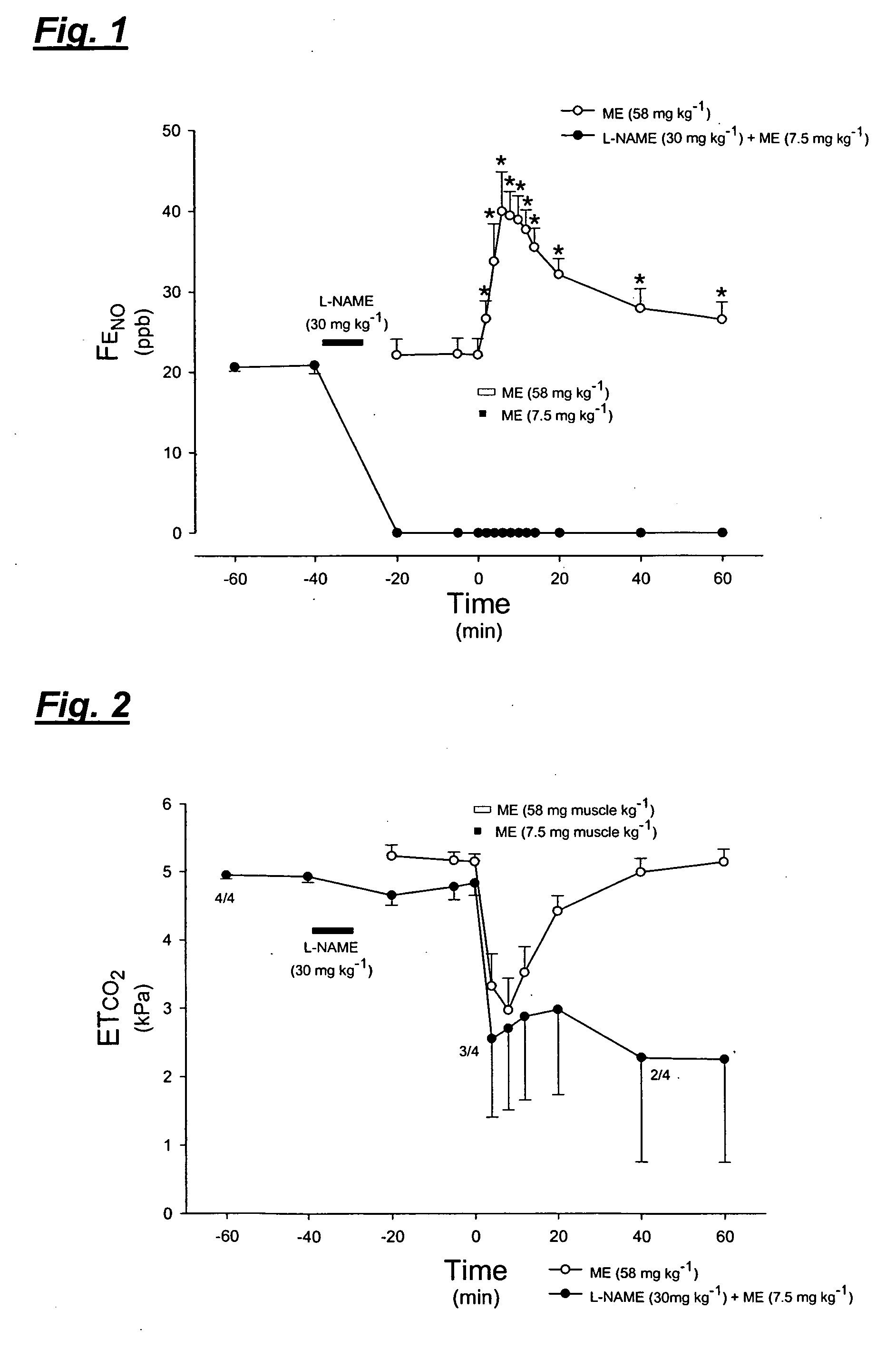
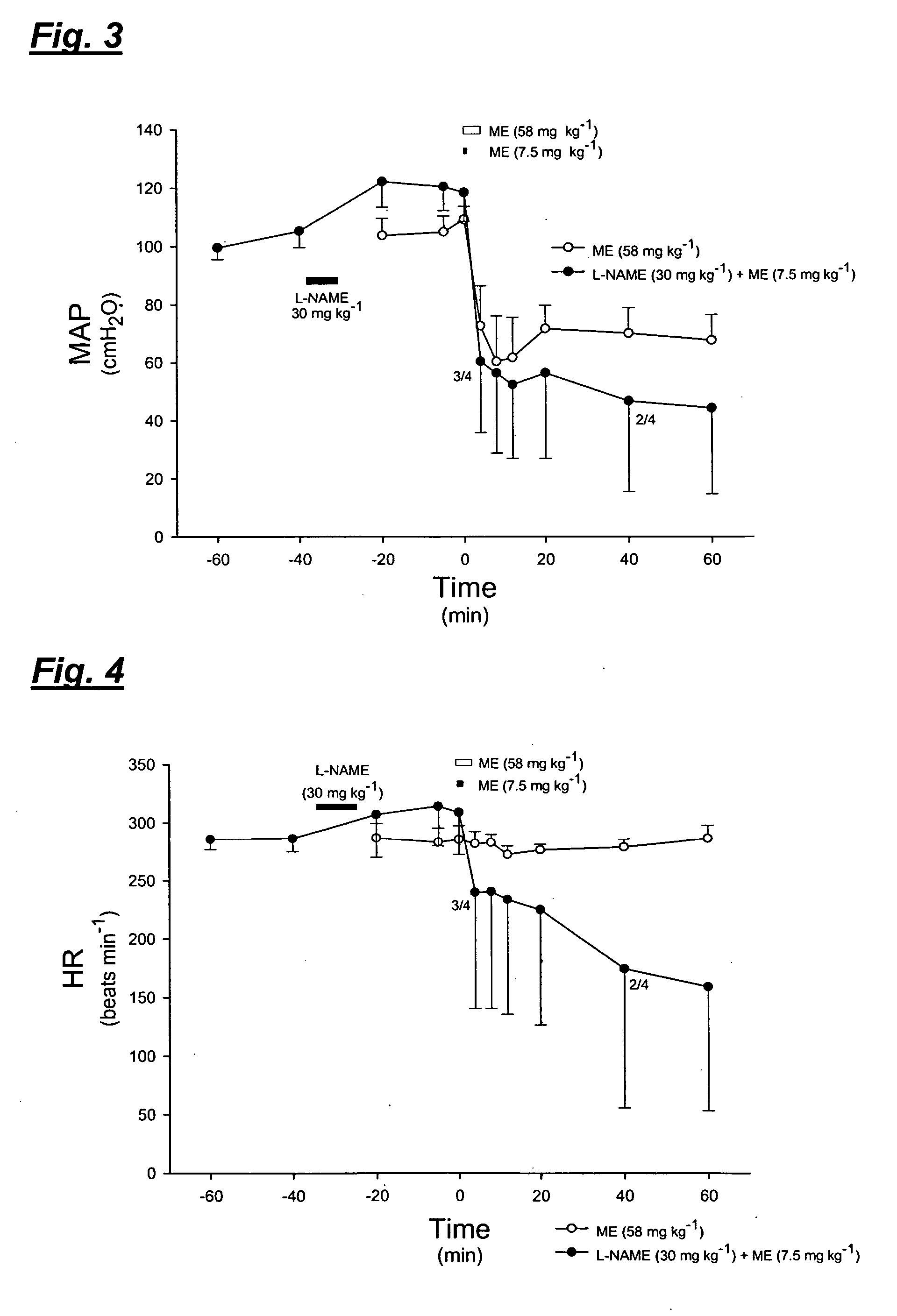
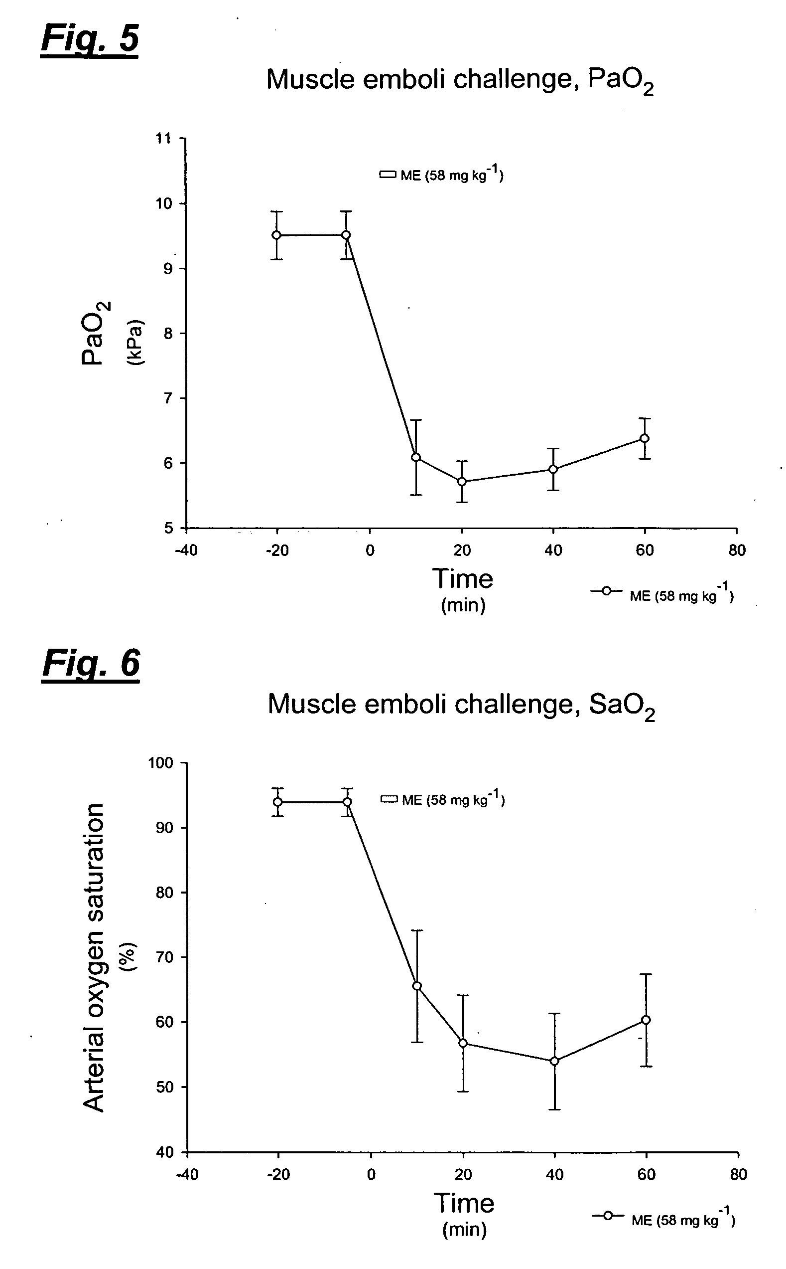
[0079] The inventive method was confirmed by the present inventors in vivo, in an animal model, using muscle homogenate to simulate thromboembolism. Experiments testing a blood clot homogenate and thrombin infusion were also performed.

Anaesthesia and Initial Surgical Procedures

[0080] The experiments were approved by the local animal ethics committee. Male white New Zealand rabbits (n=20, body weight 2.456±0.086 kg) were anaesthetised via an ear vein with sodium pentobarbital, 6 mg ml−1 in normal saline, 40-60 mg kg−1. The animals were placed in supine position and tracheotomised just below the cricoid cartilage to allow mechanical ventilation using a tracheal cannula with an outer diameter of 5 mm. The animals were ventilated by a Harvard Apparatus rodent ventilator (model 683, Harvard Apparatus, South Natick, Mass., USA). The ventilator was supplied with NO-free air using a charcoal filter (110×11 cm). Ventilation rate was 40 min−1 at constant volume where the tidal volume was i...


PUM


Abstract

Pulmonary embolism in a human patient can be detected or confirmed by monitoring the level of endogenous NO in expired air, comparing the measured level with baseline values previously obtained for the same patient or baseline values representing a healthy population. The method can be used alone, or as a supplemental or adjunct step in traditional methods for diagnosing pulmonary embolism. Analysis of the obtained NO value, with consideration of the flow and resistance, can be used to indicate the severity of the pulmonary embolism.

Description

CROSS-REFERENCE TO RELATED APPLICATIONS [0001] This application claims the benefit of Swedish Application No. 0402220-8, filed Sep. 14, 2004 under 35 USC Section 119. FIELD OF THE INVENTION [0002] The present invention relates to steps performed in the diagnosis of pulmonary embolism, and in particular to a rapid, sensitive and non-invasive test assisting in the early detection and in the evaluation of the severity of pulmonary embolism, or functioning as one step in such diagnosis. The invention in particular relates to pulmonary thromboembolism, and the early detection thereof. The invention further encompasses methods for the pre- and post-operative surveillance or monitoring of mammals, preferably human patients, as well as methods for observing the pulmonary condition of such patients during operations. BACKGROUND OF THE INVENTION Embolism [0003] An embolus is a foreign object, a quantity of air or gas, a fat globule, a bit of tissue or tumour, or a piece of a thrombus that ci...

Claims


Application Information

Patent Timeline
IPC IPC(8): G01N33/48A61BA61B5/08A61B5/083
CPCA61B5/08Y10T436/177692A61B5/0836A61B5/082
Inventor AGVALD, PERLINNARSSON, DAGADDING, CHRISTOFERGUSTAFSSON, LARSNILSSON, KRISTOFER
Owner AEROCRINE AB
Learn More