Molecular marker for diagnosing idiopathic inflammatory myopathy and application thereof

A molecular marker and idiopathic technology, applied in the field of medicine and biology, can solve problems such as modification, time-consuming, and inability to translate proteins

Active Publication Date: 2019-09-24
CHINA JAPAN FRIENDSHIP HOSPITAL
View PDF5 Cites 2 Cited by
  • Summary
  • Abstract
  • Description
  • Claims
  • Application Information

AI Technical Summary

Problems solved by technology

However, the construction of a cDNA expression library is not only time-consuming and technically difficult, but also relies on bacteria to express recombinant proteins. This type of protein expression system cannot perform post-translational modifications on proteins, and some autoantibodies that can recognize post-translational modified proteins cannot be identified. come out

Method used


Image

  • Molecular marker for diagnosing idiopathic inflammatory myopathy and application thereof
  • Molecular marker for diagnosing idiopathic inflammatory myopathy and application thereof
  • Molecular marker for diagnosing idiopathic inflammatory myopathy and application thereof

Examples


Embodiment 1

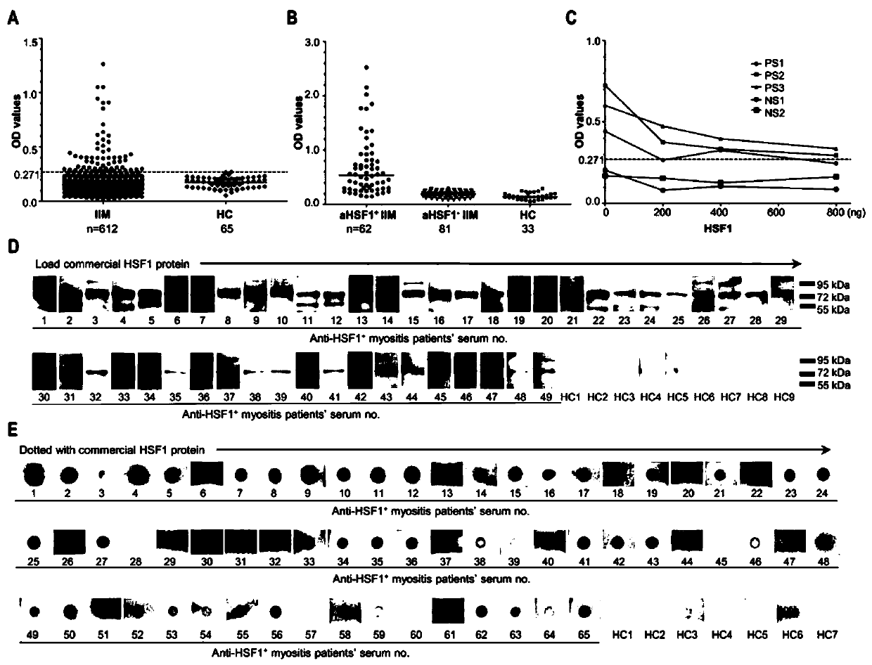
[0091] Example 1. Screening for highly specific antibodies in IIM and determining the presence of HSF1 antibodies

[0092] application Human Protein Microarray v5.1 (Life Technologies, USA) detected the distribution of antibodies in the sera of 10 DM patients and 10 HCs, and performed statistical analysis. The specific method is:

[0093] ① Blocking, configure blocking solution (50mM HEPES, 200mM NaCl, 0.01% Triton X-100, 25% glycerol, 20mM reduced glutathione, 1.0mM DTT, 1X Synthetic Block), put the chip in the chip incubation box and block for 1h at 4°C ;

[0094] ② For serum sample incubation, add the prepared sample diluent (1:500; 10 cases of DM patients, 10 cases of HCs) into the chip incubation box, and incubate at 4°C for 1.5h;

[0095] ③Secondary antibody incubation, Alexa Add 647-conjugated goat anti-human IgG fluorescent secondary antibody to the chip incubation box, and incubate at 4°C in the dark for 1.5h;

[0096] ④ Drying, use a chip centrifuge to centrif...

Embodiment 2

[0100] Embodiment 2, ELISA detection anti-HSF1 antibody

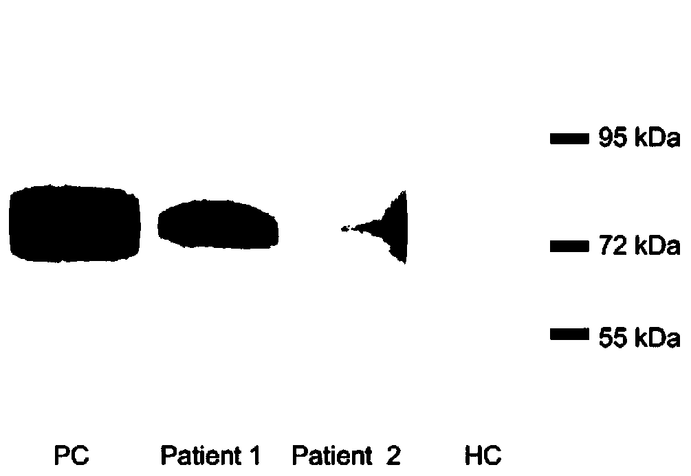
[0101] The commercially available recombinant HSF1 protein (Abcam, Cambridge, UK) was diluted and coated in the reaction wells (200ng / well) of a polystyrene plate (Thermo Scientific, Roskilde, Denmark), and the level of anti-HSF1 antibody in serum was detected by ELISA. At the same time, another commercially available recombinant HSF1 protein (OriGene, Maryland, USA) ELISA was used to detect the level of anti-HSF1 antibody in serum. Serial dilution ELISA (1:2) was used to determine the optimal dilution ratio of serum, and ELISA blocking experiment was used to determine the specificity of ELISA for detecting anti-HSF1 antibody. All serum samples were tested twice. Before detecting serum anti-HSF1 antibody by ELISA, the diluted serum of anti-HSF1 positive IIM patients (3 cases) and HCs (2 cases) was incubated with recombinant HSF1 protein (Abcam, Cambridge, UK) overnight at 4°C, and then the anti-HSF1 antibody was detect...

Embodiment 3

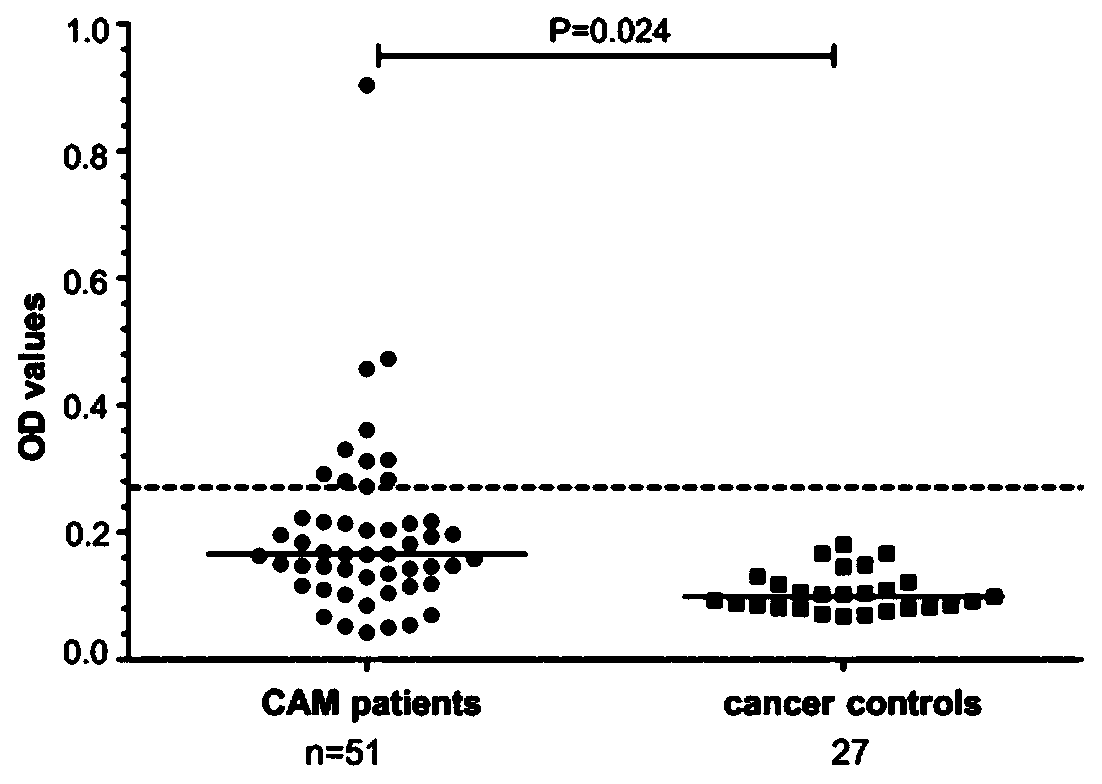
[0104] Example 3, clinical features of anti-HSF1 positive IIM patients

[0105] Anti-HSF1 positive IIM patients had a higher proportion of CAM (16.9% vs 7.3%, P=0.008), and common weight loss (P=0.046) and pruritus (P=0.039). Meanwhile, anti-HSF1 antibody was correlated with anti-TIF1γ antibody (P=0.046), hyperglobulinemia (P<0.001), elevated C-reactive protein (P=0.039) and elevated erythrocyte sedimentation rate (ESR, P=0.006). We did not find that anti-HSF1 antibodies were associated with other typical IIM clinical features, such as skin involvement, pulmonary interstitial lesions, and elevated muscle enzymes (Table 2). In addition, the karyotype of ANA in anti-HSF1-positive IIM patients' serum was mainly speckled or cytoplasmic granular.

[0106] Table 2 Clinical characteristics of anti-HSF1 antibody positive IIM patients

[0107]

[0108]

[0109] HSF1, heat shock transcription factor 1; IIM, idiopathic inflammatory myopathy; PM, polymyositis; DM, dermatomyositis; A...


PUM


Abstract

The invention relates to a molecular marker for diagnosing idiopathic inflammatory myopathy and an application thereof. The molecular marker is an autologous heat shock factor 1 (HSF1) antibody in a serum. The antibody is IgG. The idiopathic inflammatory myopathy is polymyositis (PM) or dermatomyositis (DM). The diagnosis is to distinguish the idiopathic inflammatory myopathy from a healthy population or to predict a disease progress of the idiopathic inflammatory myopathy (IIM).

Description

technical field [0001] The invention belongs to the technical field of medicine and biology, and specifically relates to a molecular marker for diagnosing idiopathic inflammatory myopathy and its application. Background technique [0002] Idiopathic inflammatory myopathy (Idiopathic inflammatory myopathy, IIM) is a group of rare heterogeneous autoimmune diseases with proximal muscle weakness, abnormal serum muscle enzymes, inflammatory infiltration of skeletal muscle and extramuscular involvement as the main clinical manifestations. Including polymyositis (polymyositis, PM), dermatomyositis (dermatomyositis, DM) and inclusion body myositis (inclusion body myositis, IBM)[ 1 ]. Autoantibodies of IIM include myositis-specific autoantibodies (MSAs) and myositis-associated autoantibodies (MAAs), which can be found in 60-80% of IIM patients. has significant value [ 2 , 3 ]. MSAs are associated with specific clinical syndromes and are only seen in IIM; whereas MAAs can be foun...

Claims


Application Information

Patent Timeline
IPC IPC(8): G01N33/68
CPCG01N33/6893G01N2800/24
Inventor 彭清林王国春张亚妹卢昕
Owner CHINA JAPAN FRIENDSHIP HOSPITAL
Why Eureka
Try Eureka