Optical examination of biological tissue using non-contact irradiation and detection

a biological tissue and non-contact technology, applied in the field of optical examination of biological tissue using non-contact irradiation and detection, can solve the problems of subject's head, breast or limb, reaches the limit of convenience and accessibility, and the path cannot be approximated by a straight line, so as to reduce the effective numerical aperture and reduce the light collection efficiency

Inactive Publication Date: 2006-03-16
NONINVASIVE TECH
View PDF14 Cites 317 Cited by
  • Summary
  • Abstract
  • Description
  • Claims
  • Application Information

AI Technical Summary

Benefits of technology

[0014] According to one aspect, an optical system for examination of biological tissue includes a light source, a light detector, optics and electronics. The light source generates a light beam, transmitted to the biological tissue, spaced apart from the source. The light detector is located away (i.e., in a non-contact position) from the examined biological tissue and is constructed to detect light that has migrated in the examined tissue. The electronics controls the light source and the light detector, and a system separates the reflected photons (e.g., directly reflected or scattered from the surface or superficial photons) from the photons that have migrated in the examined tissue. The system prevents detection of the “noise” photons by the light detector or, after detection, eliminates the “noise” photons in the detected optical data used for tissue examination.
[0016] The optical system, including its electronics, may comprise a time resolved spectroscopic (TRS) system, a phase modulation system (PMS), a phase array system, or a continuous wave (CW) system. In each case, the detector is located away from the examined tissue and there is no optical fiber in contact with the tissue surface. Conventionally, this reduces the light collection efficiency (reduces the effective numerical aperture), which in prior art was considered as a barrier to an effective spectrophotometric system. Thus, the present invention discards the conventional concept.
[0017] According to another aspect, an optical system for examination of brain tissue of a subject undergoing a security check includes a light source, a light detector, optics and electronics. The light source generates a light beam, transmitted to the head of the subject spaced apart from the source. The light detector is located away from the head and is constructed to detect light that has migrated in the tissue being examined. The electronics controls the light source and the light detector, and a system separates the reflected photons from the photons that have migrated in the examined tissue to prevent detection of the reflected photons by the light detector or eliminate after detection the reflected photons in the detected optical data used for tissue examination.
[0024] 1. Time Resolution System (TRS). This is a system in which time resolution of specular and diffusive photons is made on a time basis. A pulsed oscillator at 50 MHz activates a series of laser diodes at the desired wavelength with 10% duty ratio and illuminates the desired targets, (prefrontal cortex, or PFC, breast, muscle) through the appropriate optical system. The emergent photons are detected through wide band PMT that allows discrimination of reflected and diffusive photons. The decay of intensity is followed and processed with either time correlated single photon counting (TCSPC) or a boxcar detector. No blocking filters are needed to eliminate overload due to reflected photons in this system.
[0033] The second type, or metabolic signal, is generally based on the breakdown of ATP and formation of ADP and inorganic phosphate. As waste products, ADP and phosphate act as stimulants of respiration for neuronal mitochondria and are responsible for increased oxygen intake from circulating hemoglobin through the oxygen gradient that exists between neuronal mitochondria and the adjacent capillary bed. Thus the oxyhemoglobin of the capillary bed becomes deoxygenated to an extent, depending upon the activation of respiration, the blood flow through the capillaries, and generally, the ratio of capillary volume to mitochondrial volume. This second signal is thus a good measure of brain energy use.
[0035] In the case of breast cancer detection, the convenient activation processes described for the brain are not available for the hemoglobin signal. The spatial distribution of blood volume and hemodynamic signals are the two intrinsic signals available. Thus, cancer detection using intrinsic signals depends upon the different localization of hemoglobin deoxygenation due to increased metabolic activity of the cancer and increased blood volume signal due to the increased blood vessel content of the tumor. This proposal emphasizes the TRS system as having particular advantages of time resolution of specular reflection and maximizing the separation between shallow and deep photon migration patterns as well as having the capability to measure different biomedical components at different wavelengths or combinations thereof: hemoglobin, water, lipid, melanin and fluorescent beacons as well. The spatial distribution of blood volume, the oxygenation states of hemoglobin, lipid and water are the “intrinsic” signals available. Thus, cancer detection using intrinsic signals depends upon the localized hemoglobin deoxygenation due to increased metabolic activity of the cancer and increased blood volume signal due to the increase blood vessel content of the tumor. In addition, high sensitivity may be acquired by the use of increased water content and an altered lipid content to increase the sensitivity / specificity of cancer detection. Regarding the fluorescent beacons, the most promising is an activation signal obtained by the intravenous delivery of indocyanine green dye (ICG), or cardiogreen, at approximately 0.2 mg / kg. There will then be incremental fluorescence emission at 830 nm in response to 805 nm excitation in the tumor volume. The use of fluorescence signal with an appropriate secondary filter minimizes the possible interference due to specular reflected photons. Use of this method will increase as molecular beacons specific for particular cancers become both approved by the FDA, and available for clinical studies.

Problems solved by technology

However, the visible or IR radiation is strongly scattered and absorbed in biological tissue, and the migration path cannot be approximated by a straight line, making inapplicable certain aspects of cross-sectional imaging techniques.
The congestion of the subject's head, breast or limb due to a large number of optical coupling devices, particularly for in-magnet imaging of breast cancers, reaches the limit of convenience and accessibility.
Furthermore, the uncontrolled contact positions of such fibers to the tissue, particularly the breast, have become one of the principal sources of irreproducibility in data acquisition.
While compression, matching fluids, and probes are currently used, many problems could be solved if the test object could be viewed from afar in a non-intrusive, untethered fashion, particularly if the source position and detector acquisition could be consistently aimed or rapidly scanned over the tissue for multi-site data acquisition.
In these optical systems, however, the subject has to wear the optical coupler or probe.

Method used


Image

  • Optical examination of biological tissue using non-contact irradiation and detection
  • Optical examination of biological tissue using non-contact irradiation and detection
  • Optical examination of biological tissue using non-contact irradiation and detection

Examples


Embodiment Construction

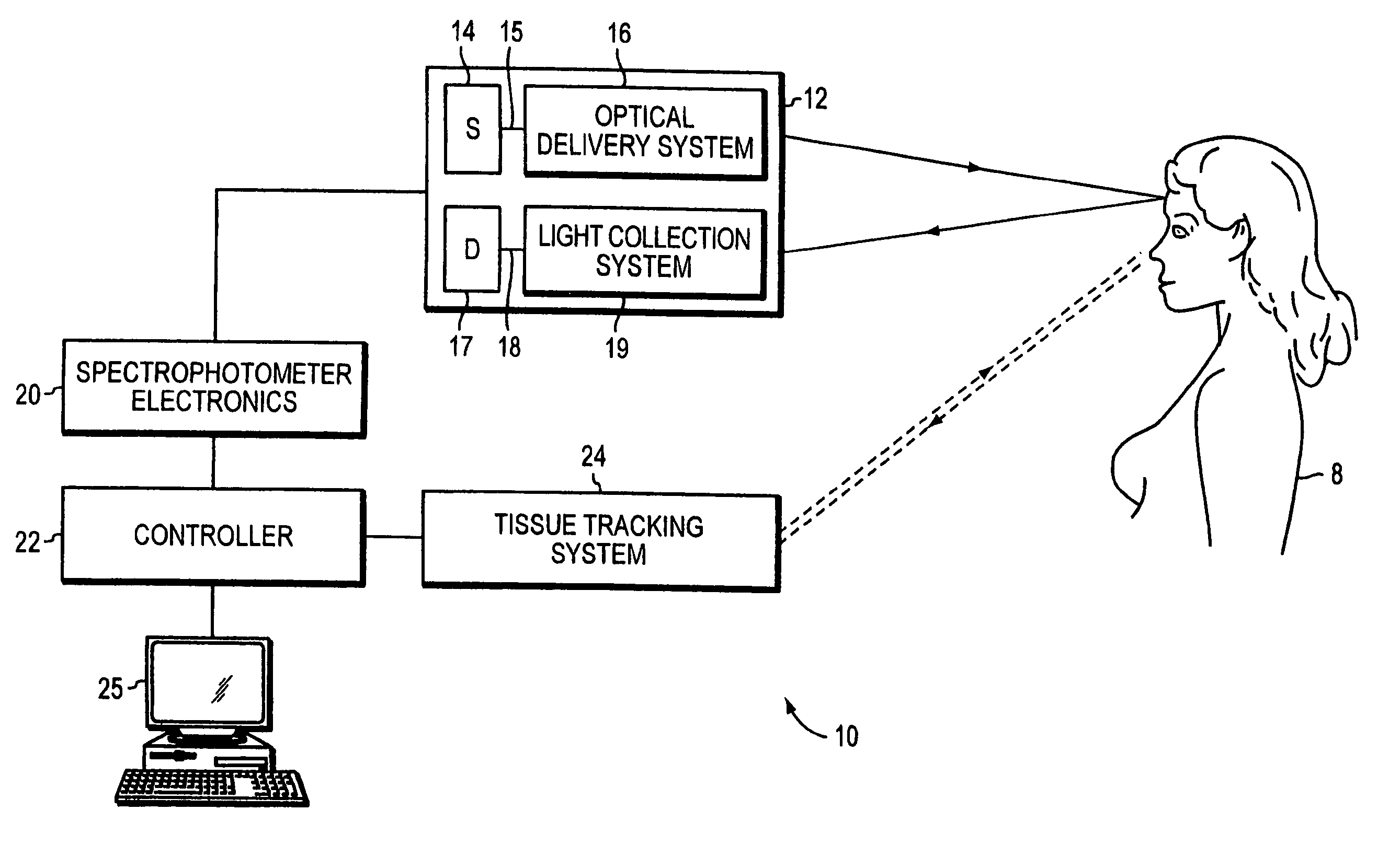
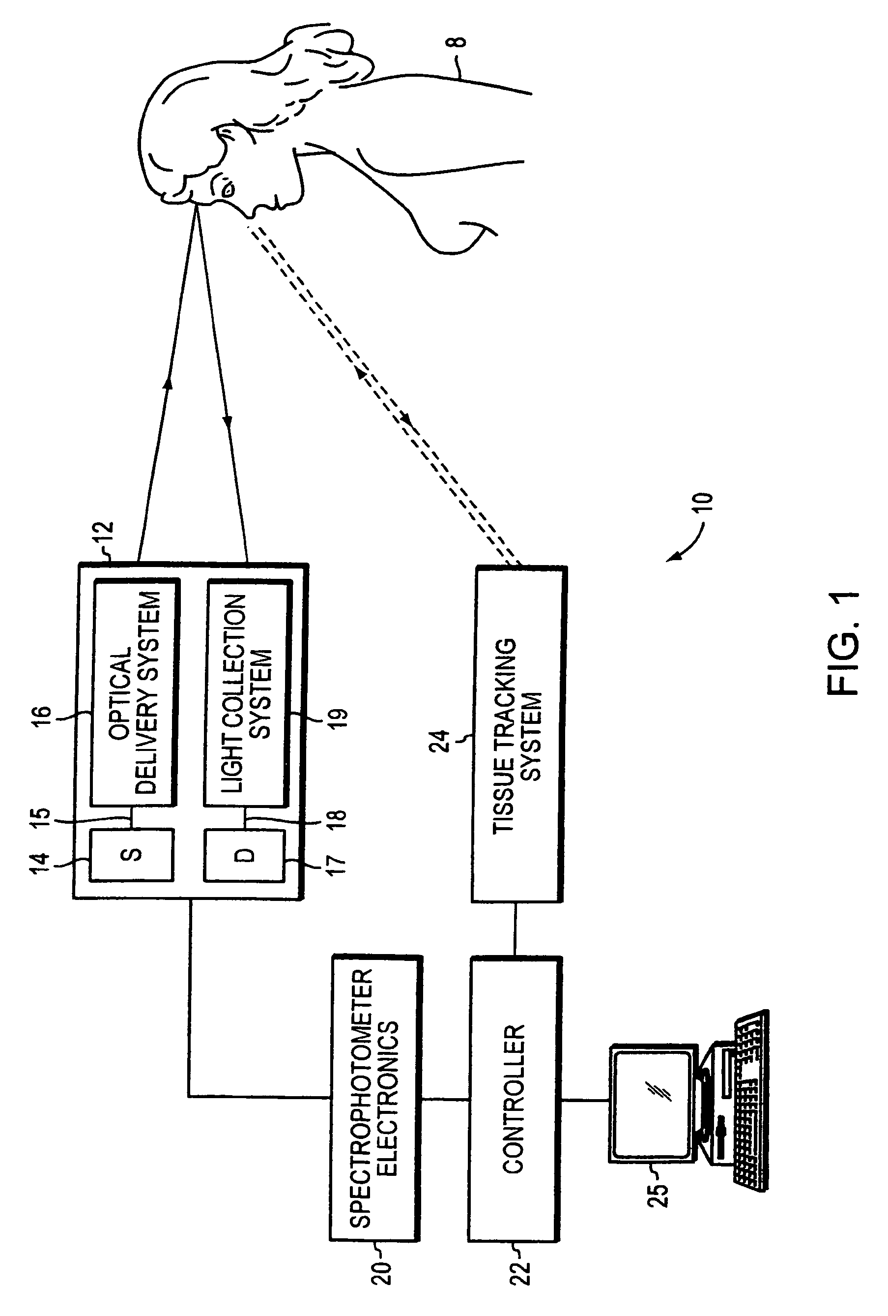
[0063] The described systems and methods use non-contact (i.e., remote) or contact light coupling in four different embodiments. The first embodiment is a spectroscopic system providing contactless (i.e., non-contact) light irradiation and light detection of photons emanating from the tissue surface. This specification primarily describes this type of optical coupling for tissue examination and imaging. In another embodiment, the optical system utilizes remote irradiation and a contact light collection system touching the tissue surface. In the third embodiment, the optical system uses a contact delivery system (being in contact with the tissue surface) and a remote light collection system. The last embodiment is an optical system utilizing contact irradiation and contact light collection.

[0064]FIG. 1 shows schematically a non-contact optical system 10 including a non-contact optical probe 12, spectrophotometer electronics 20, a system controller 22, a tissue tracking system 24 and...


PUM


Abstract

An optical system for examination of biological tissue includes a light source, a light detector, optics and electronics. The light source generates a light beam, transmitted to the biological tissue, spaced apart from the source. The light detector is located away (i.e., in a non-contact position) from the examined biological tissue and is constructed to detect light that has migrated in the examined tissue. The electronics controls the light source and the light detector, and a system separates the reflected photons (e.g., directly reflected or scattered from the surface or superficial photons) from the photons that have migrated in the examined tissue. The system prevents detection of the “noise” photons by the light detector or, after detection, eliminates the “noise” photons in the detected optical data used for tissue examination.

Description

[0001] This application is a continuation-in-part of U.S. application Ser. No. 10 / 752,440, filed on Jan. 5, 2004, entitled “Examination of Biological Tissue using Non-Contact Optical Probes,” which claims priority from U.S. Provisional Application 60 / 438,229, filed on Jan. 4, 2003; and is also a continuation-in-part of U.S. application Ser. No. 10 / 618,579, filed on Jul. 10, 2003, entitled “Examination and Imaging of Brain Cognitive Functions,” all of which are incorporated by reference. This application is also a continuation-in-part of U.S. application Ser. No. 09 / 383,476, filed on Aug. 26, 1999, entitled “Sensing and Interactive Drug Delivery,” which is incorporated by reference.[0002] The present invention relates to in vivo non-invasive examination or imaging of biological tissue including tissue models used to study pathological tissue conditions and diseases including the effects of drug agents thereon. BACKGROUND OF THE INVENTION [0003] X-ray or γ-ray radiation, optical radia...

Claims


Application Information

Patent Timeline
Patent Type & Authority Applications(United States)
IPC IPC(8): A61B5/05A61B6/00
CPCA61B5/0059A61B5/0091A61B5/415A61B5/7285A61B5/4312A61B5/6834A61B5/418A61B5/0037A61B5/0068
Inventor CHANCE, BRITTON
Owner NONINVASIVE TECH
Why Eureka
Try Eureka