Differential protein expression patterns related to disease states

Inactive Publication Date: 2006-03-30
NEOGENOMICS INC
View PDF0 Cites 29 Cited by
  • Summary
  • Abstract
  • Description
  • Claims
  • Application Information

AI Technical Summary

Benefits of technology

[0011] The present invention is a method for determining a pattern of protein expression for a disease or an altered biological state. A method for determining an altered pattern of protein expression comprising: a) collecting a biological sample from an individual having an altered biological state; b) performing a two-dimensional (2D) electrophoretic separation of a plurality of proteins in the biological sample to produce a sample 2D gel pattern; c) coloring the sample 2D gel pattern a first color; d) superimposing the sample 2D gel pattern over a control 2D gel pattern colore

Problems solved by technology

However, proteonomic testing for diagnostic purposes remains in its infancy.
Detection of abnormalities in the genome of an individual can reveal the risk or potential risk for individuals to devel

Method used


Image

  • Differential protein expression patterns related to disease states
  • Differential protein expression patterns related to disease states
  • Differential protein expression patterns related to disease states

Examples


example 1

Differential Protein Expression Patterns in Breast Cancer

[0063] Breast ductal fluid samples were collected by nipple aspiration. The nipple aspirate fluid (NAF) samples were taken from 12 unilateral breast cancer patients, 4 normal women, and two mammogram negative women with a history of breast cancer in their family and where onset of disease had begun at the same age as their age when the samples were taken.

[0064] Each sample was first diluted with the addition of cold RPMI buffer containing an EDTA-free protease inhibitor cocktail. The diluted nipple aspirate fluid was aliquoted into 1.5 ml microfuge tubes in 100 μl portions and frozen in liquid nitrogen before analysis.

[0065] NAF samples were prepared for protein analysis by first washing with trichloroacetic acid (TCA) followed by two washes with acetone. This washing allowed for greater sensitivity of protein separation in the nipple aspirate fluid as compared to previous sample preparation methods, with more than 1200 pro...

example 2

The Identification of Acetyl-LDL Receptor as a Biomarker for Breast Cancer

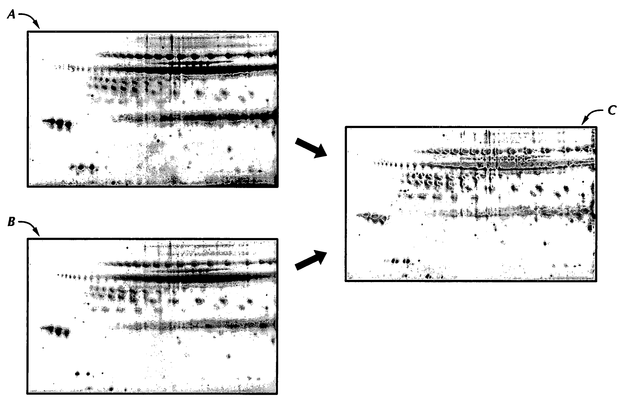
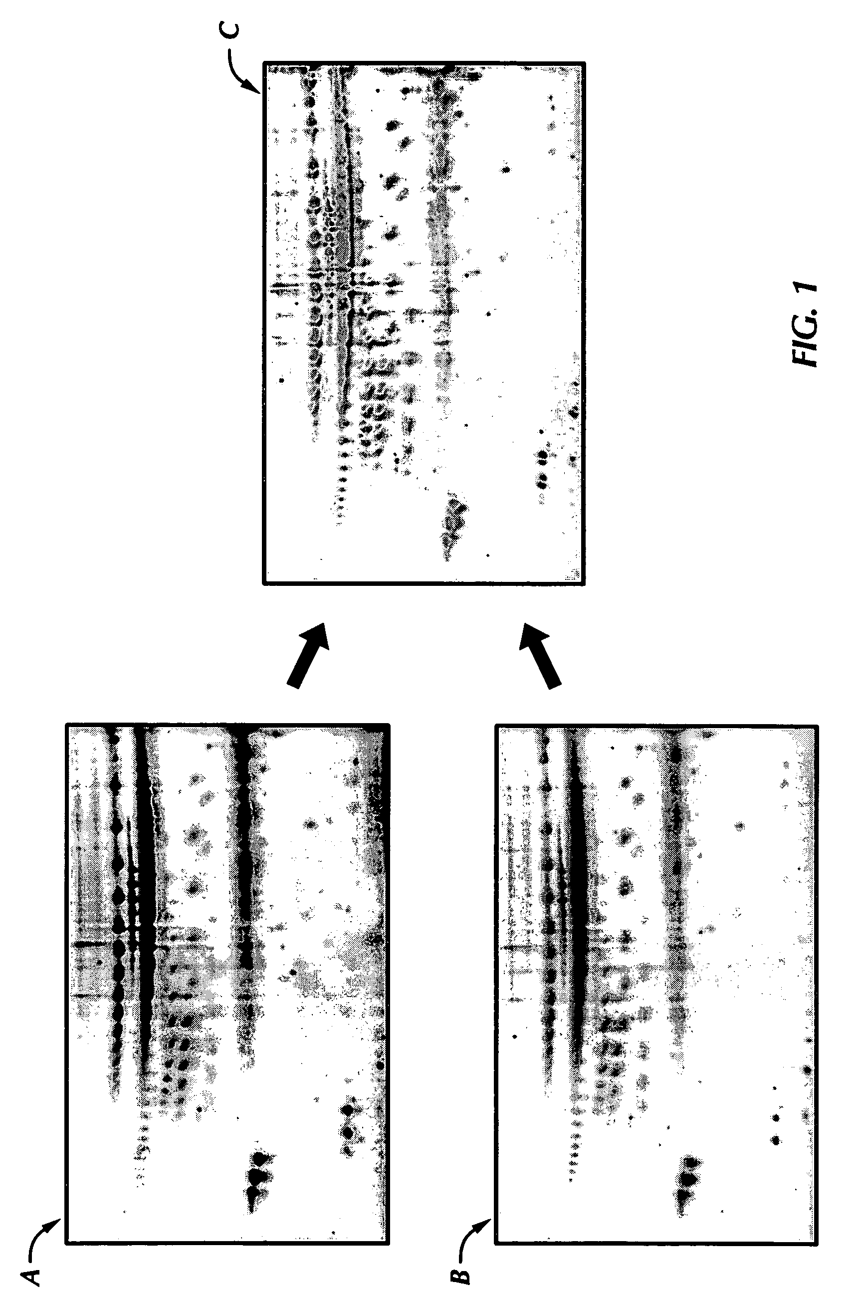
[0085]FIG. 4 identifies the three major protein spots seen in all of the control NAF samples that were reduced in ten of the twelve unilateral breast cancer patients. These protein spots were excised, in-gel digested with trypsin, subjected to mass fingerprinting analysis by matrix-assisted laser desorption ionization-time of flight mass spectrometry (MALDI-TOF MS) and expert database searching.

[0086] Following differential expression analysis, these three major protein spots were carefully excised from the gel for identification. Excised gel spots were destained by washing the gel spots twice in 100 mM NH4HCO3 buffer, followed by soaking the gel spots in 100% acetonitrile for 10 minutes. The acetonitrile was aspirated and a trypsin solution added to the gel spots.

[0087] A small volume of a trypsin solution (approximately 5-15 μg / ml trypsin) was added to the destained gel spots and incubated for 3 hours at ...

example 3

The Acetyl-LDL Receptor in Normal and Diseased Breast

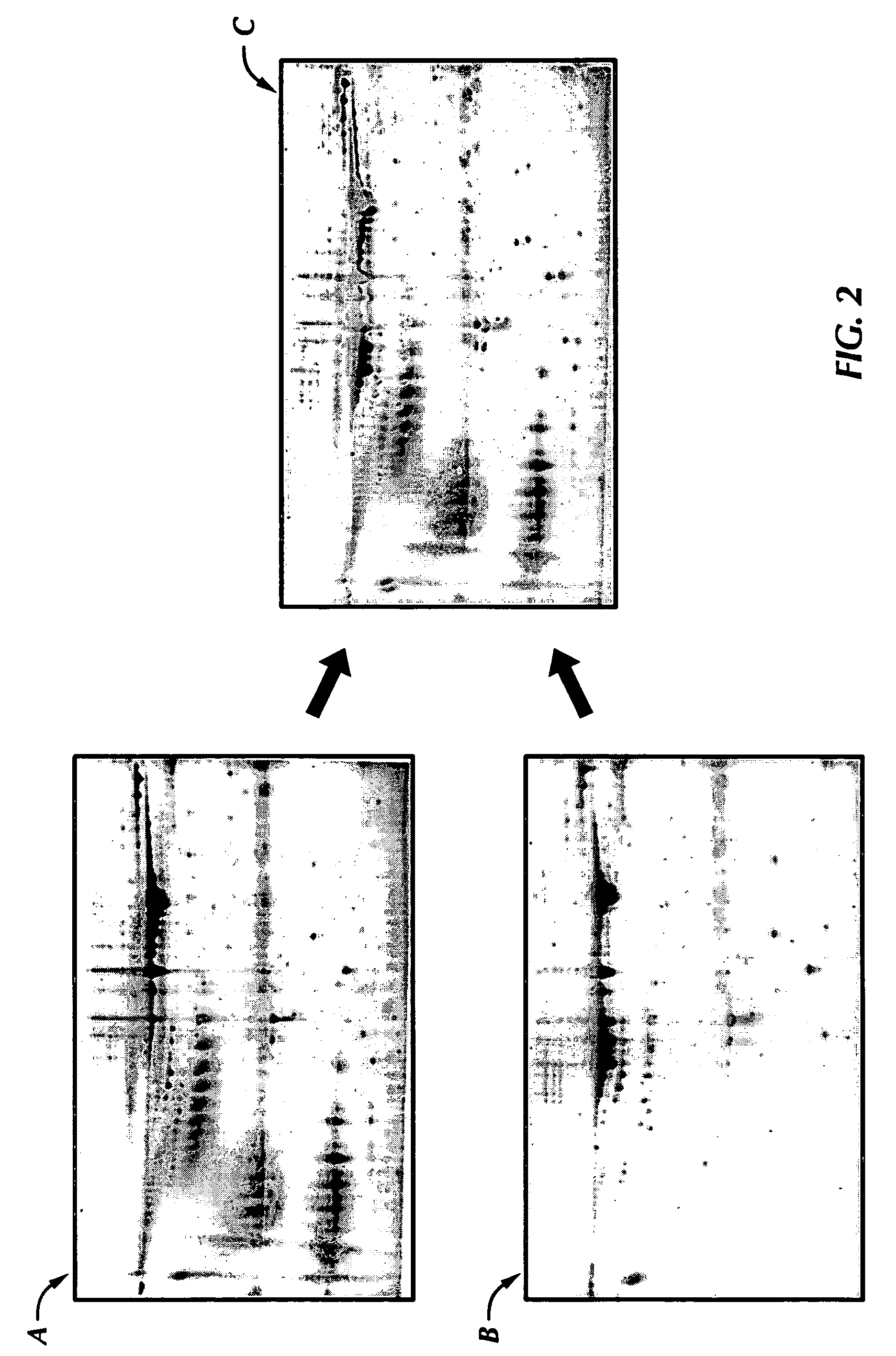
[0092] Levels of the acetyl LDL receptor were elevated in normal breasts and down-regulated in the nipple aspirate fluid sample from the cancerous breast (see FIG. 5). NAF samples were collected from both breasts of four normal women and tested for acetyl-LDL receptor concentration. The acetyl-LDL receptor concentration in the normal breasts ranged from about 8,279 ppm to about 18,669 ppm with a 95% lower confidence limit of 6,073 ppm. The mean concentration of acetyl-LDL receptor in the eight control breasts was 12,581 ppm with a standard deviation of 3,956 ppm. Thus, a normal value of acetyl-LDL receptor protein in control NAF samples was determined to be equal to or more than 8,625 ppm (the mean value minus one standard deviation) or more than or equal to 6,073 ppm (the 95% lower confidence limit of the concentration of the acetyl-LDL receptor in control breasts).

[0093] NAF samples of both breasts of the twelve unilateral bre...


PUM


Abstract

The present invention is a method for determining protein expression profiles in disease or an altered biological state. The method is based on the use of two-dimensional (2D) gel electrophoresis where the gel images are assigned two different colors. The gels are then compared by an overlay procedure that allows for identification and quantification of unique proteins by determining which colors are detected in the superimposed images and the density of those colors.

Description

CROSS REFERENCE TO RELATED APPLICATIONS [0001] This application claims priority to U.S. Provisional Patent Application Ser. No. 60 / 614,315 filed Sep. 29, 2004 and entitled “Differential Protein Expression Patterns Related to Disease States” by inventors Ira L. Goldknopf, et al.BACKGROUND OF THE INVENTION [0002] 1. Field of the Invention [0003] The invention relates to a method for determining biomarkers and protein expression patterns and using those biomarkers and protein expression patterns to define diseases or altered biological states. The method is based on use of proteomic analysis to identify biomarkers and protein expression patterns that define disease states or alterations in normal biological processes. [0004] 2. Description of the Related Art [0005] Proteomics is a new field of medical research wherein proteins are identified and linked to biological functions, including roles in a variety of disease states. With the completion of the mapping of the human genome, the id...

Claims


Application Information

Patent Timeline
IPC IPC(8): G01N33/574G06K9/00
CPCG01N33/57415G01N2550/00G01N33/6896G01N33/6842
Inventor GOLDKNOPF, IRA L.SHETA, ESSAM A.BLACK, MELANIE B.WILSON, CHRIS W.PARK, HELEN R.
Owner NEOGENOMICS INC
Why Eureka
Try Eureka