Method and apparatus for performing maxillary sinus elevation

a maxillary sinus and elevation technology, applied in the field of osseous regeneration of the maxillary sinus, can solve the problems of limited dental implants placement in the anterior lower jaw, inferior dental bridge alternatives in comparison to dental implants, and considerable challenges in the replacement of the maxillary posterior teeth

Inactive Publication Date: 2006-04-20
HOCHMAN MARK N
View PDF19 Cites 119 Cited by
  • Summary
  • Abstract
  • Description
  • Claims
  • Application Information

AI Technical Summary

Benefits of technology

[0027] A method and device is disclosed that provides a new approach to the placement of dental implants when inadequate bone mass exists in pat

Problems solved by technology

Initially the placement of dental implants were limited to the anterior lower jaw, as this region provided sufficient bone quantity, quality and strength to support and hold a dental implant having an effective length.
The previous alternative of dental bridges is now considered an inferior alternative in comparison to dental implants.
To that end, the replacement of the maxillary posterior teeth have presented a considerable challenge because, after the loss of maxillary posterior teeth the quality and quantity of the remaining supporting bone may be insufficient to support implants properly or reliably.
Once a tooth is removed from the complex, the surrounding alveolar bone is frequently resorbed because of the lack of physical stimulation and support of the teeth.
This leads to a loss of bone mass and a corresponding reduction in the effective height and thickness of the bone of the maxillary complex.
In addition, with the loss of teeth from the upper jaw (and the maxillary complex) the compensatory enlargement of the maxillary sinus occurs that also reduces the vertical height of the remaining bone of the maxillary complex.
These effects further compromise the option for dental implants to be used.
If the membrane is not properly dissected from the bone, bone augmentation may not occur, or may not be sufficient.
Unintentional perforation of the subantral membrane may also lead to undesirable short and long-term consequences.
A lack of integrity of the membrane can also lead to the migration of regenerative bone materials leading to long-term chronic infections.
This technique is fraught with many risks and complications because of the limitations of healing potential in the maxillary sinus.
While, this technique is safer, an overzealous use of an osteotome during the placement of the regenerative material can result in the perforation of the subantral membrane with disadvantages discussed above.
The new technique also uses an inferior approach to the membrane via the alveolar

Method used


Image

  • Method and apparatus for performing maxillary sinus elevation
  • Method and apparatus for performing maxillary sinus elevation
  • Method and apparatus for performing maxillary sinus elevation

Examples


Embodiment Construction

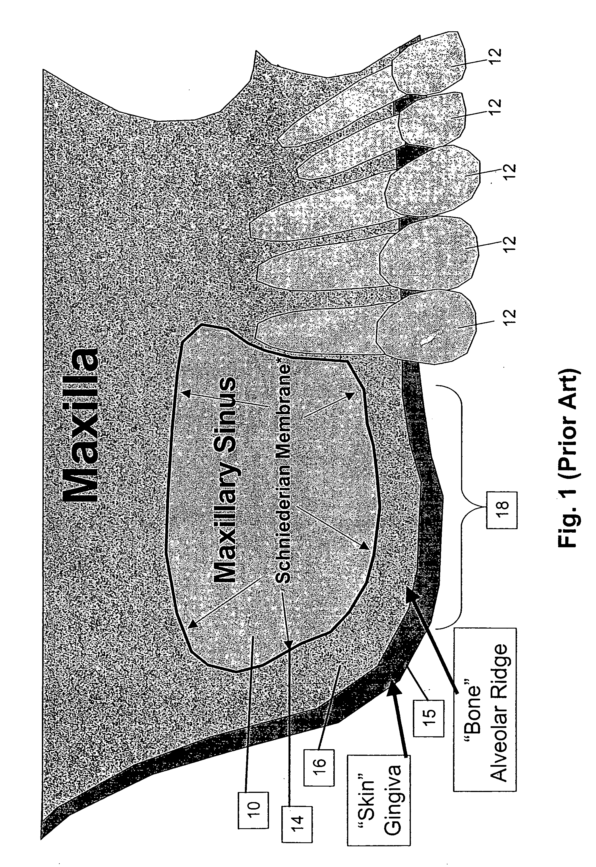
[0041]FIG. 1 shows a typical cross section of the maxillary sinus 10. Several teeth 12 are seen in the Figure in a normal configuration. The maxillary sinus 10 is separated from the mouth by the alveolar ridge 16 formed of bone. The bone is covered on top by the subantral membrane 14 and on the bottom by the gingiva 15. As shown in the Figure, because this person has lost several molars, the floor of the maxillary sinus has been depressed or lowered in the area 18. As a result, the alveolar ridge 16 in this region has lost considerable bone mass and quality. Frequently, the bone in this area is porous and is not strong enough to support an implant of sufficient length and an associated final dental prosthesis. Typically, in a health person the ridge 16 in this area may be about 8 to 14 mm however, in persons who have lost their molars, the thickness of the ridge 16 may be no more than about 4 mm.

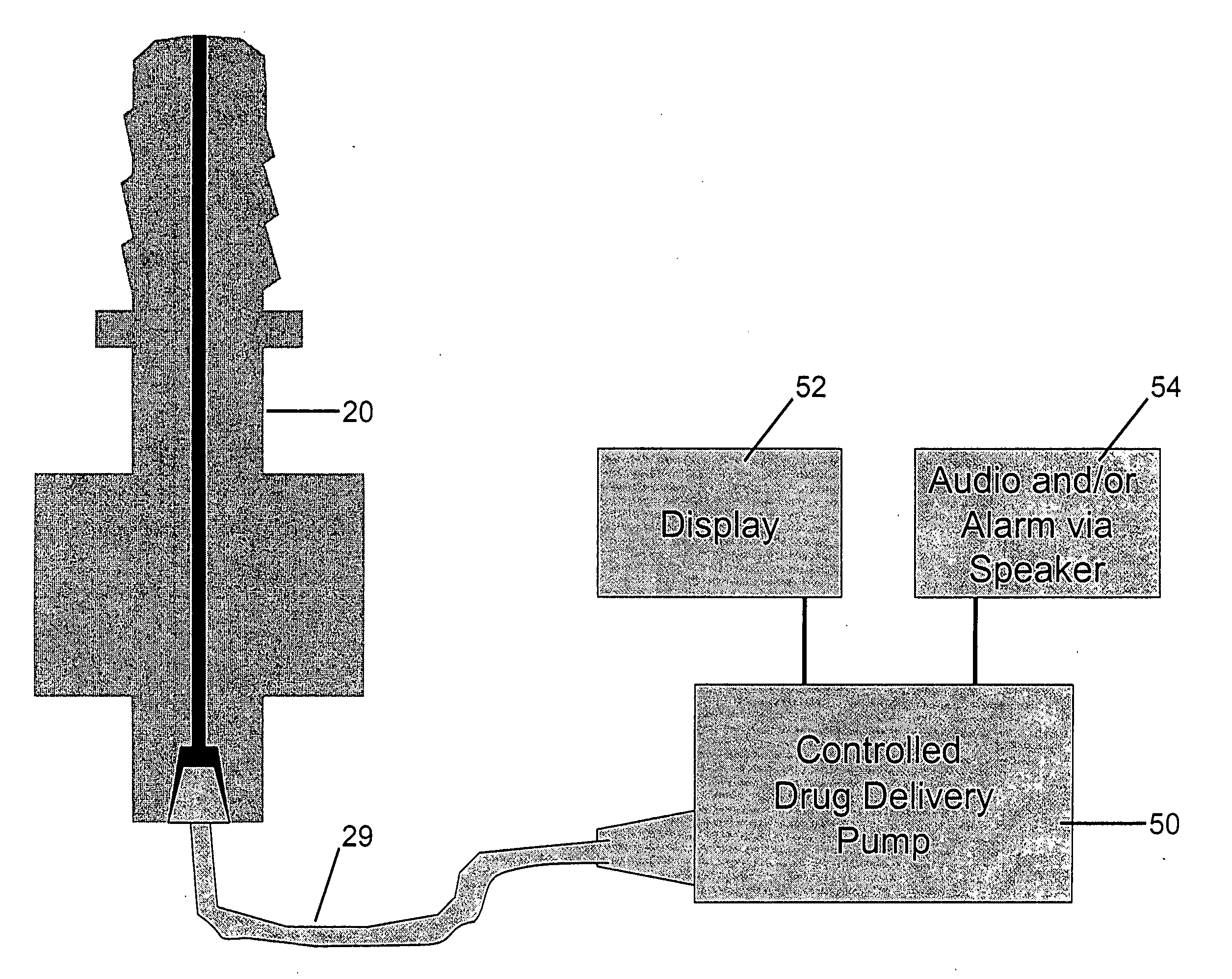
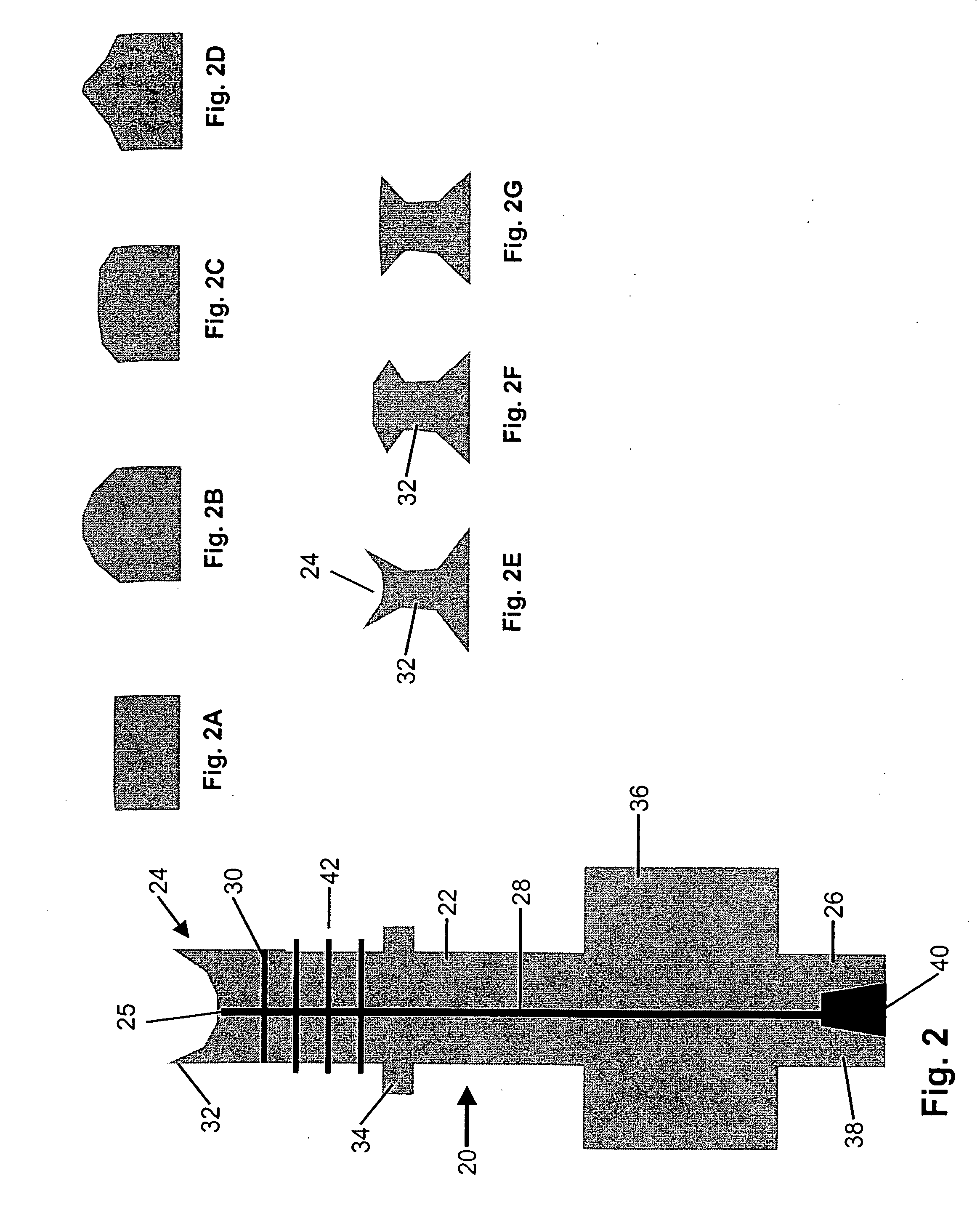
[0042]FIG. 2 shows a first embodiment of a sleeve 20 constructed in accordance with thi...


PUM


Abstract

A method and apparatus is disclosed for providing implants in the upper jaws of a person. A sleeve is inserted through the alveolar ridge to the maxillary sinus. The sleeve is used to raise the subantral membrane and form a cavity. A filler, such as a bone growth stimulant is injected through the sleeve into the cavity. In the process, the sleeve also can cut and/or condense the bone around itself so that the bone can hold an implant. Optionally, the bone growth stimulant is also introduced into the bone surrounding the sleeve. During the injection, the pressure within the sleeve or the cavity is monitored to detect and prevent the rupture of the subantral membrane.

Description

RELATED APPLICATIONS [0001] This application claims priority to U.S. provisional application Ser. No. 60 / 619,542 filed Oct. 15, 2004 and incorporated herein by reference.BACKGROUND OF THE INVENTION [0002] A. Field of Invention [0003] This invention relates generally to improvements to the delivery of drugs, particularly for osseous regeneration for the maxillary sinus. More specifically this invention provides a method and device to the elevation of the floor of the maxillary sinus to increase the amount of bone available based on using hydro-dissection to raise the subantral membrane floor of the sinus (a.k.a. the Schneiderian membrane) allowing placement of osseous regenerative materials. [0004] B. Description of the Prior Art [0005] Dental implants have been used in dentistry for about 20 years. They offer a tremendous benefit to patients by allowing the replacement of missing teeth. Prof. Per-Ingvaar Branemark introduced the use of titanium dental implants that showed that predi...

Claims


Application Information

Patent Timeline
IPC IPC(8): A61C8/00
CPCA61C8/0033A61C8/0092
Inventor HOCHMAN, MARK N.
Owner HOCHMAN MARK N
Why Eureka
Try Eureka