Method for the rapid image processing of medical images

Inactive Publication Date: 2006-05-04
SIEMENS AG
View PDF5 Cites 1 Cited by
  • Summary
  • Abstract
  • Description
  • Claims
  • Application Information

AI Technical Summary

Benefits of technology

[0015] In a preferred development of the present method, the second part of the image processing algorithm, which is executed on the master processor, is implemented via a command extension available with standard processors for the rapid parallel signal processing, such as in MMX, SSE, SSE2. The combined use of this command extension of the master processor and the use of pixel shade units on the graphics processor allow a very rapid calculation of the post processing algorithms on this standard compon

Problems solved by technology

The main problem of image processing is the high processing speed, as the doctor requires the images in real-time where possible for instrument navigation in the case of interventions, particularly with the use of a catheter.
With current master processors, the processing speed needed for this purpose cannot be achieved with the above image resolutions.
The development of

Method used


Image

  • Method for the rapid image processing of medical images
  • Method for the rapid image processing of medical images

Examples


Embodiment Construction

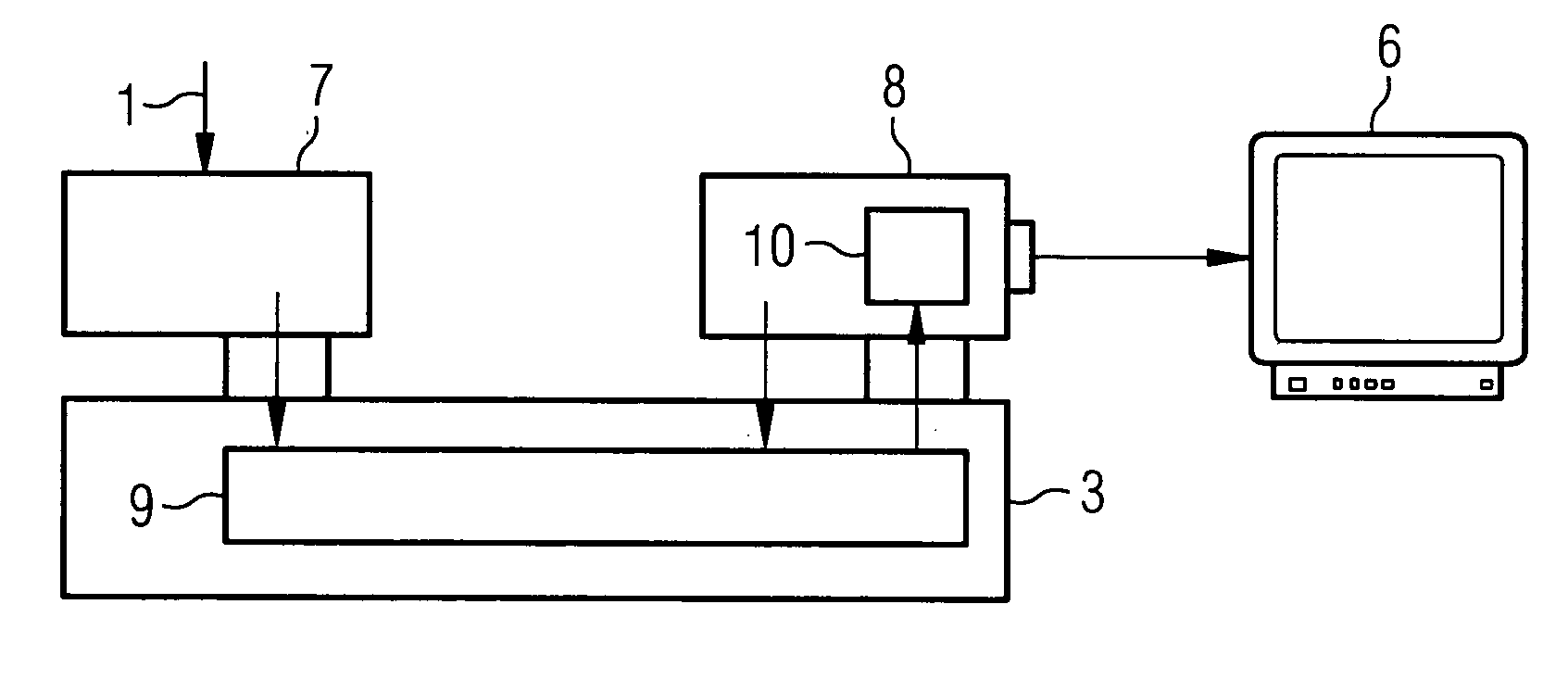
[0019]FIG. 1 shows an example for the ratio with the image processing of medical images which are hitherto present with the x-ray imaging. The raw data obtained by the x-ray detector 1 is directly fed to a digital signal processor 2 (DSP) which carries out the complete image processing. The digital signal processor 2 is additionally provided at a conventional PC3, by means of which the command input is effected. The image data processed by the digital signal processor 2 (DSP) is fed to a special graphics card 4 by means of a direct link 5, by means of which the processed images are displayed on a monitor 6. The master processor of the PC 3 does not take part in the image processing, but can however receive the processed image data from the digital signal processor 2, in order to store these for instance for a later display or further processing.

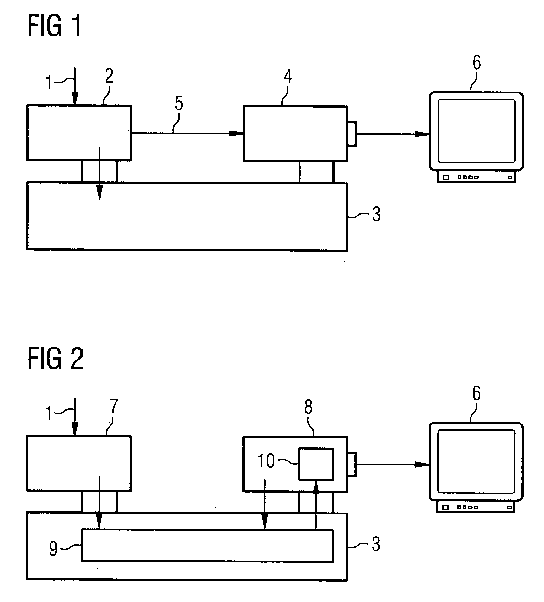
[0020] Contrastingly, FIG. 2 shows the conditions present in the implementation of the present method. In this example, the raw data is acq...


PUM


Abstract

The present invention relates to a method for the rapid image processing of medical images, in particular fluoroscopic image recordings using image-modifying image processing algorithms on an image computer. With this method, a first part of the image processing algorithms is executed on a graphics processor of a graphics card and a second remaining part is executed on a master processor of the image computer. This division, for which standard hardware components are to be used, allows the omission of complex special developments of the hardware for the image computer.

Description

CROSS REFERENCE TO RELATED APPLICATIONS [0001] This application claims priority to the German application No. 10 2004 051 568.9, filed Oct. 22, 2004 which is incorporated by reference herein in its entirety. [0002] 1. Field of the Invention [0003] The present invention relates to a method for the rapid image processing of medical images, in particular fluoroscopic image recordings, using image-modifying image processing algorithms on an image processor. [0004] 2. Background of Invention [0005] Medical-specific imaging methods such as computer tomography, x-ray angiography or magnetic resonance tomography for instance, require complex image processing of the images recorded using the corresponding modalities. This image processing is intended on the one hand to improve the image quality, for example by noise suppression, and on the other hand to highlight structures in the images essential for the respective diagnosis, for instance by means of edge sharpening or filtering. SUMMARY OF...

Claims


Application Information

Patent Timeline
IPC IPC(8): G06F15/16G06T15/00
CPCG06T1/20G06T15/005
Inventor DREXLER, CHRISTOPHERHORNEGGER, JOACHIMMACK, MARTIN
Owner SIEMENS AG
Why Eureka
Try Eureka