
Animal model systems for viral pathogenesis of neurodegeneration, autoimmune demyelination, and diabetes

a model system and neurodegeneration technology, applied in the field of animal model systems for the pathogenesis of neurodegeneration, autoimmune demyelination, diabetes, can solve the problems of cns invasion, significant women's health issue, and damage to myelin and/or neurons, and achieve the effect of reducing the severity of a diseas

Inactive Publication Date: 2006-06-15
CARANTECH
View PDF0 Cites 21 Cited by
  • Summary
  • Abstract
  • Description
  • Claims
  • Application Information

AI Technical Summary

Benefits of technology

"The present invention provides non-human animal model systems for viral pathogenesis of neurodegeneration, autoimmune demyelination, and diabetes. These models can be used for studying disease mechanisms and identifying and characterizing candidate therapeutics for these diseases. The non-human primate animal model systems are infected with herpesvirus and show similarities to the human disease states, making them useful for research and development of treatments. The invention also provides a wide range of herpesvirus variants that can be used to infect non-human primates. Overall, the invention provides valuable tools for research and development of treatments for neurological and autoimmune diseases."

Problems solved by technology

MS affects women twice as often as men, and thus also represents a significant women's health issue.
CNS invasion by T cells following viral infection, whether due to mimicry or to clear the acute infestation, may also damage the myelin and / or neurons, through either direct cytotoxicity to cells harboring the virus or production of pro-inflammatory products that create a toxic environment within the CNS and activation of macrophages or microglia (bystander damage).
It is also widely perceived that viral infections may trigger MS attacks.
One difficulty in establishing direct relationships between viral exposures and MS is that appropriate in vivo experimental systems for validation of such associations are lacking.
Only inbred susceptible strains subsequently develop an unrelenting and severe progressive demyelinating disease with what is believed to be bystander damage to myelin.
It is important to understand that although these models have provided the first (and only existing) insights into the relationships between autoimmune CNS demyelination and viral infections, they are still insufficient for proving direct association of MS with any of the viruses that ubiquitously infect humans without adverse consequences.
CNS complications of TMEV infections are under the restrictive control of genetic influence and it is difficult to extrapolate their mechanisms to outbred human populations.
Many of these disease models require intracranial injection of viruses in neonatal animals, an artificial situation that similar to EAE does not mimic natural exposure of humans to pathogens.
As for other viruses, however, evidence for a direct link of causality between HHV6-A and disease pathogenesis has been lacking.
Because >95% of the general population is exposed to the virus during infancy, it is difficult to envision HHV6 infection as a sole cause for MS prevalence (approximately 1:1,000 for Caucasions in the United States) in the absence of additional factors of pathogenesis.
Yet, there is still no direct evidence of an association between any viral exposures and common forms of MS.
These observation however, has not been confirmed by subsequent attempts (Coates et al., Nat Med 4:537-8 (1998)), and could not be formally confirmed by immunohistochemistry.
Thus, detection of viral sequences in the CNS is not sufficient for proof of pathogenicity.
However, a large number of subsequent studies that examined IgG / IgM reactivity in serum and / or CSF, presence of HHV6 DNA or viral transcripts in serum, CSF and brain, peripheral T cell proliferative responses to HHV6, or virus recovery in culture have not unequivocally confirmed these results.

Method used


Image

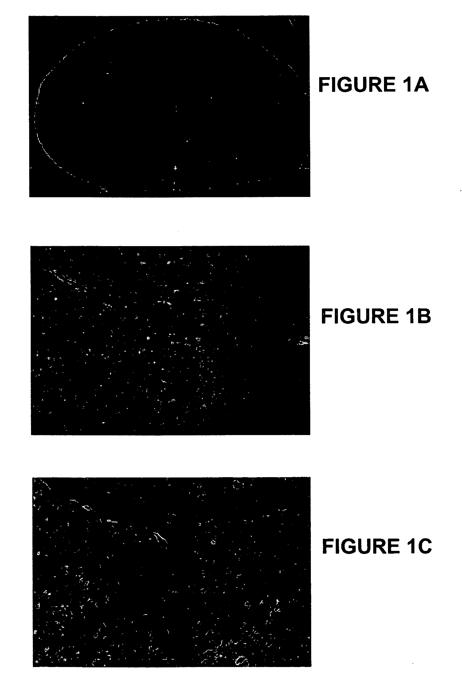
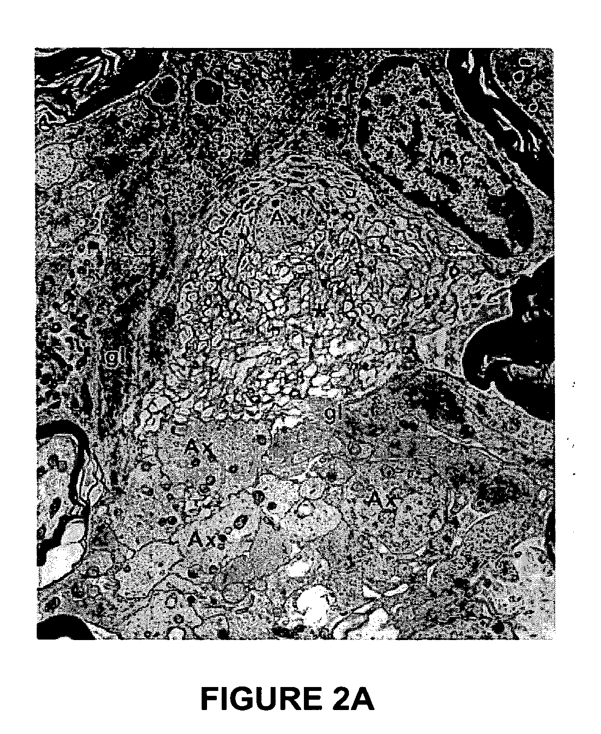
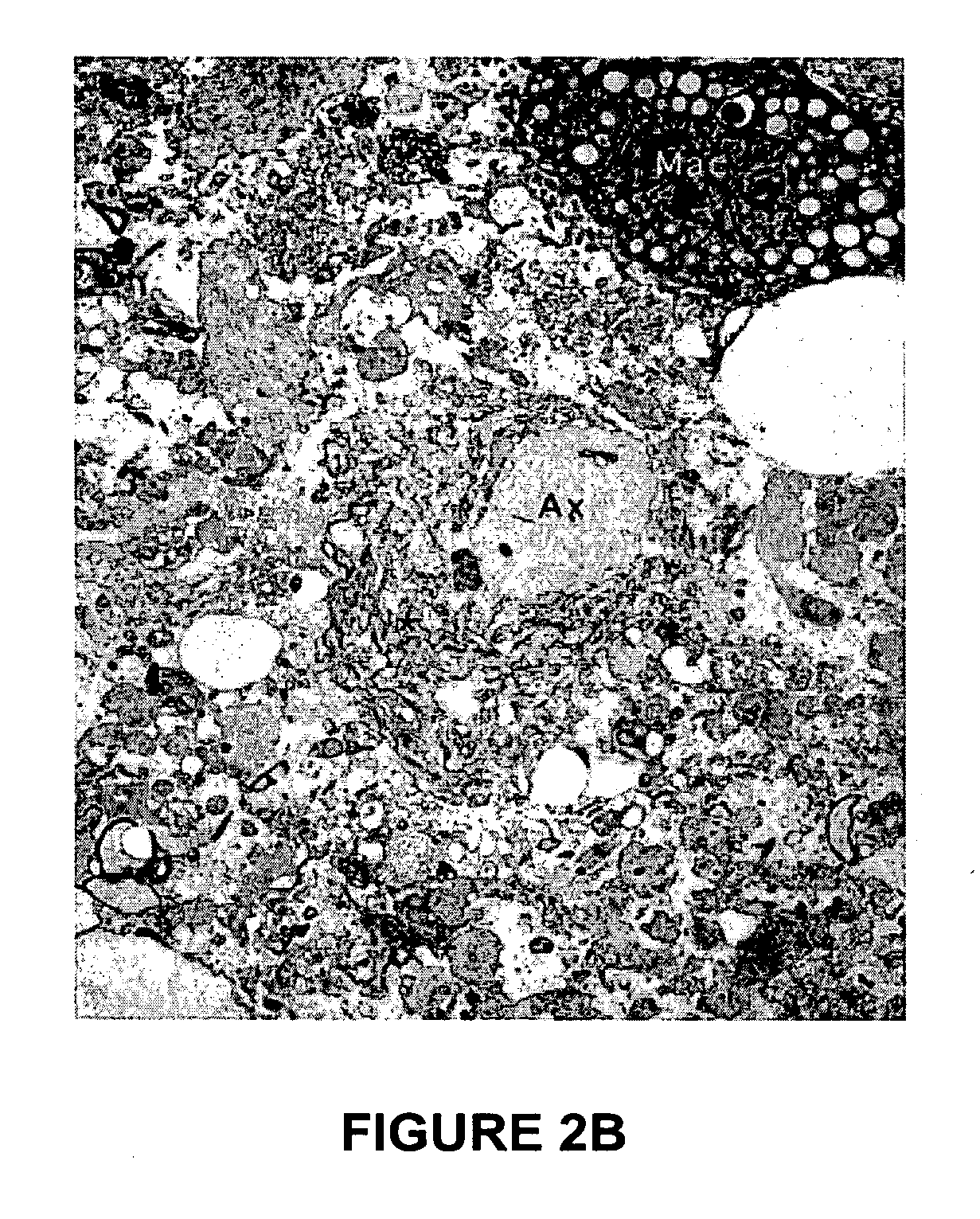
  • Animal model systems for viral pathogenesis of neurodegeneration, autoimmune demyelination, and diabetes
  • Animal model systems for viral pathogenesis of neurodegeneration, autoimmune demyelination, and diabetes
  • Animal model systems for viral pathogenesis of neurodegeneration, autoimmune demyelination, and diabetes

Examples


example 1

Infection of Marmoset Cells In Vitro

[0146] This Example demonstrates the susceptibility of marmoset lymphocytes (PBMC) to infection in vitro with HHV6 variants A and B.

[0147] Common marmosets are susceptible to infection by herpesviruses (Provost et al., J Virol 61:2951-2955 (1987); Jenson et al., J Gen Virol 83:1621-1633 (2002); Ramer et al., Comp Med 50:59-68 (2000); Farrell et al., J Gen Virol 78(6):1417-1424 (1997); Cox et al., J Gen Virol 77(6):1173-1180 (1996); Wedderburn et al., J Infect Dis 150:878-882 (1984); Johnson et al., Proc Natl Acad Sci USA 78:6391-6395 (1981); de-The et al., Intervirology 14:284-291 (1980); Ablashi et al., Biomedicine 29:7-10 (1978); Falk et al., Int J Cancer 17:785-788 (1976)), and express a CD46 molecule that is highly homologous to the human HHV6 receptor. Murakami et al., Biochem J 330:1351-1359 (1998). Using trans-well co-cultures with HHV6-infected human T cell lines, it was demonstrated that marmoset lymphocytes (PBMC) can be infected in vi...

example 2

Infection of Marmosets In Vivo

[0148] This Example demonstrates the susceptibility of C. jacchus marmosets to in vivo infection with HHV6 variants A and B.

[0149] Seven adult marmosets were infected with HHV6 in vivo using various protocols (summarized in Table 3 below): (1) intravenous inoculation of the animal's own PBMC infected ex vivo with HHV6-A or HHV6-B (as verified by IFA and PCR), followed by intravenous injection of a cell lysate containing identical live virus variant 6-8 weeks later; (2) two intravenous injections of viral lysates from MOLT3 HHV6-B-infected cultures at 5 weeks interval; and (3) one inoculation of HSB2 cells infected with HHV6-A (HHV6-A+ HSB2), followed by injection of either infected or uninfected cells 3 months later.

TABLE 3Infection of Common Marmosets with HHV6 VariantsAgeClinicalEuthanasiaInflam-Animal ID(yrs)SexInfusion 1 (day 0)Infusion 2 (day)signs(day)mationDemyelination190-947F8 × 106 HHV6-A+· PBMCHHV6-A+ HSB2 Lysate (42)Yes68++125-9FHHV6-B+·...

example 3

Immuno-Reactivity to Viral Antigens

[0154] This Example discloses methodology for monitoring T cell and antibody responses to HHV6-Antigens.

[0155] Methods were developed to serially monitor T cell and antibody responses to viral antigens. Preliminary evidence indicated that marmosets in the colony were naïve to HHV6, and that antibody reactivity appeared after inoculation. (See, FIG. 8.) No significant T cell reactivity was detected in PBMC or lymphoid organs. The presence of HHV6 DNA was monitored serially by nested PCR methodology. Consistent with the known tropism of HHV6 variants, HHV6-B, but not HHV6-A, was detected in the blood of infected animals. (See, FIG. 9.)

[0156] Importantly, while clear serum (IgG antibody) reactivity to HHV6-infected cells was observed to appear during the weeks following inoculation and was readily detected using FACS analysis, no IgG reactivity at all was detected in any of these sera by standard ELISA methods utilizing purified viral lysates coate...


PUM

PropertyMeasurementUnit
solubleaaaaaaaaaa
full-length aaaaaaaaaa
fluorescence activated cell sorting analysisaaaaaaaaaa

Abstract

Provided are non-human animal model systems for viral pathogenesis of neurodegeneration, autoimmune demyelination, and autoimmune diseases such as diseases of the central nervous system, including multiple sclerosis (MS), and diabetes. Such non-human animal model systems may be suitably employed for the study of diseases such as MS and diabetes and for the identification and characterization of candidate therapeutic compounds and compositions for the treatment of such diseases. Also provided herein are markers and methods for the detection, in patients susceptible to autoimmune disease, of autoimmune diseases of the central nervous system such as progressive multifocal leukoencephalopathy (PML) following treatment with one or more therapeutic agent as exemplified herein by the therapeutic agent natalizumab. Exemplary animal model systems comprise marmosets infected with a herpesvirus such as HHV6-A and HHV6-B, transgenic mouse and zebrafish animal model systems wherein the transgene encodes CD46, and methods for monitoring the risks of patients having MS, diabetes and other auto-immune disorders treated with anti-adhesion molecules such as natalizumab.

Description

CROSS-REFERENCE TO RELATED APPLICATIONS [0001] This application claims priority under 35 U.S.C. § 119(e) to U.S. Provisional Patent Application No. 60 / 618,277, filed Oct. 12, 2004, and to co-pending U.S. Provisional Patent Application No. 60 / 720,676, filed Sep. 26, 2005, each of which is incorporated herein by reference in its entirety.GOVERNMENT SUPPORT [0002] Certain aspects of the presently disclosed inventions were developed with support from a National Multiple Sclerosis Society Pilot Grant No. PP0916. The Government may have certain rights to some aspects of those inventions.BACKGROUND OF THE INVENTION [0003] 1. Technical Field of the Invention [0004] The present invention relates generally to viral pathogenesis and autoimmune diseases such as diseases of the central nervous system, including multiple sclerosis (MS), and diabetes. More specifically, provided herein are non-human animal model systems for viral pathogenesis of neurodegeneration and autoimmune demyelination. Such...

Claims


Application Information

Patent Timeline
Patent Type & Authority Applications(United States)
IPC IPC(8): A01K67/027
CPCA01K67/027A01K67/0275A01K2217/05A01K2227/105A01K2227/106A01K2227/40A01K2267/03A01K2267/0325A01K2267/0337A01K2267/0356A01K2267/0362C07K14/70596C12N15/8509C12N2710/16511A61P25/00A61P25/28A61P35/00A61P37/02A61P43/00
Inventor GENAIN, CLAUDE
Owner CARANTECH
Learn More