Device and method for inducing vascular injury and/or blockage in an animal model

a technology of which is applied in the field of devices and methods for inducing vascular injury and/or blockage in animal models, can solve the problems of direct photoionization of the tissue at the focus and thermoelastic damage to the local surrounding tissue, and achieve the effect of facilitating injury

Inactive Publication Date: 2006-06-29
U S GOVERNMENT REPRESENTED BY THE DEPT OF VETERANS AFFAIRS
View PDF19 Cites 31 Cited by
  • Summary
  • Abstract
  • Description
  • Claims
  • Application Information

AI Technical Summary

Benefits of technology

[0021] The present invention provides a device and method for optically inducing precision vascular injury and / or blockage deep in the tissue of an animal. No exogenous agents are required to facilitate the injury, as the laser light interacts directly with the endogenous tissue and fluids. In an exemplary embodiment, a tightly focused beam of high intensity, ultrashort, laser pulses drives nonlinear interactions between the laser light and the tissue at the focus of the beam, producing photodisruption at the targeted vasculature. These nonlinear interactions result in both direct photoionization of the tissue at the focus and thermoelastic damage to the local surrounding tissue. As a result of these damage mechanisms, a localized hemorrhage, thrombosis, or breach of the blood-brain barrier is formed within a single targeted vessel, providing an in vivo animal model for vascular injury or blockage. Because of the nonlinear nature of the laser-tissue interaction, photodisruption can be targeted deep to the surface of the tissue without extensive collateral damage to tissue surrounding the targeted vessel.

Problems solved by technology

These nonlinear interactions result in both direct photoionization of the tissue at the focus and thermoelastic damage to the local surrounding tissue.

Method used


Image

  • Device and method for inducing vascular injury and/or blockage in an animal model
  • Device and method for inducing vascular injury and/or blockage in an animal model
  • Device and method for inducing vascular injury and/or blockage in an animal model

Examples


example 1

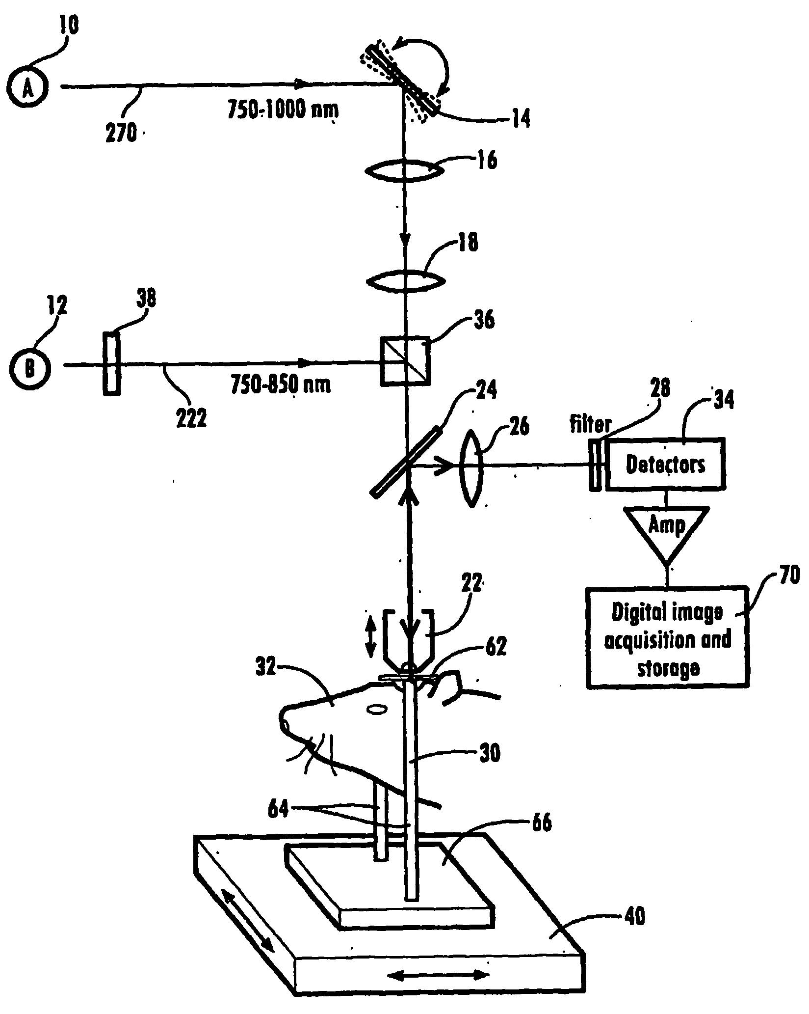
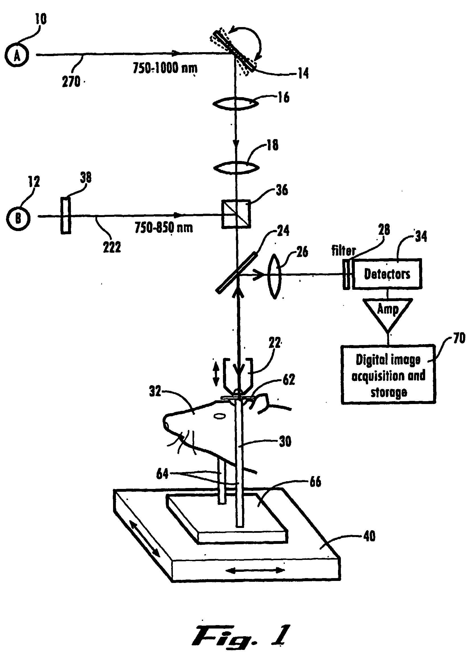
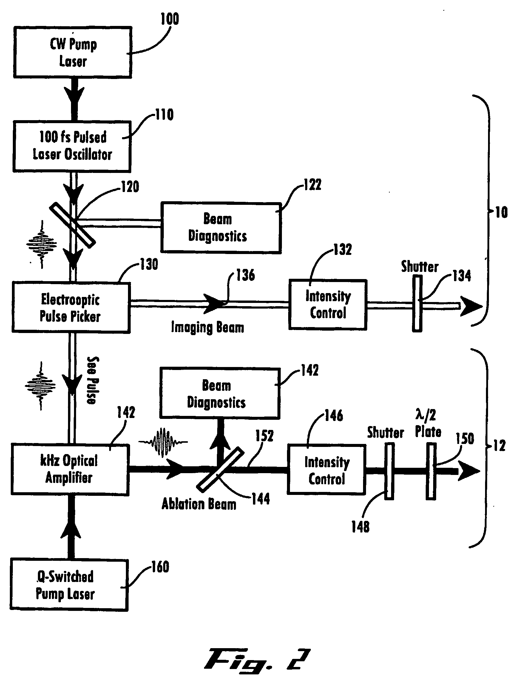
[0050] Sprague-Dawley rats, 100-300 grams in weight, were prepared in order to provide optical access to neuronal blood flow. The animals were anesthetized with urethane and craniotomies, roughly 4 millimeters by 4 millimeters in extent, were performed over parietal cortex to create a cranial window. The dura was removed and a metal frame 62 was glued to the skull. The exposed brain surface was covered with 1.0 to 1.5% low gelling temperature agarose(W / V) in artificial cerebral spinal fluid (ACSF). A glass coverslip was clipped in place on top of the agarose to maintain pressure over the brain and to provide optical access. Water soluble fluorescein-isothiocyanate dextran was injected intravenously to label the blood plasma for the purpose of targeting and imaging vessels. Alternatively, a thinned-skull preparation can be performed to gain optical access to the neuronal blood flow. The metal frame is then mounted to a kinematic mount and computer-controlled translation stage at the ...

example 2

[0058] Using the experimental set-up (animal preparation, system configuration and measurement) described in Example 1, photodisruption was performed on vessels in the cortex ranging in depth from the surface to ˜175 micrometers in depth. The resulting vascular disruption was found to divide into three categories, which are listed below in order of the approximate severity and size of the damage to the microvessel:

[0059] Thrombosis

[0060] Near the threshold energy for vascular disruption and with a limited number of pulses, photodisruption results in extremely limited extravasation of plasma. TPSLM images were taken at various time points before, during and after vascular photodisruption. In an exemplary experiment, the vessel is intact before irradiation. After irradiation with 10 pulses of 0.3 microjoules, a small amount of extravasated plasma could be visualized as fluorescence outside the vessel walls, however, the vessel lumen remained unobstructed. Extravasated fluorescence c...


PUM


Abstract

Ultrashort laser pulses are used to induce photodisruptive breakdown in vasculature in an animal to controllably produce hemorrhage, thrombosis or breach of the blood-brain barrier in individual, specifically-targeted blood vessels. Damage is limited to the targeted vessels such that neighboring vessels exhibit no signs of vascular damage, including vessels directly above and directly below the targeted vessel. Ultrashort laser pulses of lower energy are also used to observe and quantify the baseline and altered states of blood flow. Observation and measurement may be performed by TPLSM, OCT or other known techniques, providing a real-time, in vivo model for the dynamics and effects of vascular injury.

Description

RELATED APPLICATIONS [0001] This application claims the benefit of priority of U.S. Provisional Application Ser. No. 60 / 432,371 filed Dec. 11, 2002, the disclosure of which is incorporated herein by reference in its entirety.GOVERNMENT RIGHTS [0002] Pursuant to 35 U.S.C. §202(c), it is hereby acknowledged that the U.S. Government has certain rights in the invention described herein, which was made in part with funds from the National Institute of Neurological Disorders and Stroke, Grants No. R01-NS41096 and No. R01-NS043300-01A1.FIELD OF THE INVENTION [0003] The present invention relates to a device and method for inducing vascular injury and / or blockage in animal models for the study of vascular disease, and more particularly an optical device and method for producing laser-induced hemorrhage, thrombosis, and breach of the blood-brain barrier in specifically targeted individual blood vessels with micrometer precision. BACKGROUND OF THE INVENTION [0004] With the average lifespan and...

Claims


Application Information

Patent Timeline
Patent Type & Authority Applications(United States)
IPC IPC(8): A61B18/18A61BA61B18/20
CPCA61B18/20A61B18/203A61B2017/00057A61B2018/00452
Inventor FRIEDMAN, BETHKLEINFELD, DAVIDLYDEN, PATRICKDNISHIMURA, NOZOMISCHAFFER, CHRISTOPHERBSCHROEDER, LEE FREDERICKTSAI, PHILBERT
Owner U S GOVERNMENT REPRESENTED BY THE DEPT OF VETERANS AFFAIRS
Why Eureka
Try Eureka