Activation of calcium-mediated cell functions in cells and tissues, including aggregation of human platelets, by nanosecond pulsed electric fields

a technology of nanosecond pulsed electric fields and cell functions, applied in electrotherapy, therapy, etc., can solve the problems of allergic reactions, high cost of treatment, and contamination of platelet rich plasma (prp) with infectious agents, and achieve the effect of increasing intracellular calcium and increasing intracellular calcium in the cell

Active Publication Date: 2006-07-20
EASTERN VIRGINIA MEDICAL SCHOOL +1
View PDF1 Cites 21 Cited by
  • Summary
  • Abstract
  • Description
  • Claims
  • Application Information

AI Technical Summary

Benefits of technology

[0008] In one or more embodiments of the invention, at least one nsPEF is applied to the cells. The cells may be suspended in a medium or present as part of a tissue. The cells may be any prokaryotic or any eukaryotic cells, including but not limited to fat cells, bone cells, vascular cells, muscle cells, cartilage cells, stem cells or a combination thereof. The cells may also be abnormal cells, including cancer cells, such as leukemia cells or fibrosarcoma cells.
[0009] In one or more aspects of the invention, the cells are human platelets, whereby activation and aggregation of the platelets is induced. In other aspects, the cells are muscle cells, whereby contractions are induced in the muscle cells. In other aspects, the cells are nerve cells, whereby neurotransmitter release is induced in the nerve cells.
[0010] The invention also provides a method for increasing intracellular calcium in cells comprising applying at least one nsPEF to the cells, whereby intracellular calcium in the cells is increased. The at least one nsPEF has a pulse duration of at least about 100 picoseconds and no more than about 1 microsecond and an electric field strength of at least about 10 kV / cm and no more than about 350 kV / cm. In one or more embodiments, the cells are human platelets, whereby activation and aggregation of the platelets is induced.
[0011] Also provided in the invention is a method for activating and aggregating human platelets comprising applying at least one nsPEF to the platelets, whereby the platelets are activated and induced to form aggregates. The at least one nsPEF has a pulse duration of at least about 100 picoseconds and no more than about 1 microsecond and an electric field strength of at least about 10 kV / cm and no more than about 350 kV / cm. In one aspect, the at least one nsPEF has a pulse duration of about 10 nanoseconds and an electric field strength of about 125 kV / cm. In another aspect, the at least one nsPEF has a pulse duration of about 60 nanoseconds and an electric field strength of about 30 kV / cm. The platelets may be suspended in a medium or included in a tissue.
[0012] At least another aspect of the invention provides a method for treating an injury, trauma, or the loss of blood in a subject, comprising applying at least one nsPEF to platelets at the site of injury, trauma, or blood loss, whereby the platelets are activated and induced to form aggregates. The at least one nsPEF has a pulse duration of at least about 100 picoseconds and no more than about 1 microsecond and an electric field strength of at least about 10 kV / cm and no more than about 350 kV / cm.
[0013] The invention also provides a method of treating an injury, trauma, or the loss of blood in a subject, comprising applying at least one nsPEF to autologous platelets, whereby the platelets are activated and induced to form aggregates. The activated and aggregated platelets are then applied to the site of injury, trauma, or blood loss. The at least one nsPEF has a pulse duration of at least about 100 picoseconds and no more than about 1 microsecond and an electric field strength of at least about 10 kV / cm and no more than about 350 kV / cm. The blood loss in a subject may be related to a bleeding disorder resulting from inactive platelets or low platelet counts. The blood loss may also be related to a platelet disorder such as congenital afibrinogenemia, Glanzmann's thrombasthenia, gray platelet syndrome, and Hermansky-Pudlak syndrome.

Problems solved by technology

However, the use of animal products could cause allergic reactions or cause possible contamination of platelet rich plasma (PRP) with infectious agents.
The use of recombinant thrombin or a peptide that mimics thrombin action could be used as an alternative to animal-derived thrombin; however, this type of treatment is expensive and could also give rise to allergic reactions.
The disadvantages of cauterization are that the tissues could be burned and vessels are sealed by coagulation.
This procedure induces tissue necrosis, resulting in inflammation, pain, and scarring.

Method used


Image

  • Activation of calcium-mediated cell functions in cells and tissues, including aggregation of human platelets, by nanosecond pulsed electric fields
  • Activation of calcium-mediated cell functions in cells and tissues, including aggregation of human platelets, by nanosecond pulsed electric fields
  • Activation of calcium-mediated cell functions in cells and tissues, including aggregation of human platelets, by nanosecond pulsed electric fields

Examples


example 1

nsPEFs Induce Increases in Intracellular Calcium Using Human Neutrophils

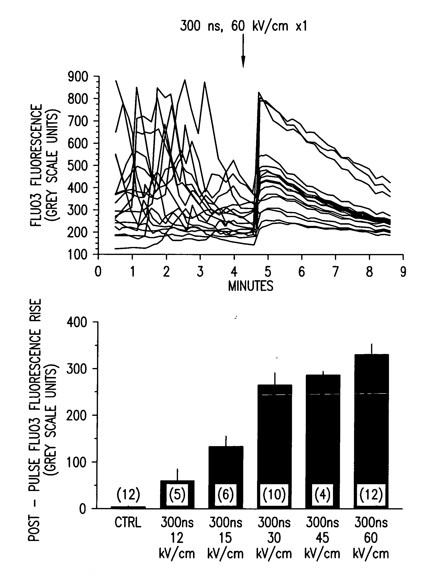
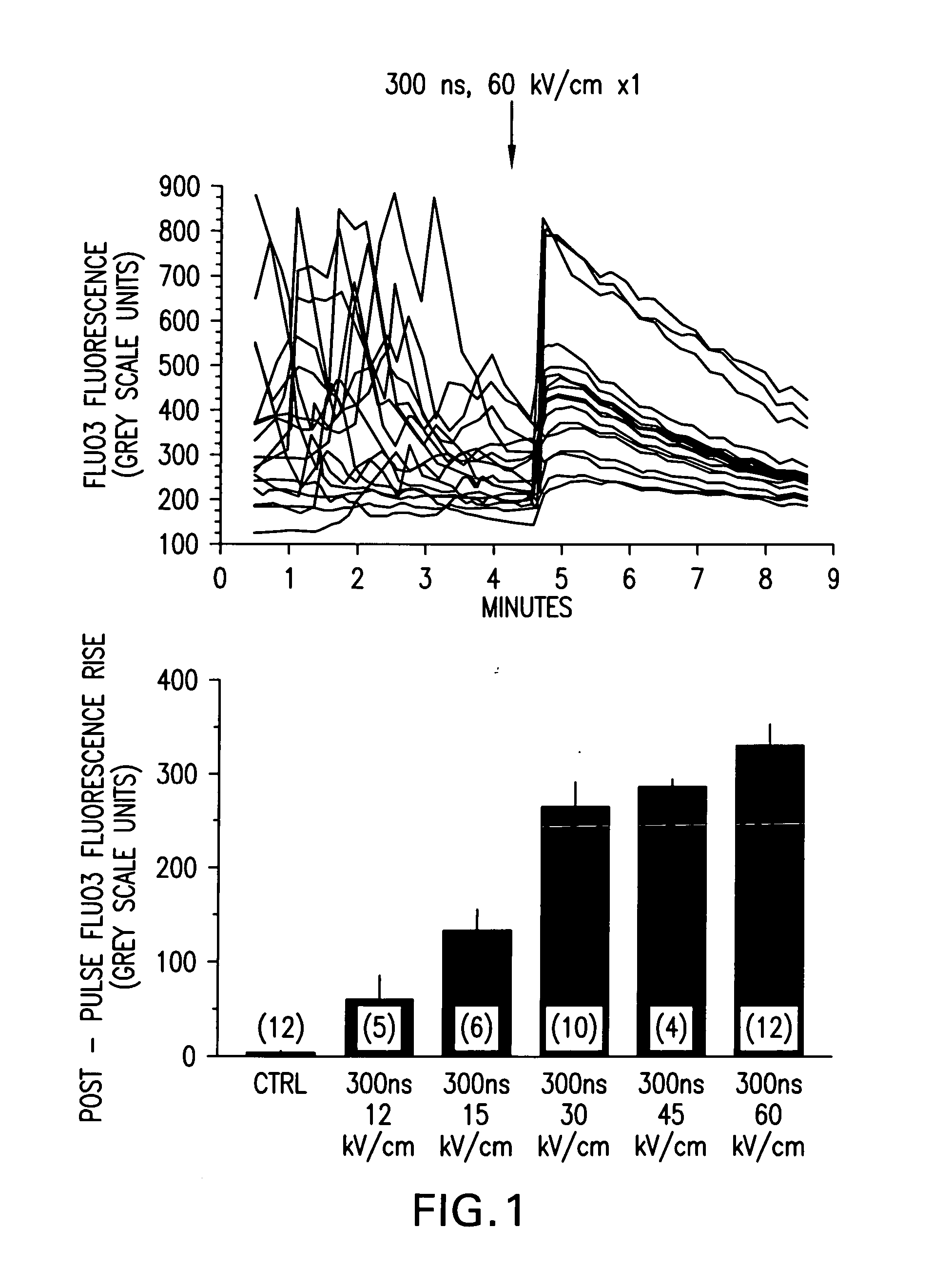
[0054]FIG. 1 demonstrates qualitative [Ca2+]i responses of polymorphonuclear leukocytes (PMNs) to nsPEF applications. Fluo3 labeled PMNs were examined by fluorescence microscopy before and following a single 300 ns, 60 kV / cm pulse application (upper panel). Each line is the grey-scale intensity (averaged over a spot (17 pixel diameter) within the cell) representation of intracellular Fluo3 fluorescence observed in one cell in the microscopic field. The highly variable Fluo3 fluorescence observed from cell-to-cell is due to changes in [Ca2+]i as the cells moved on the pulsing electrode surface. Application of a single nsPEF to the cells resulted in a rapid, variable increase in [Ca2+]i that waned in most cells over 4-5 minutes, loss of spontaneous fluctuations in [Ca2+]i and immediate loss of cellular movement. Lower panel shows dose-response relationship between intensity of the single pulse and the mean±S.E.M....

example 2

Investigation of nsPEF-Induced Apoptosis and Caspase Activation in Order to Determine Whether Calcium-Dependent or Calcium-Independent Cellular Mechanisms were Being Activated During Apoptosis

Materials and Methods

[0056] Cell culture—Non-transformed HL-60 and Jurkat cells were obtained from and cultured as recommended by American Type Culture Collection (ATCC, Rockville, Md.) as previously described (Beebe, S. J., et al. (2002). IEEE Trans. Plasma Sci. 30:1 Part 2, 286-292; Beebe, S. J., et al. (2003). FASEB J. 17, 1493-1495). Cells were removed from log phase growth and suspended in Hanks Balanced Salt Solution (HBSS) in the presence or absence of Ca2+ and Mg2+. When calcium was omitted, cells were washed several times in buffer without added calcium. In other experiments, the calcium chelator BAPTA was added to chelate intracellular calcium. Cells were loaded with BAPTA-AM and washed in HBSS before the addition of agonists.

[0057] Administration of nsPEF—Cell suspensions (7.7×10...

example 3

Investigation of nsPEF Stimulation at Electric Fields Below the Threshold for Apoptosis in Order to Explore Non-Apoptotic Internal Calcium Responses to nsPEF

[0063] The effects of nsPEFs on the release of internal calcium and activation of calcium influx in HL-60 cells were also investigated using real-time fluorescent microscopy with Fluo-3 and fluorometry with Fura-2. nsPEFs induced an increase in intracellular calcium levels that was seen in all cells. With pulses of 60 ns duration and electric fields between 4 and 15 kV / cm, intracellular calcium increased 200 nM-700 nM, respectively, above basal levels (˜100 nM) while the uptake of propidium iodide was absent. This suggests that increases in intracellular calcium were not due to plasma membrane electroporation. nsPEF and the purinergic agonist UTP induced calcium mobilization in the presence and absence of extracellular calcium with similar kinetics and appeared to target the same IP3- and thapsigargin-sensitive calcium pools in...


PUM


Abstract

Methods for inducing calcium mobilization in cells through the application of nanosecond pulsed electric fields (“nsPEFs”) are provided. The invention also provides a method of increasing intracellular calcium in cells through the application of nsPEFs. In one embodiment of the invention, the cells are human platelets, whereby activation and aggregation of the platelets is induced. Methods for treating an injury, trauma, or loss of blood in a subject, through the application of nsPEFs are also provided.

Description

REFERENCE TO RELATED APPLICATIONS [0001] This application claims the benefit of U.S. Provisional Application No. 60 / 636,695, filed Dec. 17, 2004, the disclosure of which is incorporated herein by reference.BACKGROUND OF THE INVENTION [0002] Electric fields can be used to manipulate cell function in a variety of ways. One specific cell mechanism that can be affected by electric fields is calcium mobilization within a cell. Calcium signaling, an important cell function, is responsible for a variety of cellular responses and actions. The release of internally stored calcium can stimulate responses to agonists, activate growth and respiration, cause the secretion of neurotransmitters, activate transcription mechanisms, cause the release of a variety of hormones, produce muscle contractions, and initiate release of key factors in the apoptosis pathway (Berridge, M. J., Bootman, M. D., Lipp, P. (1998) Nature. 395, 645-648). This calcium mobilization also triggers the influx of calcium fro...

Claims


Application Information

Patent Timeline
Patent Type & Authority Applications(United States)
IPC IPC(8): A61N1/32
CPCA61N1/0412A61N1/0424A61N1/325
Inventor BLACKMORE, PETER F.BEEBE, STEPHEN J.BUESCHER, E. STEPHENSCHOENBACH, KARL H.
Owner EASTERN VIRGINIA MEDICAL SCHOOL
Why Eureka
Try Eureka