Tethered capsule endoscope for Barrett's Esophagus screening

a technology of esophagus and endoscope, which is applied in the field of apparatus and a method for diagnostic imaging within the body lumen, can solve the problems of inability to control the rate at which the camera-capsule moves through the gastrointestinal tract, the resolution of the procedure is currently impractical, and the cost of the procedure is thus relatively high, so as to reduce the distortion of the image

Active Publication Date: 2006-08-31
UNIV OF WASHINGTON
View PDF12 Cites 195 Cited by
  • Summary
  • Abstract
  • Description
  • Claims
  • Application Information

AI Technical Summary

Benefits of technology

[0016] The scanner can include either a single mode optical fiber, a dual cladding optical fiber, a single mode optical fiber used for conveying light from the light source, and at least one multimode collection optical fiber for conveying light reflected from an inner surface of a lumen to the at least one light sensor, or a waveguide comprising a microelectromechanical system (MEMS) device. In one preferred embodiment, the scanner comprises either a waveguide or an optical fiber that is driven by the actu...

Problems solved by technology

However, the numbers of people that are likely candidates for esophageal screening and the current cost associated with the practice of flexible endoscopy performed by a physician compared to the reimbursement associated with such mass screenings make this solution currently impractical.
Only a doctor can perform an examination of the esophagus using a conventional flexible endoscope, and the procedure is thus relatively expensive.
However, there is no...

Method used


Image

  • Tethered capsule endoscope for Barrett's Esophagus screening
  • Tethered capsule endoscope for Barrett's Esophagus screening
  • Tethered capsule endoscope for Barrett's Esophagus screening

Examples


Embodiment Construction

Exemplary Application of Present Invention

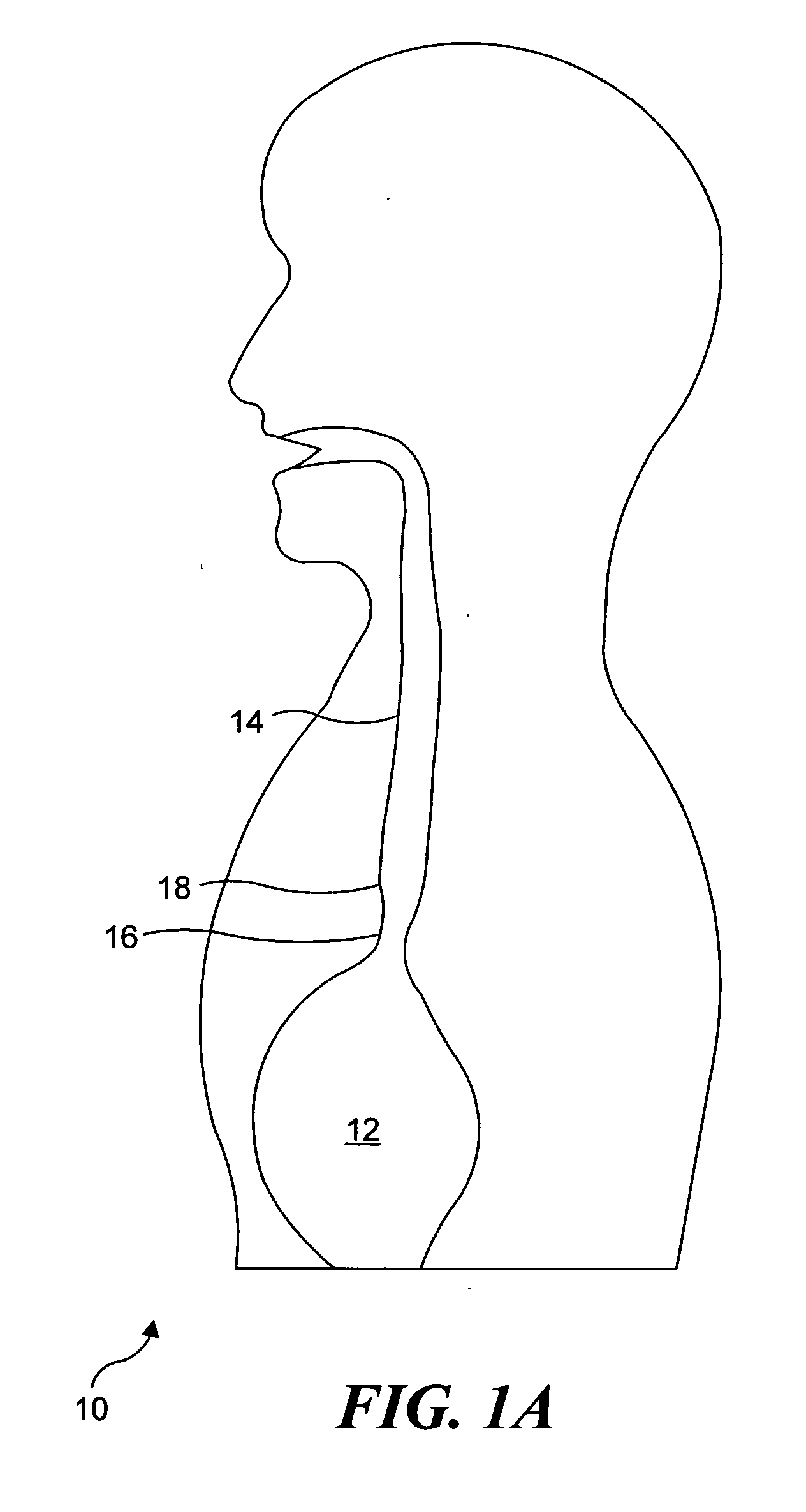
[0053] Although the present invention was initially conceived as a solution for providing relatively low cost mass screening of the general population to detect Barrett's Esophagus without requiring interaction by a physician, it will be apparent that this invention is also generally applicable for use in scanning, diagnoses, rendering therapy, and monitoring the status of therapy thus delivered to an inner surface of almost any lumen in a patient's body. Accordingly, although the following discussion often emphasizes the application of the present invention in the detection of Barrett's Esophagus, it is intended that the scope of the invention not in any way be limited by this exemplary application of the invention.

[0054]FIG. 1A includes a schematic illustration 10 showing a stomach 12, an esophagus 14, and a lower esophageal sphincter (LES) 16. LES 16 normally acts as a one-way valve, opening to enable food swallowed down the esophagus ...


PUM


Abstract

A capsule is coupled to a tether that is manipulated to position the capsule and a scanner included within the capsule at a desired location within a lumen in a patient's body. Images produced by the scanner can be used to detect Barrett's Esophagus (BE) and early (asymptomatic) esophageal cancer after the capsule is swallowed and positioned with the tether to enable the scanner in the capsule to scan a region of the esophagus above the stomach to detect a characteristic dark pink color indicative of BE. The scanner moves in a desired pattern to illuminate a portion of the inner surface. Light from the inner surface is then received by detectors in the capsule, or conveyed externally through a waveguide to external detectors. Electrical signals are applied to energize an actuator that moves the scanner. The capsule can also be used for diagnostic and/or therapeutic purposes in other lumens.

Description

FIELD OF THE INVENTION [0001] The present invention generally pertains to apparatus and a method for diagnostic imaging within a body lumen using a scanning endoscope that is housed in a capsule, and more specifically, pertains to apparatus and a method for imaging an internal wall of the lumen with an imaging capsule to detect a medical condition, where the capsule is electronically coupled to an external data receiver through a tether lead that is also used to control the location of the capsule within the lumen. BACKGROUND OF THE INVENTION [0002] Barrett's Esophagus (BE) is a condition of the esophagus that is pre-cancerous, a precursor to cancer of the esophagus. The standard practice for diagnosing Barrett's Esophagus is using flexible endoscopy, often with the esophageal lumen insufflated with air. A normal esophagus is usually light pink in color, while the stomach appears slightly darker pink. Barrett's Esophagus usually manifests itself as regions of slightly darker pink co...

Claims


Application Information

Patent Timeline
IPC IPC(8): A61B1/00
CPCA61B1/0008A61B1/00172A61B1/043A61B1/07A61B1/2733A61B5/0068A61B5/0071A61B5/0075A61B5/0084A61B5/1455A61B5/14556A61B5/4233A61B1/0638A61B1/00148A61B1/041A61B1/00097A61B1/000094A61B1/00055A61B1/00087A61B1/015A61B1/0661A61N5/0603A61N5/062A61N2005/0609
Inventor SEIBEL, ERIC J.KIMMEY, MICHAELJOHNSTON, RICHARD S.
Owner UNIV OF WASHINGTON
Why Eureka
Try Eureka