
Methods for use of apoptotic cells to deliver antigen to dendritic cells for induction or tolerization of T cells

a technology of dendritic cells and apoptotic cells, which is applied in the field of targeted antigen presentation, can solve the problems of ineffective antigen delivery to the mhc class i processing compartment, unfavorable use of powerful adjuvants, and inefficient antigen delivery to the dendritic cell induction or tolerization, etc., and achieve the effect of efficient delivery of specific antigens

Inactive Publication Date: 2006-11-16
THE ROCKEFELLER UNIV
View PDF1 Cites 3 Cited by
  • Summary
  • Abstract
  • Description
  • Claims
  • Application Information

AI Technical Summary

Benefits of technology

This patent describes a method for delivering antigens to dendritic cells and inducing T cell responses. The method involves inducing apoptotic cells to undergo apoptosis, allowing them to access phagocytic dendritic cells and be internalized. The dendritic cells are then exposed to the apoptotic cells expressing antigens, resulting in the uptake and processing of the antigens. This method can be used to generate antigen-specific cytotoxic T lymphocytes for the treatment of chronic infections and tumors. The patent also describes the use of liposomes and heat shock proteins to enhance the uptake and processing of antigens by dendritic cells. Overall, this patent provides a more efficient and effective way to induce immunity and tolerance to chronic infections and tumors.

Problems solved by technology

However, these prior approaches still pose a number of limitations on the development of effective therapeutic treatments.
Further, osmotic lysis of dendritic cell pinocytic vesicles may be difficult to perform in vivo, and may result in inefficient antigen delivery to the MHC class I processing compartment.
Also, the use of powerful adjuvants may be undesirable for some clinical applications.
These prior methods, however, do not recognize forms of cell death or the processing pathways antigens from dead or dying cells access in the dendritic cell system.
The mechanism of internalization of apoptotic cells by dendritic cells, however, is different than in macrophages, indicating that results in the macrophage system are not necessarily predictive of dendritic cell responses.
In addition, Rubartelli et al. does not show presentation of engulfed material and therefore speculates as to the fate of such material.

Method used


Image

  • Methods for use of apoptotic cells to deliver antigen to dendritic cells for induction or tolerization of T cells
  • Methods for use of apoptotic cells to deliver antigen to dendritic cells for induction or tolerization of T cells
  • Methods for use of apoptotic cells to deliver antigen to dendritic cells for induction or tolerization of T cells

Examples


example 1

Use of Apoptotic Cells to Deliver Antigen to Dendritic Cells and Induce Class I-Restricted CTLs

Materials & Methods

[0119] Generation of mononuclear subsets. Peripheral blood mononuclear cells [PBMCs] were isolated from blood by sedimentation in Ficoll-Hypaque [Pharmacia Biotech]. T cell-enriched [ER+] and T cell-depleted [ER−] populations were prepared by resetting with neuraminidase treated-sheep red blood cells, as previously described (37). T cells were purified from ER+ cells by removal of monocytes, NK cells and MHC class II+ cells (37). Monocytes were obtained from ER− cells by plastic adherence. Dendritic cells were prepared from ER− cells cultured for 7 days in the presence of GM-CSF and IL-4, followed by 4 days in monocyte conditioned medium (42,43).

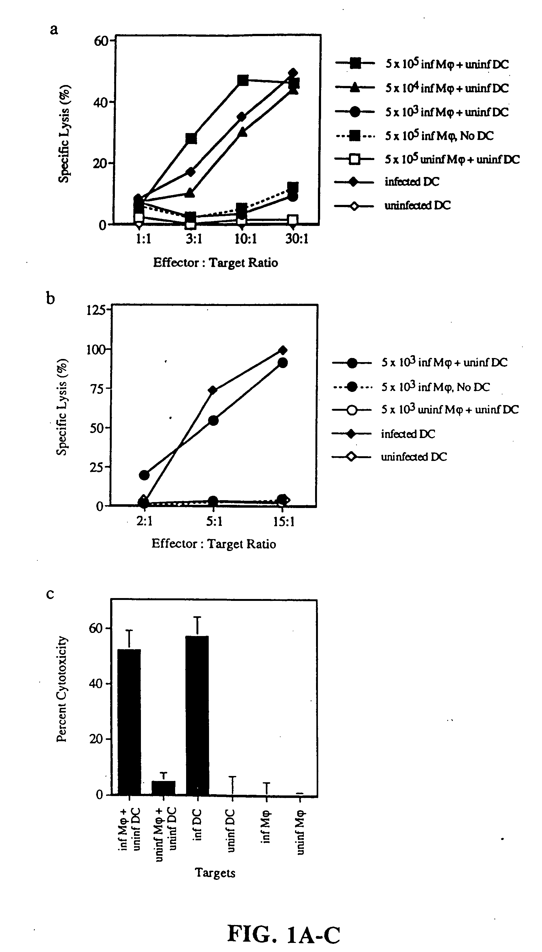
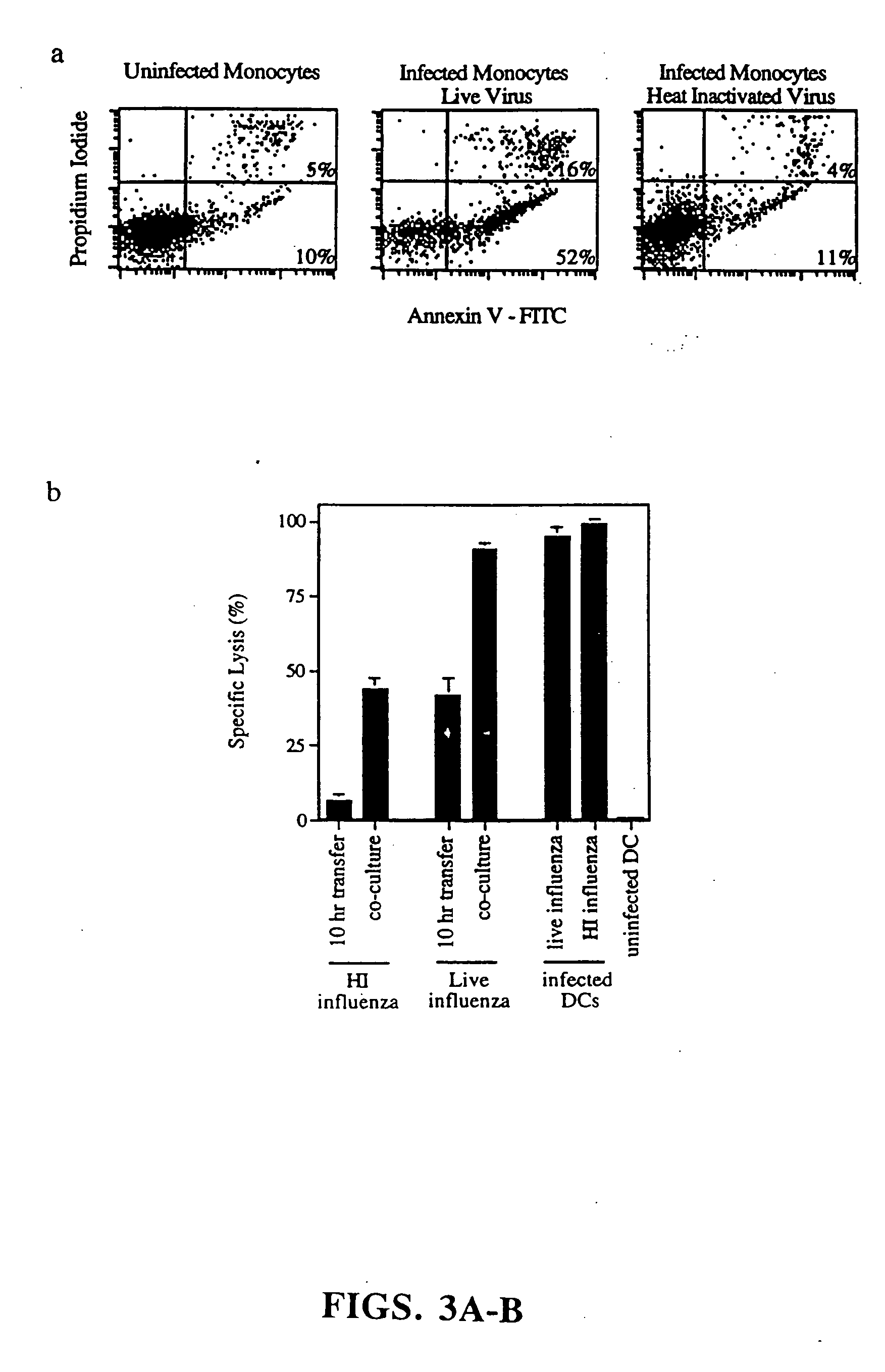
[0120] Induction and detection of apoptosis. Monocytes were infected with influenza virus in serum free RPMI. Initial time course studies established that after 5 hours, monocytes began to express early markers for apoptosis a...

example 2

Apoptotic Transfected 293 Cells Serve as Antigenic Material for ‘Cross-Priming’ of CD8+ T Cells

[0138] The methods and materials unless otherwise specified were the same as in Example 1.

[0139] The following methods were used to carry out the experiments shown in FIG. 6. 293 cells, a human kidney epithelial cell line, were transfected with a construct encoding the matrix gene from Influenza A (filled triangles). After 2 days in culture, 1×10⁴ transfected 293 cells were added to fresh wells and were UV-B irradiated in order to induce apoptotic death. DCs and T cells were then added to these wells and after 7 days, responding T cells were assayed for influenza-specific cytolytic activity using matrix peptide pulsed T2 cells as targets.

Results

[0140] The results demonstrate that a transfected tumor cell line can serve as a donor apoptotic cell, allowing for the transfer of antigen to the uninfected dendritic cell and the effective induction of antigen-specific CD8+ CTLs. Controls incl...

example 3

CD8+ T-Cells and not CD4+ Cells are Responsible for Infuenza-Specific Cytotoxicity

[0141] The following methods were used to carry out the experiments shown in FIG. 7. Generation of CD8+ CTLs requires CD4+ T-cell help. Purification of CD8+ and CD4+ T cells after CTL induction, established that CD8+ cells were responsible for the influenza-specific cytolytic activity. Influenza infected allogeneic monocytes were co-cultured with DCs and T-cells. After 7 days, subpopulations of T cells were purified and tested for cytolytic activity (37). Effector: Target ratio=15:1.

Results

[0142] Given that the lysis of T2 cells, an HLA-A2.1+ cell line, was dependent on the matrix peptide, which is specific for HLA-A2.1, it was expected that the effectors were MHC class I-restricted (see Example 1). To confirm this, highly purified CD4+ and CD8+ subpopulations were isolated at the end of a 7 day culture period. As demonstrated in FIG. 7, CTL activity was detected only in the CD8+ fraction.


PUM

PropertyMeasurementUnit
distanceaaaaaaaaaa
pore sizeaaaaaaaaaa
pharmaceutical compositionaaaaaaaaaa

Abstract

This invention relates to methods and compositions useful for delivering antigens to dendritic cells which are then useful for inducing antigen-specific cytotoxic T lymphocytes and T helper cells. This invention also provides assays for evaluating the activity of cytotoxic T lymphocytes. According to the invention, antigens are targeted to dendritic cells by apoptotic cells which may also be modified to express non-native antigens for presentation to the dendritic cells. The dendritic cells which are primed by the apoptotic cells are capable of processing and presenting the processed antigen and inducing cytotoxic T lymphocyte activity or may also be used in vaccine therapies.

Description

[0001] This application claims priority to U.S. provisional application Ser. No. 60 / 075,356 filed Feb. 20, 1998, Ser. No. 60 / 077,095 filed Mar. 6, 1998; and Ser. No. 60 / 101,749 filed Sep. 24, 1998 all of which are incorporated herein by reference.[0002] This invention was made with United States Government support under National Institutes of Health grant AI-39516, AI-13013 and AI 39672. The United States Government has certain rights in this invention.FIELD OF THE INVENTION [0003] This invention relates to targeted antigen presentation in the immune system. In particular, this invention relates to the use of apoptotic cells to deliver antigens to dendritic cells for induction or tolerization of antigen-specific T cell responses. The apoptotic cell-mediated antigen delivery system described herein has a wide range of preventive, diagnostic and therapeutic applications. BACKGROUND OF THE INVENTION [0004] CD8+ cytotoxic T lymphocytes (CTLs) play a critical role in immune defense again...

Claims


Application Information

Patent Timeline
Patent Type & Authority Applications(United States)
IPC IPC(8): A61K39/00A61K35/12A61K35/26A61K39/145A61K39/385A61K48/00A61P31/00A61P35/00C12N5/02C12N5/0784
CPCA61K39/0008A61K39/001A61K39/145A61K39/385A61K48/00C12N2760/16134A61K2039/5158A61K2039/57A61K2039/605C12N5/0639C12N2501/48A61K2039/5154A61K39/12A61P31/00A61P35/00Y02A50/30A61K39/4615A61K39/4622A61K39/464838
Inventor ALBERT, MATTHEWBHARDWAJ, NINASTEINMAN, RALPHINABA, KAYODARNELL, ROBERT
Owner THE ROCKEFELLER UNIV
Learn More