
Method and apparatus for photothermal modification of x-ray images

a technology of x-ray images and photothermal modification, which is applied in the field of imaging and non-destructive testing through xradiation, can solve problems such as interference in the production of images, and achieve the effect of increasing the volume of heated objects

Inactive Publication Date: 2006-11-23
DIEBOLD ROGER M
View PDF16 Cites 30 Cited by
  • Summary
  • Abstract
  • Description
  • Claims
  • Application Information

AI Technical Summary

Benefits of technology

[0016] Second Mechanism: Ordinary thermal expansion increases the volume of the heated object resulting in a larger object, which, depending on the resolution of the x-ray apparatus will show up in the image as a change in the size of the object.
[0019] The first effect of the absorption of a short burst of optical radiation is a temperature increase and a consequent increase in the dimensions through ordinary thermal expansion in the absorbing region of the body. The temperature gradient gives rise to a corresponding density gradient the size of which is determined by both the size of the temperature gradient and the thermal expansion coefficient of the material heated. When a short burst of radiation first is absorbed, the temperature and density gradients at the interface between the strong and weakly absorbing regions of the body are large, and localized over a short distance. As time progresses, the heat deposited from the optical source diffuses over a progressively longer distance so that the temperature and density gradients become smaller, but are spread over a larger region of space; finally, for long times, the temperature in the body equilibrates and the density gradients disappear.
[0022] In the present invention, the object of irradiation of the body with pulses of optical radiation is to produce density gradients in the body demarking the presence of differences in optical absorption so that such differences can be recorded in the x-ray image. For example, in examination of mammary tissue it is known (see Oraevsky et al.) that radiation with a wavelength of approximately one micron is absorbed more strongly by blood than by mammary tissue. In Oraevsky's photoacoustic experiments, a pulsed 1.06 μm laser with a few nanoseconds duration is fired at a breast, or a phantom of a breast. The optical radiation is diffused strongly by the mammary tissue, but on reaching a tumor that is highly vascularized and hence possesses a high blood content, the radiation is preferentially absorbed by the blood leading to a heating and a pressure increase at the site of the tumor. The rapid pressure increase in the volume where optical absorption takes place causes an outward going pressure wave to be launched that can be detected by an array of transducers located a short distance from the breast permitting an acoustic image to be produced. It is important to note that in the present invention and in photoacoustic detection, the optical radiation is strongly diffused by the breast tissue; however, the directionality of the optical radiation is of no consequence, it is nevertheless absorbed. The difference in absorption between tumors with their high blood content and healthy tissue at near infrared wavelengths provides reasonably good contrast for images formed in both the photoacoustic method and the present invention.
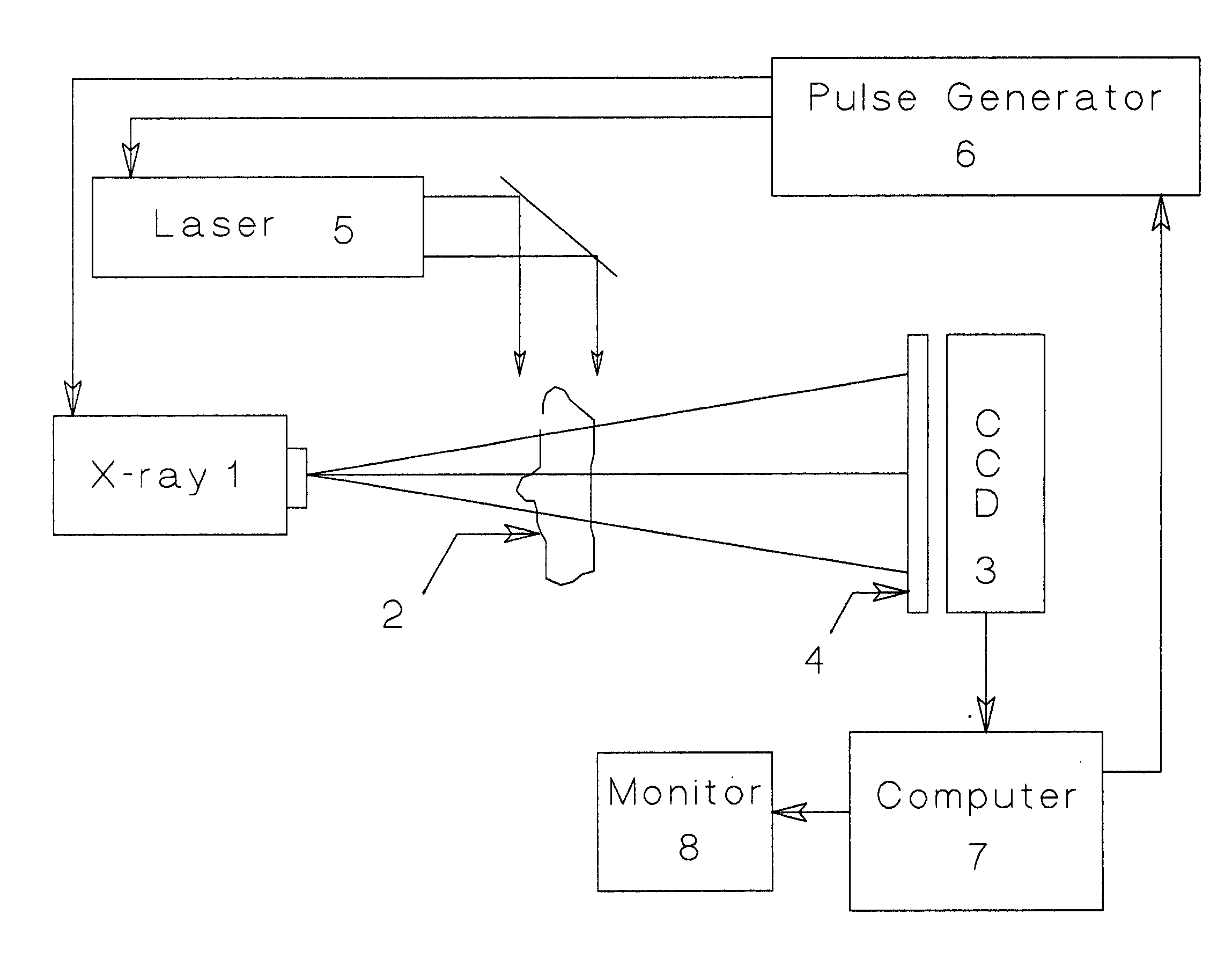
[0028] In a preferred embodiment of the invention, an image, or number of images are acquired and added when the optical and x-ray pulses are synchronized to provide the maximum change in the image. Then, a second image or set of images is acquired and added without the optical pulses. Subtraction of the two images gives a difference image that highlights the photothermal effects and minimizes the features of the image that are not affected by the absorption of optical radiation. The same result as modulating the x-ray source can be obtained with a continuous x-ray source by gating the signal to the image forming device with, for instance, a gated image intensifier.

Problems solved by technology

Objects placed in one arm of the interferometer modify the phase of the x-radiation in that arm only, resulting in the registration of the phase changes experienced by the x-rays passing through the body at the point where the two beams of x-rays are combined and interfere to produce an image.

Method used


Image

  • Method and apparatus for photothermal modification of x-ray images
  • Method and apparatus for photothermal modification of x-ray images
  • Method and apparatus for photothermal modification of x-ray images

Examples


Embodiment Construction

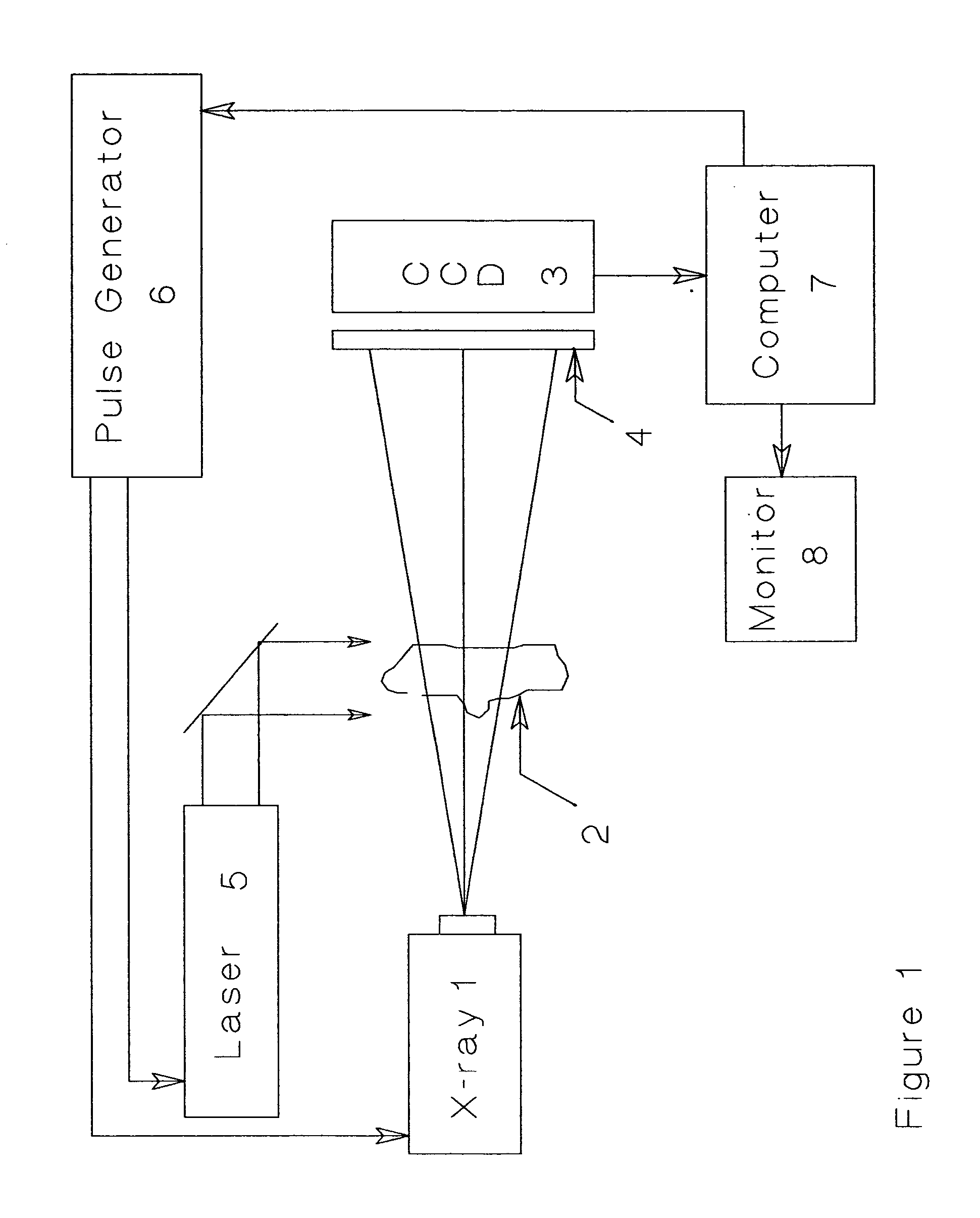
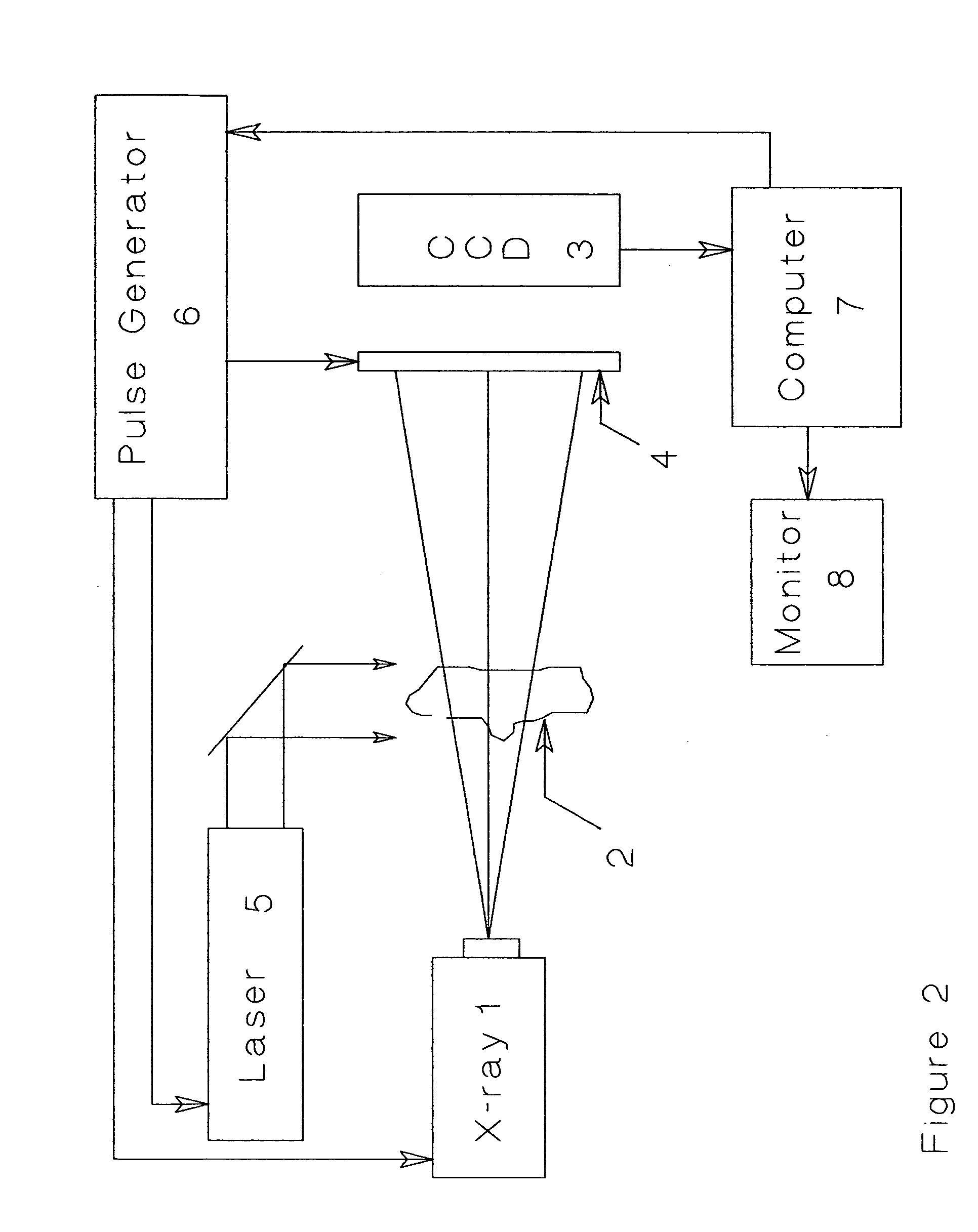
[0035] Referring now to the drawing, the elements comprising a preferred embodiment of the apparatus comprising the invention consists of an x-ray source 1, a body to be examined 2, and a CCD camera or equivalent imaging forming device 3 for recording the x-ray intensity pattern after the x-rays traverse the body, with or without the use of a phosphor screen 4 that converts x-ray photons into radiation (typically visible) suitable for detection by the CCD.

[0036] For the purpose of the present invention optical radiation is defined as laser radiation from the ultraviolet and visible to and the near infrared regions of the spectrum, microwaves, and radio-frequency radiation i.e. any region of the electromagnetic spectrum where absorption contrast between the object of interest and its surroundings is maximal. Additionally, gated image intensifier shall refer to a device that converts x-ray photons to visible photons (with gain) that can be gated on and off electronically, or a device...


PUM

PropertyMeasurementUnit
diameteraaaaaaaaaa
wavelengthaaaaaaaaaa
diameteraaaaaaaaaa

Abstract

An x-ray image of a body can be modified by absorption of laser radiation that causes thermal gradients to be generated in portions of the body. If an object within the body has a higher optical absorption than the surrounding medium, the effect of absorption of the laser radiation is to cause the production of thermal gradients. Thermal gradients give rise to density gradients, which modify an x-ray image through changes in x-ray index of refraction at the site of the thermal gradient. The overall effect of the laser heating is to produce an x-ray contrast mechanism wherein the x-ray image becomes sensitive to differences in the optical absorption within a body. An application of the invention is for detection of tumors that are highly vascularized, using a laser operating in the near infrared.

Description

BACKGROUND OF THE INVENTION [0001] The present invention relates generally to imaging and non-destructive testing through use of x-radiation. The laser produces thermal gradients wherever there is optical contrast, i.e. different optical absorption coefficients, between objects within a body and the surrounding material in the body. The method has application to non-destructive testing where a body scatters optical radiation (so that no clear image can be made), but which has differential absorption between parts within the body whose image is sought. One application of the method is to tissue imaging such as x-ray mammography where tissue scatters optical radiation strongly, so that a clear optical image cannot be formed, but which does not completely absorb the optical radiation. X-rays penetrate tissue and can form a sharp image. In the case of tissue, the method makes the x-ray image sensitive to the presence of blood, blood vessels, and tumors, all of which have significant opt...

Claims


Application Information

Patent Timeline
Patent Type & Authority Applications(United States)
IPC IPC(8): G01N23/04
CPCG01N23/04G01N23/041
Inventor DIEBOLD, ROGER M.
Owner DIEBOLD ROGER M
Learn More