
Method and device for image reconstruction

Inactive Publication Date: 2006-12-21
SIEMENS HEALTHCARE GMBH
View PDF7 Cites 3 Cited by
  • Summary
  • Abstract
  • Description
  • Claims
  • Application Information

AI Technical Summary

Benefits of technology

[0007] An object of the present invention is to provide a method for image reconstruction as well as an associated image processing device with which an increase of the computing capacity and an optimized resource utilization are possible.
[0017] In the inventive method, the reprogrammable FPGA (in which, for example, the algorithm to be used is loaded at the start of an acquisition sequence) can thus completely utilize its respective available resources for the calculation. If another algorithm is necessary for a subsequent examination or, respectively, image reconstruction, this can be calculated after a suitable reprogramming of the FPGA or FPGAs, for example given a system restart. The image computer system (as is present, for example, in a clinic) functions with fewer processors overall, so costs can be saved. Moreover, the images are available faster for post-processing or medical assessment. This acceleration of the image reconstruction can also be meaningful when it must be decided as quickly as possible during an examination whether a further, possibly more precise, examination or an examination concerning a different body region must be conducted. In this case, the patient does not have to wait long until the reconstructed images are available to the doctor and he or she can decide whether the examination must be continued. Finally, the efficient utilization of the examination apparatus can be improved.
[0022] According to the invention, the access to the primary memory can be implemented as a direct memory access. By circumventing the processor or the processors of the image computer system, it is thereby possible to achieve a faster memory access, such that the total time necessary for the image data generation from the acquired data to the return delivery of the finished image data to the primary memory of an image computer system is further shortened.
[0023] According to the invention, the algorithm to be used can be loaded at the start of an image acquisition sequence. It is thus possible to change the algorithm as often as needed (for example at the start of an acquisition sequence) and to accordingly reprogram the FPGA.
[0024] The image data can be calculated basically without time delay. With the inventive method it is possible to already view finished calculated images practically “on the fly” during the time in which a measurement with a medical examination apparatus is still running and, if applicable, to adapt the implementation of the measurement dependent on the image exposures already present. For example, during the workflow of a data acquisition procedure (scan) a decision can be made about further measurements to be effected that can then immediately follow. It is also possible to detect incorrect settings or quality problems in the examination apparatus that lead to image exposures that cannot be evaluated or to image exposures that do not show the desired structures in the desired manner, such that the measurement parameters can be changed if necessary, or the measurement can be terminated. Unnecessary measurements or later repetitions of measurements are thus avoided with the use of the inventive method.
[0034] Multiple FPGAs of a component can be cascaded and / or arranged in parallel. Cascaded FPGAs of a module or component receive the image (insofar as it was already calculated by preceding FPGAs) and the raw data still to be calculated (transferred from the preceding FPGA), and relay their finished data or still-existing raw data to the next FPGA. For example, three or four FPGAs can be connected in series, with each FPGA adding its contribution to the unfinished image and forwarding this improved image. After passage through the cascaded FPGAs, the finished image is stored in turn in an image storage. FPGAs arranged in parallel can implement parallel calculation operations insofar as the underlying algorithm incapable of being conducted by means of parallel calculations. In this case, no preceding calculation results are necessary, or steps can follow from preceding calculation results that enable a parallel calculation in the next calculation stage, for which the FPGAs arranged in parallel are then used. A combination of cascaded components or a parallel arrangement of components or, if applicable, different components is possible to achieve an optimal utilization of the existing computation capacities.

Problems solved by technology

Such a general purpose processor is not designed for the requirements that are specifically present in image reconstruction, such that (depending on the algorithm or algorithms used for the reconstruction) the reconstruction can take a considerable amount of time.
Usage of such processors that are optimized (with regard to their speed) for specific tasks also enables only an insufficient utilization of the existing resources; in static systems, because the problem generally exists that such processors can inevitably utilize only a partial range of their resources for an algorithm to be executed.
This leads to the situation that the image computer that is charged with the reconstruction must embody a very high number of processors, the existing logic of which is only insufficiently utilized.
High costs and undesirably long wait times thereby arise for a user who would like to view the images that are not yet available during or even shortly after the examination.

Method used


Image

  • Method and device for image reconstruction
  • Method and device for image reconstruction
  • Method and device for image reconstruction

Examples


Embodiment Construction

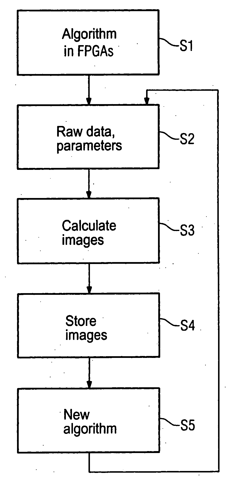
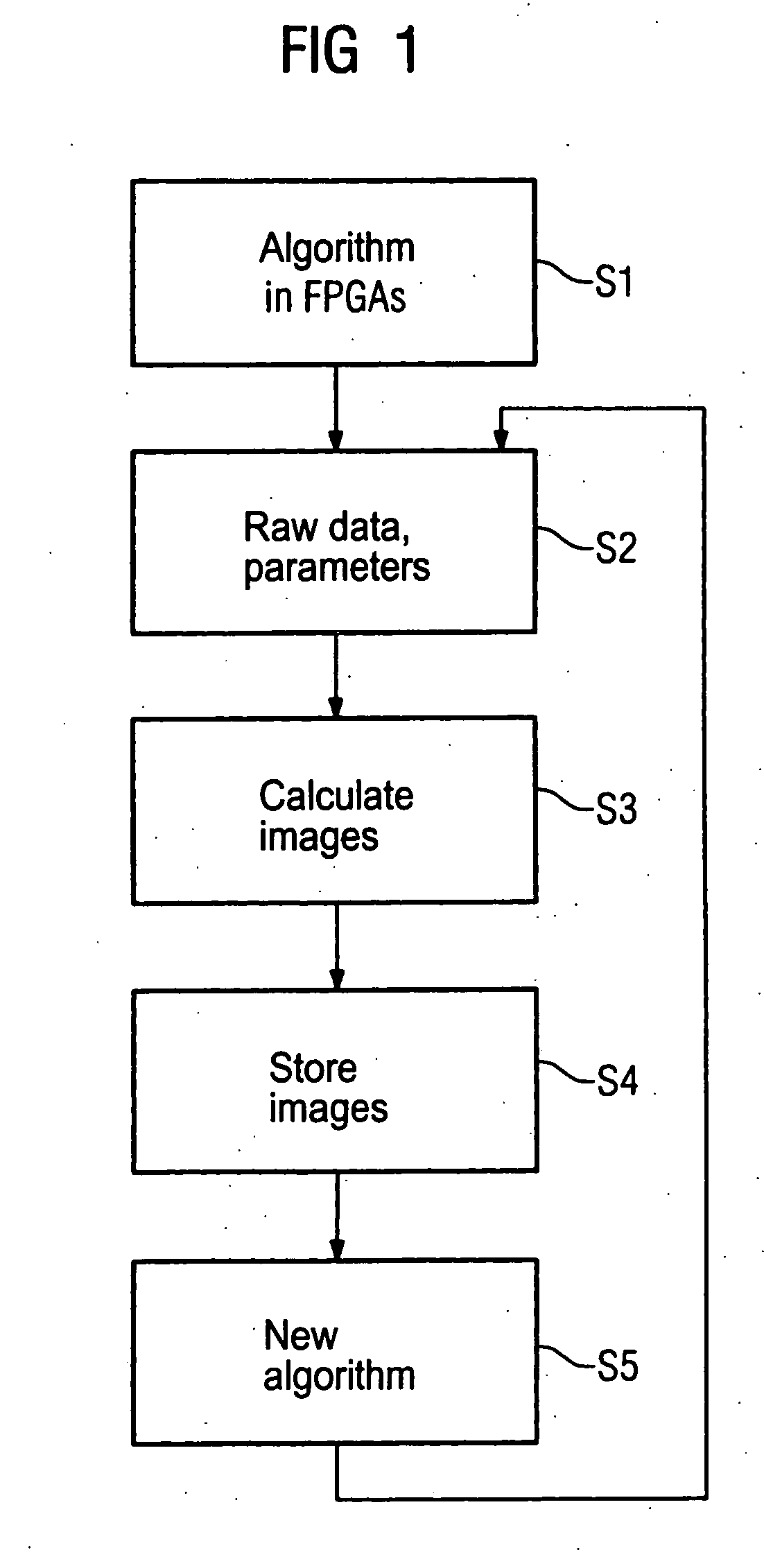
[0040]FIG. 1 is a flowchart of an embodiment of the inventive method. In the inventive method for image reconstruction from raw data acquired with an imaging medical examination apparatus, in a step S1 an algorithm to be used for the image reconstruction is initially loaded in at least one reprogrammable FPGA. This loading need not necessarily ensue at the sequence start; but can ensue at any time in the course of a sequence. In step 52, the reprogrammable FPGA, or multiple reprogrammable FPGAs access the raw data acquired by the medical examination apparatus and also access parameters that have been predetermined for the image reconstruction, such as calculation parameters, which are to be understood for the present purpose in the broadest sense (thus also including parameters with regard to a desired representation format and the like). This access ensues via a data connection, preferably via an access to a peripheral bus of an image computer system or the like.

[0041] If the raw ...


PUM


Abstract

In a method and processing device for image reconstruction from raw data acquired with a medical imaging examination apparatus, in particular from data of magnetic resonance tomography, an algorithm to be used for the image reconstruction is loaded into at least one reprogrammable FPGA, the reprogrammable FPGA, accesses via a data connection, the acquired raw data as well as calculation parameters provided for the image reconstruction and reconstruction image data are calculated from the raw data by the FPGA, dependent on the calculation parameters.

Description

BACKGROUND OF THE INVENTION [0001] 1. Field of the Invention [0002] The present invention concerns a method for image reconstruction from raw data acquired with an imaging medical examination apparatus, in particular a magnetic resonance tomography apparatus, as well as an image processing device that implements such a method. [0003] 2. Description of the Prior Art [0004] In the use of imaging medical examination devices such as, for example, magnetic resonance tomography systems or computed tomography systems or the like, raw data are initially acquired from which images are created in the framework of an image reconstruction using software. Such images are suitable for inspection by a medical-technical assistant or doctor for further processing or evaluation. [0005] In typical image reconstruction software available today in the medical field, the further processing of the initially present raw data has conventionally been implemented by a “general purpose” processor. Such a gener...

Claims


Application Information

Patent Timeline
IPC IPC(8): G06K9/00G06K9/54G06K9/60
CPCG01R33/5608
Inventor DEMHARTER, NIKOLAUS
Owner SIEMENS HEALTHCARE GMBH
Learn More