
Hollow nanoparticles of protein and drug using the same

Inactive Publication Date: 2006-12-28
JAPAN SCI & TECH CORP
View PDF1 Cites 12 Cited by
  • Summary
  • Abstract
  • Description
  • Claims
  • Application Information

AI Technical Summary

Benefits of technology

[0008] As a result of intensive study, the inventors of the present invention accomplished the present invention by successfully coexpressing a particle-forming protein with a protein forming a capsid structure. This coexpression method significantly increases the productivity of particles.
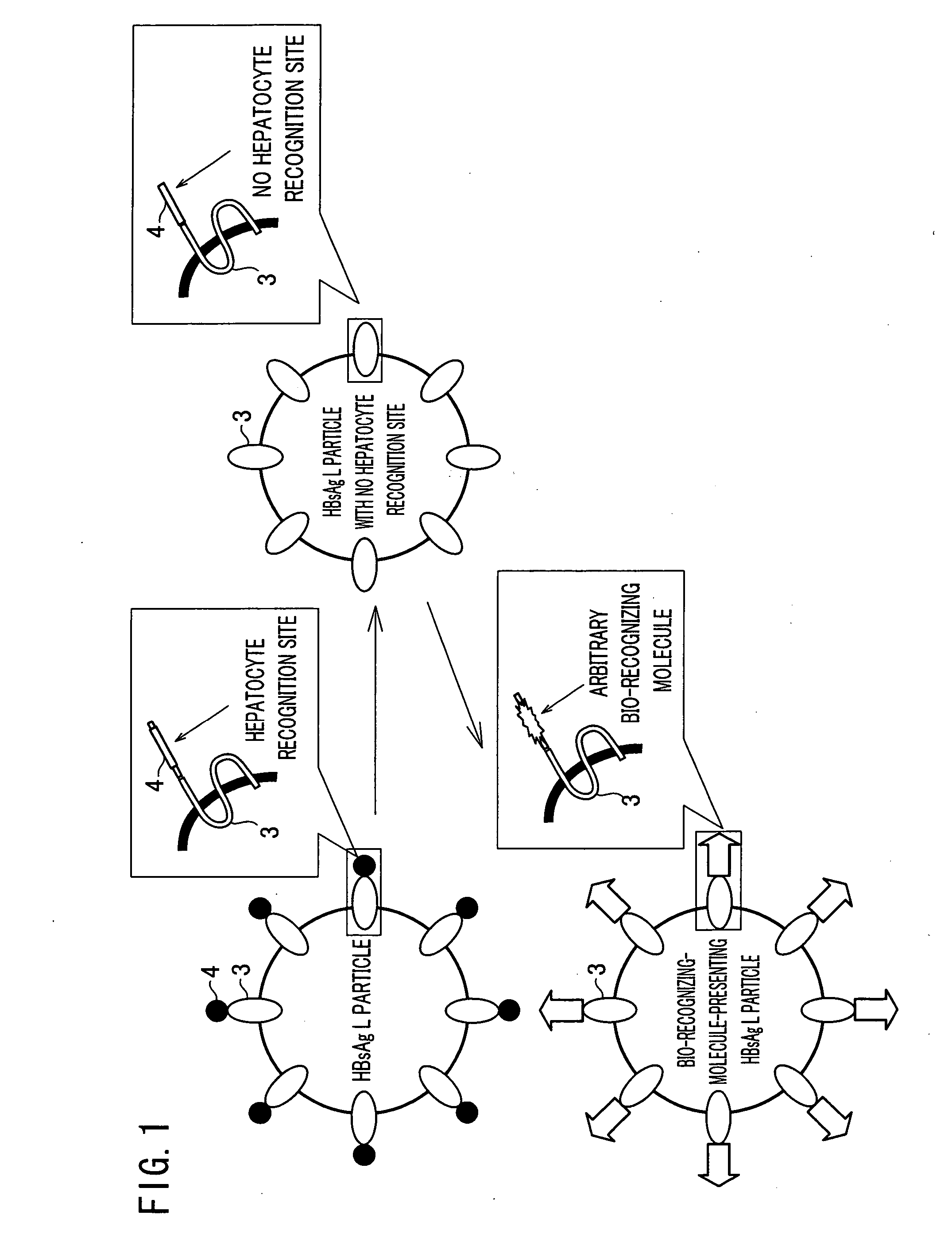
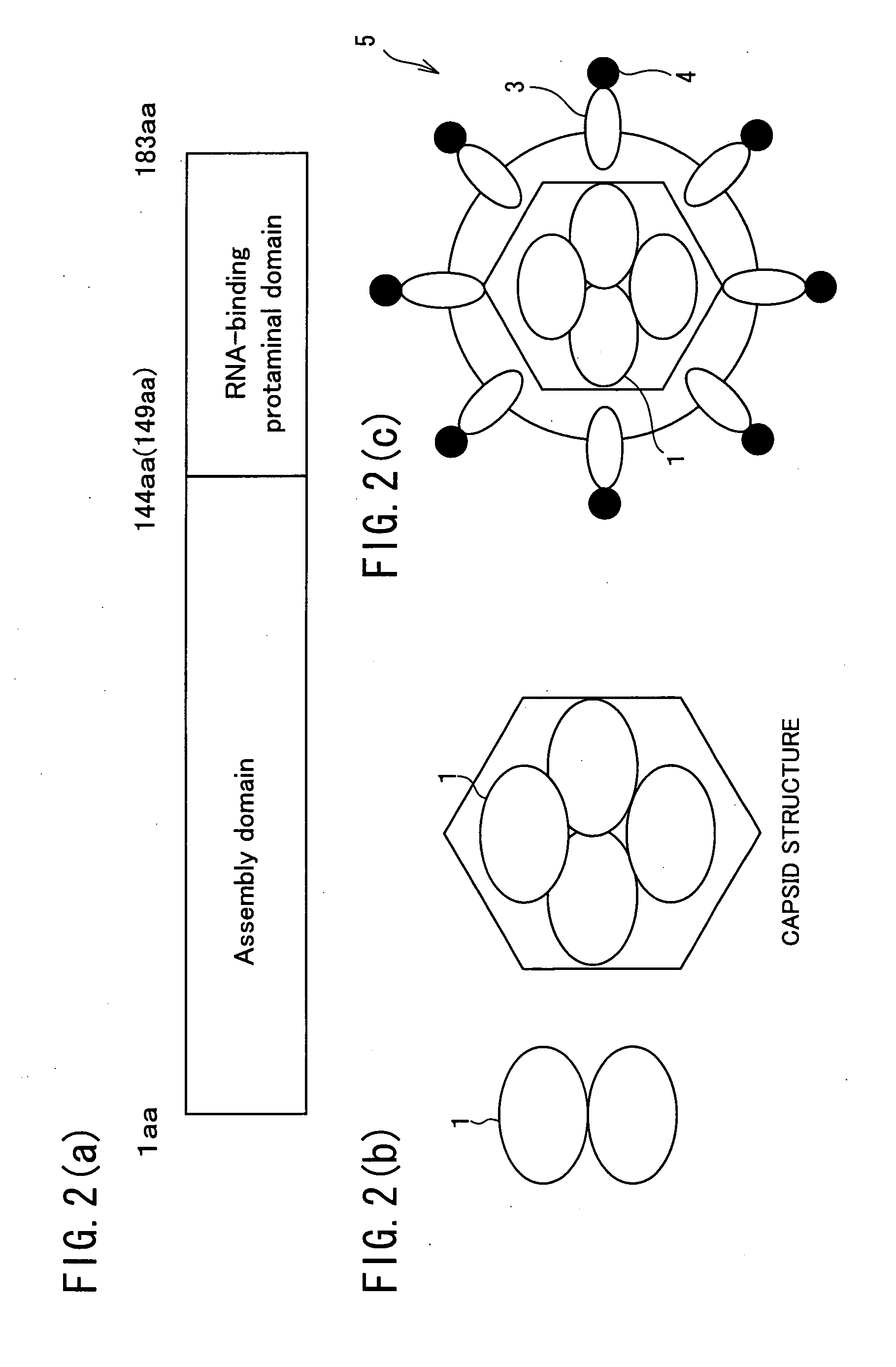
[0010] Examples of the “particle-forming first protein” include hepatitis B virus surface-antigen protein. When expressed in the eukaryotic cell, this particle-forming protein is expressed on the endoplasmic reticulum as a membrane protein and accumulates thereon before it is released as particles. The “capsid structure” refers to shells of protein that cover and protect virus genomes of virus particles. Examples of the “second protein forming a capsid structure” include hepatitis B virus core-antigen protein. When coexpressed with the first protein, the second protein interacts with the first protein, and encourages particle formation. The “interaction” here refers to an effect caused by direct contact of the first protein and the second protein. The effect can be obtained with a single first protein directly in contact with a second protein; however, a larger number is more preferable. Further, when interact with the first protein, the second protein does not necessarily have to be encapsulated in the first protein but may exist outside the first protein. With the interaction, the particle formation of the first protein is encouraged, thus increasing particle forming rate. Further, by encapsulating a target-cell substance in the particles, the protein may be used as a drug.
[0015] Such a modified hepatitis B virus surface-antigen protein, like the BTC displaying hepatitis B virus surface-antigen protein or the bFGF displaying hepatitis B virus surface-antigen protein has severe difficulty in particle forming when it is expressed alone; however, by coexpressing with the second protein forming the capsid structure, the particle forming rate will be significantly increase, thus providing a certain effect.

Problems solved by technology

However, none of the conventional gene transfer methods is sufficient to specifically transfer protein drugs to a target cell / tissue.

Method used


Image

  • Hollow nanoparticles of protein and drug using the same
  • Hollow nanoparticles of protein and drug using the same
  • Hollow nanoparticles of protein and drug using the same

Examples


example 1

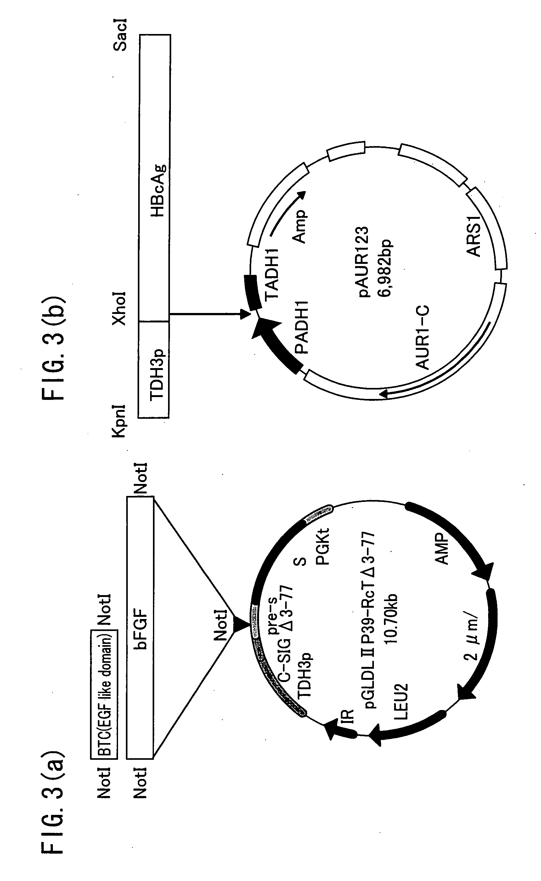
[0063] The following explains a preparation method of the modified HBsL protein. As shown in FIG. 3(a), the HBsAg L protein-expression-vector according to the Patent Document 1 above, reported by the inventor of the present invention, was expressed in yeast. The vector of pGLDL II P 39-RcT Δ 3-77 was prepared by deleting the pre-S region of pGLDLII P39-RcT(3rd amino residue to 77th amino residue, and inserting genes encoding BTC or bFGF using restriction enzyme NotI. As a result, HBsAg L protein that displays BTC as a bio-recognizing molecule, that is BTC displaying HBsAg L particles, or bFGF displaying HBsAg L particles were obtained.

example 2

Preparation of HBcAg Yeast-Expression-Plasmid

[0064] (2-1) Insertion of Promoter TDH3 into a Plasmid

[0065] A DNA fragment of the promoter TDH3 incorporated in the wild-type HBsAg expression plasmid pRS405-2-LAG was amplified by PCR method. This PCR was performed with a reaction liquid whose composition is shown in Table 1, with the use of primers of sequence numbers 3 and 4, respectively as the primers (+) and (−).

TABLE 1PCR: HBcAg10X LA PCR Buffer II (Mg2+free)8μlMgCl2 (25 mM)8μldNTP mixture (2.5 mM each)8μlDNA template (45 pg / μl)8μlPrimer (+)0.8μlPrimer (−)0.8μlTaKaRa LA Taq (5 U / μl)0.8μlOtsuka distilled water45.6μlTotal80μlPCR: TDH310X LA PCR Buffer II (Mg2+free)8μlMgCl2 (25 mM)8μldNTP mixture (2.5 mM each)8μlDNA template (10 ng / μl))8μlPrimer (+)0.8μlPrimer (−)0.8μlTaKaRa LA Taq (5 U / μl)0.8μlOtsuka distilled water45.6μlTotal80μl

[0066] The reaction liquid was divided into four portions, three of which were subjected to PCR reaction in such a manner that: after heating for 30 se...

example 3

Coexpression of HBcAg and HBsAg in Yeast

[0073] The HBsAg L protein expression vector of Example 1 and the HBcAg expression vector of Example 2 were cotransformed to each other. In other words, the vectors were transformed to S. cerevisiae AH22R-strain by the following spheroplast method (Albert et al., 1978).

[0074]S. cerevisiae AH22R-strain was inoculated on YPDA agar medium (see Table 2 for the composition) and cultivated for a few days at 30° C., and the resulting colony was subjected to preincubation overnight in 50 ml YPDA medium (see Table 2 for the composition) at 30° C. After the preincubation, the product was inoculated in 50 ml YPDA medium to obtain the condition: OD600=0.05 for the beginning of the main incubation, and then was further cultivated for 8 hours at 30° C. until OD600=0.4-0.8. Thereafter, the culture solution x 4 ml is divided into 15 ml tubes (IWAKI), and then, the fungus body was collected by centrifugation at 4° C. at 2000 rpm for 5 min using a multi-place...


PUM

PropertyMeasurementUnit
Fractionaaaaaaaaaa
Fractionaaaaaaaaaa
Fractionaaaaaaaaaa

Abstract

The subject invention is hollow nanoparticles that comprise particle-forming first proteins (e.g. hepatitis B virus surface-antigen protein), containing a bio-recognizing molecule for recognizing a specific cell, wherein at least one of the first proteins interacts with a second protein (e.g. hepatitis B virus core-antigen protein) forming a capsid structure. With this structure, the present invention provides hollow nanoparticles, that allow transfer of a substance to a specific cell or tissue, and can be manufactured with stable productivity. The present invention further provides a drug made of the hollow nanoparticles.

Description

TECHNICAL FIELD [0001] The present invention relates to hollow nanoparticles, that allow a disease-treating target-cell-substance to be encapsulated therein. The invention particularly relates to a drug allowing a disease-treating-target-cell substance encapsulated therein to be transferred to a specific cell or tissue. BACKGROUND ART [0002] In the field of medicine, there has been active research on drugs that directly and effectively act on the affected area without causing serious side effects. One area of active research is a method known as a drug delivery system (DDS), in which active ingredients of drugs or other substances are specifically delivered to a target cell or tissue, where they can exhibit their effects. [0003] One known example of conventional method of sending genes to cells is so-called a gene transfer method. In this method, genes encoding the protein are incorporated into an expression vector, and this expression vector is transferred to the target cell by an ...

Claims


Application Information

Patent Timeline
IPC IPC(8): A61K48/00A61K9/50A61K9/16C12N15/09A61K9/51A61K36/06A61K38/00A61K47/48A61P31/12A61P37/04C07K14/02C07K19/00C12N1/19C12N7/04C12N15/88
CPCA61K9/5184A61K47/48869A61K48/00A61K2039/5258B82Y5/00C12N2810/851C07K2319/33C12N7/00C12N15/88C12N2730/10122C12N2730/10123C07K14/005A61K47/6925A61P31/12A61P37/04A61K9/51A61K38/00
Inventor KURODA, SHUNICHITANIZAWA, KATSUYUKIKONDO, AKIHIKOUEDA, MASAKAZUSENO, MASAHARU
Owner JAPAN SCI & TECH CORP
Learn More