Endoscope with remote control module or camera

a remote control module and endoscope technology, applied in the field of endoscopy, can solve the problems of providing the same image quality, user still has to contend with the mass of the combined device, and weighs against the disadvantages, so as to achieve the effect of less fatigue, increased freedom of movement, and convenient operation

Inactive Publication Date: 2006-12-28
PERCEPTRONIX MEDICAL
View PDF14 Cites 124 Cited by
  • Summary
  • Abstract
  • Description
  • Claims
  • Application Information

AI Technical Summary

Benefits of technology

[0024] The endoscope of the present invention is easier to operate. Since the camera is not mounted or attach

Problems solved by technology

Another limitation of fluorescence imaging endoscopy is that, due to the very weak intensity of the emitted light, it does not provide the same image quality for morphological structure and therefore typically requires additional caution and time to guide the endoscope during the procedure.
In some instances, there may be a tether supporting the weight of the probe and camera, but the user still has to contend with the mass of the combined devices.
However, these advantages must be weighed against the disadvantages of increased cost for miniature image-capture device, which tends to cost more than a conventionally-sized image-capture device.
Additionally, whether the image-capt

Method used


Image

  • Endoscope with remote control module or camera
  • Endoscope with remote control module or camera
  • Endoscope with remote control module or camera

Examples


Embodiment Construction

[0031] While the invention may be susceptible to embodiment in different forms, there is shown in the drawings, and herein will be described in detail, specific embodiments with the understanding that the present disclosure is to be considered an exemplification of the principles of the invention, and is not intended to limit the invention to that embodiment as illustrated and described herein.

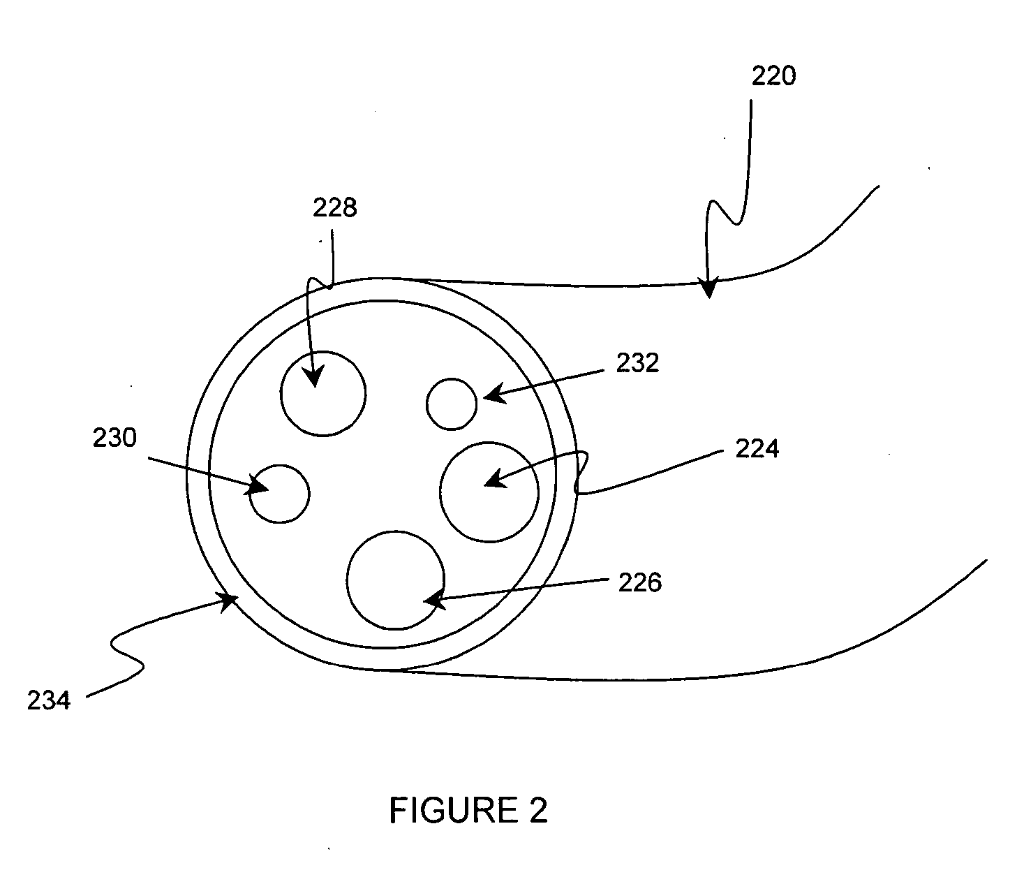
[0032] One embodiment of the present invention is shown in FIG. 3. In this embodiment, illumination components are placed at the distal end of the channel 320, as shown in cross-section in FIG. 3. Illumination source 324, located at the distal end of channel 320, generates desired interrogating radiation onto the target tissue. Illumination source 324 is preferably at least one LED, more preferably at least two LEDs. Illumination source 324 could also be a laser, a xenon lamp, a mercury lamp, tungsten halogen lamp, metal halide lamp or other light source. Power and control of illumination sou...


PUM


Abstract

An endoscope with image-capture device remote from the probe is disclosed. The endoscope assembly has a probe, an illumination source such as one or more LEDs, and an image capture device located remotely from the probe such that the user does not have to bear the weight of the image capture device. In one embodiment the image-capture device is located in a control module with an image processor.

Description

BACKGROUND OF THE INVENTION [0001] This invention relates to the field of endoscopy and more particularly in vivo imaging of cells, tissue, organs or body cavities of humans or other animals to observe, locate, diagnose, and treat disease. Generally, endoscope assemblies have at least one illumination source, the endoscopy device, and an image observation means such as an ocular or an image-capture device such as a camera. In addition, computer processing and data display options are common. [0002] Imaging is the capture of electromagnetic radiation either reflected or emitted from an object of interest such as cells, tissues, organs, or body cavities of humans or other animals, in a manner which preserves or otherwise represents the spatial distribution of said radiation at the object. In the field of medical imaging, and more particularly endoscopy, visible light is utilized to illuminate body tissues and return a diagnostic or otherwise useful image. So, endoscopes enable visual ...

Claims


Application Information

Patent Timeline
IPC IPC(8): A61B1/00
CPCA61B1/00009A61B1/00016A61B1/04A61B1/043A61B5/0075A61B5/0084A61B1/2676
Inventor GARNER, DAVID M.
Owner PERCEPTRONIX MEDICAL
Why Eureka
Try Eureka