

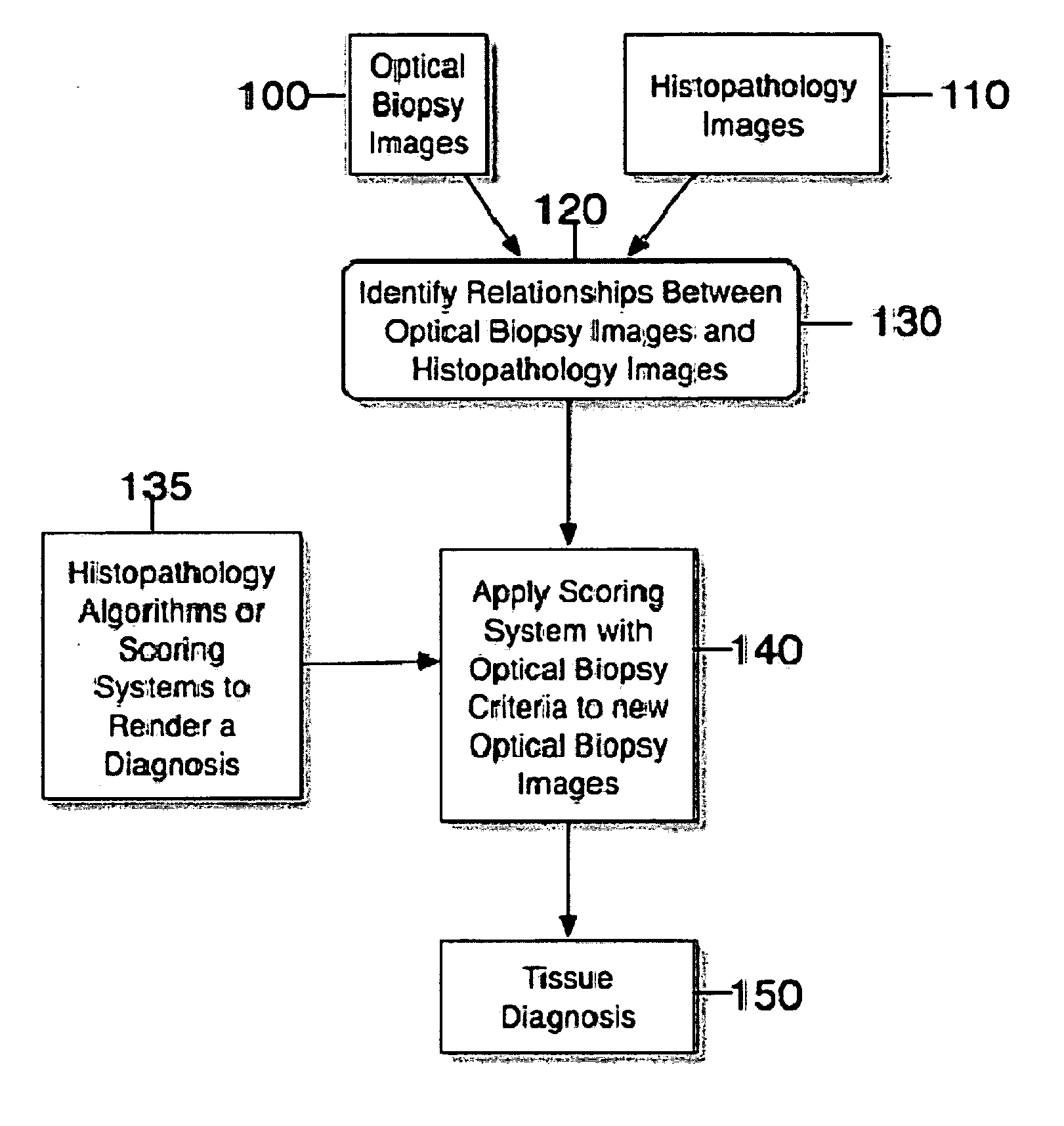
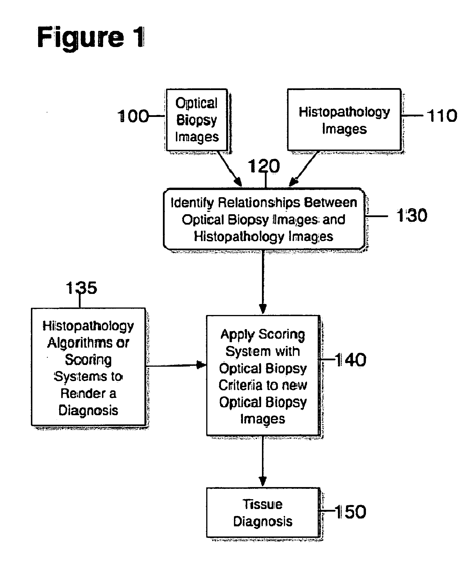
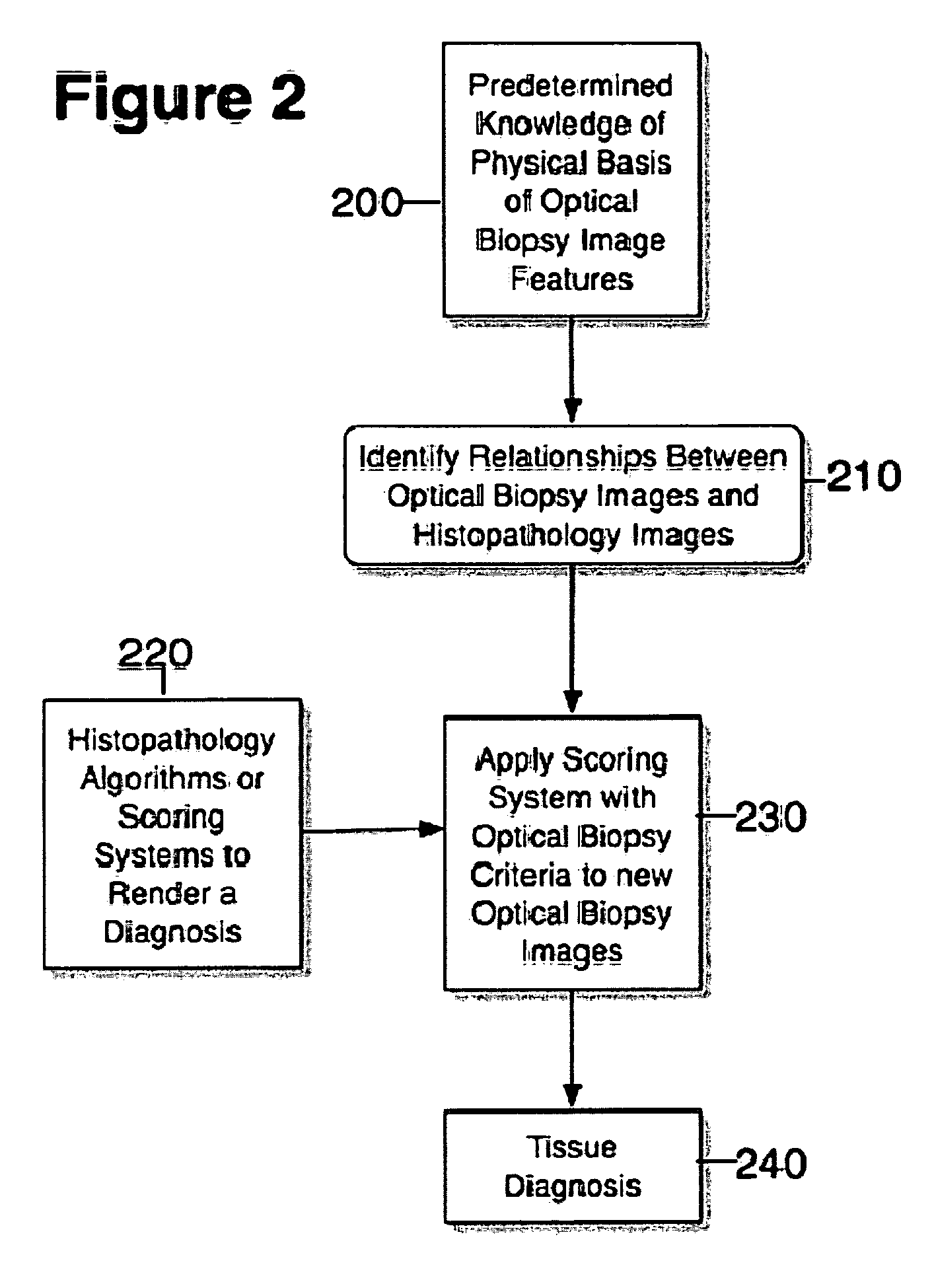
Systems. processes and software arrangements for evaluating information associated with an anatomical structure by an optical coherence ranging technique
an anatomical structure and optical coherence ranging technology, applied in the field of systems. processes and software arrangements for evaluating information associated with anatomical structures by optical coherence ranging techniques, can solve the problems of high scattering, increased costs to the health care system and to the individual patient, and differences between the data obtained by these prior art methods and the medical standards of care for diagnosis
- Summary
- Abstract
- Description
- Claims
- Application Information
AI Technical Summary
Benefits of technology
Problems solved by technology
Method used
Image

Examples
example 1
Determination of SIM at SCJ From OCT Images
[0058] i. Exemplary Design
[0059] An exemplary study for the exemplary embodiments of the present invention was a blinded, prospective trial. Its primary objective was to identify OCT image features for differentiating intestinal metaplasia at the SCJ. Patients undergoing a routine outpatient upper endoscopy were requested to participate in the study. OCT images of the SCJ were obtained during the endoscopy procedure. Two pathologists reviewed each biopsy specimen, and noted the presence of the following tissue types: gastric or oxyntic cardia, squamous mucosa, pancreatic metaplasia. The existence of intestinal metaplasia was noted by the presence of goblet cells. Image features of intestinal metaplasia were determined by creating and reviewing an OCT atlas “training set”, containing biopsy-correlated images of known tissue types. These features were then prospectively applied to a “validation set” of unknown tissue types. The sensitivity,...
example 2
Identifying High Grade Dysplasia and Intramucosal Carcinoma in OCT Images of SIM
[0081] i Exemplary Study Design
[0082] The exemplary study performed was a blinded, prospective trial. Recruited subjects were patients with BE undergoing routine endoscopic surveillance or confirmatory biopsies for IMC or HGD. OCT images of the Barrett's epithelium were obtained during endoscopy. Biopsy correlated OCT images of the esophagus were viewed and scored by a reader blinded to the tissue diagnosis. For each image the score for surface maturation and gland architecture were summed to establish a “dysplasia index”. Each biopsy specimen was independently reviewed, and a consensus diagnosis was rendered.
[0083] ii Exemplary OCT System
[0084] The exemplary OCT device which can be utilized for the exemplary embodiment of the present invention and used in the study is described in J. M. Poneros et al., “Diagnosis of specialized intestinal metaplasia by optical coherence tomography,” Gastroenterology...
PUM
 Abstract
Description
Claims
Application Information