
Skeletal musle-derived cells and methods related thereto

a technology of skeletal muscle and derived cells, applied in the direction of skeletal/connective tissue cells, biocide, peptide/protein ingredients, etc., can solve the problems of frequent and life-threatening conditions, heart failure, etc., and achieve the effect of reducing desmin expression and reducing desmin expression

Inactive Publication Date: 2007-04-19
YAEGER PETER C +2
View PDF0 Cites 0 Cited by
  • Summary
  • Abstract
  • Description
  • Claims
  • Application Information

AI Technical Summary

Benefits of technology

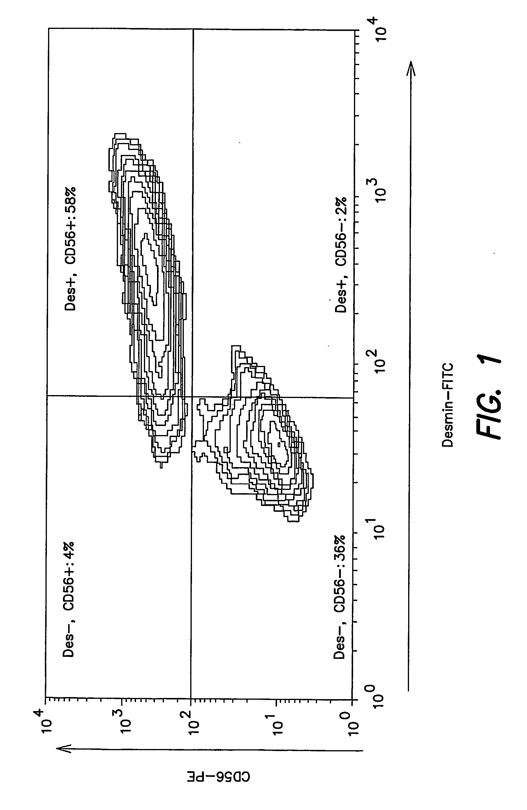
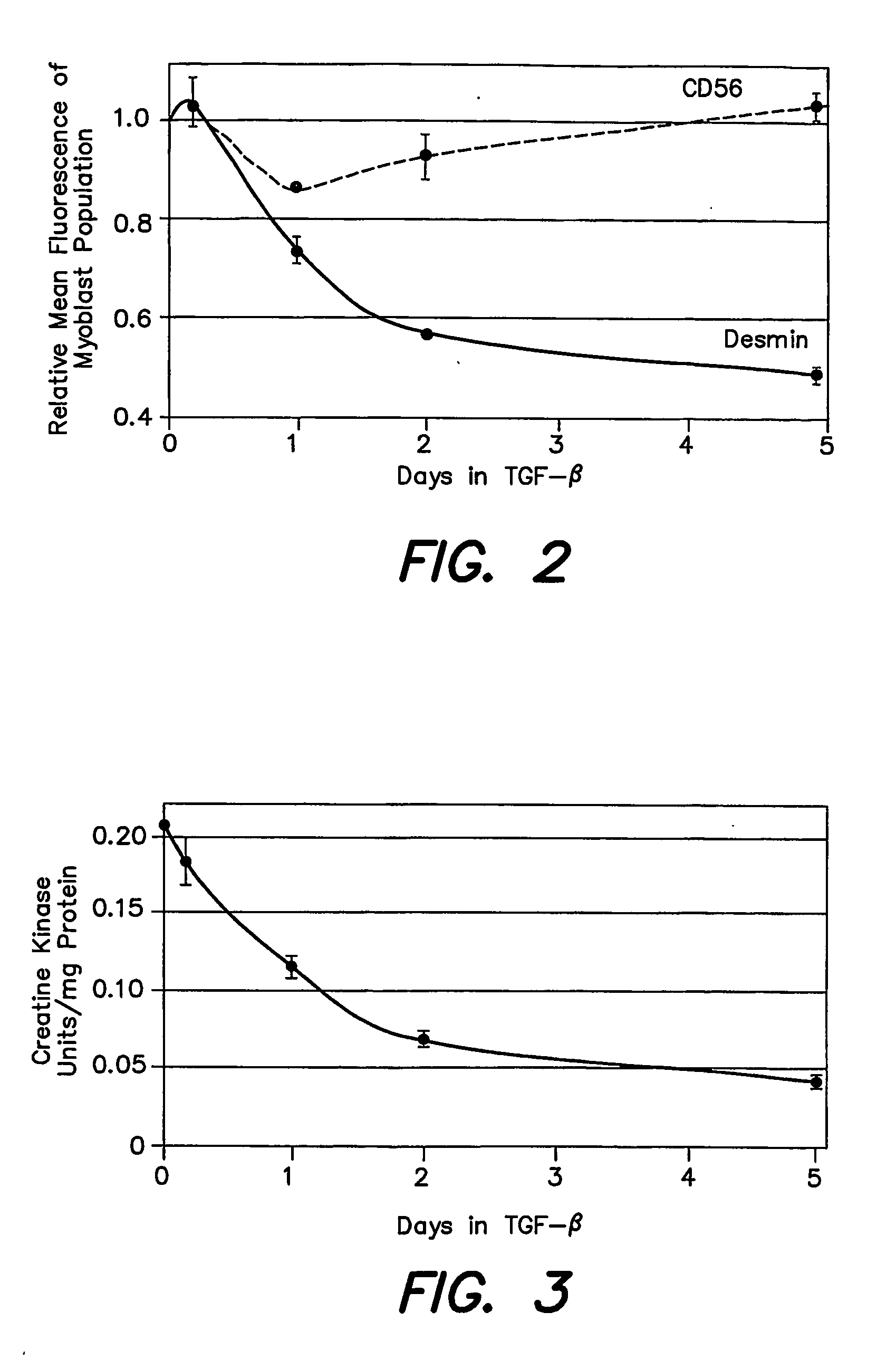
[0069] To determine the effects of TGF-β on cell growth and differentiation of HuSkMCs, cells of strain A were propagated in 2nd passage and exposed to 1 ng/ml TGF-β2 for different intervals, each extending to the termination of a 5 day culture period. Cells were then immunolabeled and analyzed by flow cytometry for quantitative detection of desmin and CD56 expression as described above. While the pattern of fluorescence intensity of the desmin-negative peak was unaffected by TGF-β2, the fluorescence intensity of the desmin-positive peak showed a progressive decline as the time of exposure to TGF-β2 increased. This change reflected a decrease in desmin expression in the myoblast population.
[0070] Quantification of the flow cytometry results (FIG. 2) showed that the average fluorescence intensity of the myoblast population of HuSkMCs exposed to TGF-β2 for five days was 48% of that for untreated cells. Approximately half the decrease in desmin expression occurred after one day of exposure to TGF-β2. The observed decrease in desmin expression in response to TGF-β2 was further suppo...

Problems solved by technology

Heart failure, mostly due to myocardial insufficiency, is a frequent and life-threatening condition, despite medical and surgical advances.
To this end, HuSkMCs may need to be propagated for several passages, since the number of cells available from biopsies is generally limited.
The challenge is not only to consistently produce a large number of cells but also to reliably characterize the identity and differentiation state of cells in culture.

Method used


Image

  • Skeletal musle-derived cells and methods related thereto
  • Skeletal musle-derived cells and methods related thereto

Examples


example 1

Derivation of HuSkMCs Strains

[0050] HuSkMCs were derived from quadriceps muscle of a 25 year old male cadaver (Strain A), rectus femoris muscle of a 77 year old female amputee (Strain B), quadricep muscle of a 36 year old female cadaver (Strain C), or vastus laterus muscle of a 45 year old male cadaver (Strain D). Cadaver tissue, provided by the National Disease Research Institute (NDRI, Philadelphia, Pa.), was procured 8 to 19 hours post-mortem. Skeletal muscle was shipped and maintained at 0-4° C. for 2-4 days in University of Wisconsin's Solution or Iscove's Modified Dulbecco's Medium (IMDM). Then muscle was trimmed of obvious connective tissue and fat and rinsed in phosphate buffered saline (PBS). The trimmed muscle, with a wet weight of at least 4 grams, was minced into pieces of approximately 1 mm3. The minced muscle was digested in type II Collagenase (Worthington, Lakewood, N.J.) at 470 U / ml, using 15-30 ml digestion solution per gram muscle, at 37° C. for 1 hour with inte...

example 2

Propagation of HuSkMCs

[0051] All cultures were propagated in a 37° C., 5% CO2, humidified environment, using collagen-I coated flasks. Medium for propagation was composed of Ham's F-12 containing GLUTAMAX™ (Invitrogen, Carlsbad, Calif.), 50 μg / ml gentamicin, 1 μg / ml amphotericin B, 15-20% FBS (Cat. No. SH30071; Hyclone, Logan, Utah), and basic fibroblast growth factor (bFGF; R&D Systems, Minneapolis, Minn.). The bFGF concentration was 5 ng / ml, except that 20 ng / ml bFGF was used for the entire propagation of Strain D and for Strain A propagation after 1st passage. The inoculation density after 1st passage was 5×10³ cells / cm2. TGF-β2 (Genzyme, Cambridge, Mass.) was added as indicated in other Examples. Cultures received fresh medium every 2-4 days. When 70-100% confluent, at a density ranging from 8×10⁴ to 1.5×10⁵ cells / cm2, cells were detached with 0.05% trypsin, 0.5 mM EDTA and the cell suspensions were subcultured, or analyzed as described below. In some cases, cells were cryopre...

example 3

Immunolabeling Procedures for Flow Cytometry

[0053] Indirect fluorescent immunolabeling was performed to detect desmin or TE7. HuSkMCs suspensions were fixed with 4% paraformaldehyde in PBS for 20 minutes at 20-25° C. Fixed cells were washed and incubated 30 minutes at 20-25° C. with mouse anti-desmin antibody (clone D33; Dako Corp, Carpenteria, Calif.) at 2.5-5.0 μg / ml in 0.1% saponin, 10% FBS in PBS (saponin permeabilization buffer (SPB)) or with mouse “anti-fibroblastantibody (clone TE7; Research Diagnostics, Flanders, N.J.) at 2.2 to 4.0 μg / ml in SPB. Cells were then washed and incubated 30 minutes at 4° C. with fluorescein isothiocyanate (FITC)-conjugated goat anti-mouse-IgG antibody (Jackson Immunoresearch, West Grove, Pa.) at 14 μg / ml in SPB.

[0054] Direct fluorescent immunolabeling was performed to detect CD56. HuSkMCs suspensions were incubated 30 minutes at 4° C. with phycoerythrin (PE)-conjugated mouse anti-CD56 antibody (clone NCAM16.2, BD BioSciences, San Jose, Calif...


PUM

PropertyMeasurementUnit
Fractionaaaaaaaaaa
Fractionaaaaaaaaaa
Timeaaaaaaaaaa

Abstract

The disclosure provides methods of propagating skeletal muscle cell (SkMC) cultures enriched in differentiation-competent myoblasts that express normal levels of CD56 and reduced levels of desmin. The methods comprise culturing SkMCs in a mitogen-rich cell culture medium supplemented with TGF-β. The disclosure also provides therapeutic methods utilizing SkMCs propagated in TGF-β, e.g., methods of treating myocardial infarction by transplantation of autologous or allogeneic SkMCs.

Description

[0001] This application claims priority to U.S. application Ser. No. 60 / 502,762, filed on Nov. 17, 2003, herein incorporated by reference in its entirety. FIELD OF THE INVENTION [0002] The present invention relates to methods of propagating skeletal muscle-derived cells, and in particular, cells intended for implantation into injured heart tissue. The invention further relates to cell culture medium compositions that contain TGF-β. BACKGROUND OF THE INVENTION [0003] Heart failure, mostly due to myocardial insufficiency, is a frequent and life-threatening condition, despite medical and surgical advances. Therapeutic application of autologous human skeletal muscle cells (HuSkMCs) to mitigate the deterioration of cardiac function resulting from myocardial infarction has shown promise in several preclinical and clinical studies (see, e.g., Atkins et al. (1999) Heart Lung Transplant., 18:1173-1180; Hutcheson et al. (2000) Cell Transplant., 9:359-368; Pouzet et al. (2001) Circulation, 102...

Claims


Application Information

Patent Timeline
IPC IPC(8): A61K35/34C12N5/06C12N5/08A61K35/12A61K38/17C12N5/077
CPCA61K35/12C12N5/0658C12N2501/15
Inventor YAEGER, PETER C.STEWART, JEFFREY D.WENTWORTH, BRUCE M.
Owner YAEGER PETER C
Learn More