
Applications of HIFU and chemotherapy

a technology applied in the field of hifu and chemotherapy, can solve the problems of no improvement in the mortality rate of radiation treatments on the breast, significant short and long-term side effects of radiation therapy, damage to the skin,

Inactive Publication Date: 2007-04-19
UST INC
View PDF10 Cites 49 Cited by
  • Summary
  • Abstract
  • Description
  • Claims
  • Application Information

AI Technical Summary

Benefits of technology

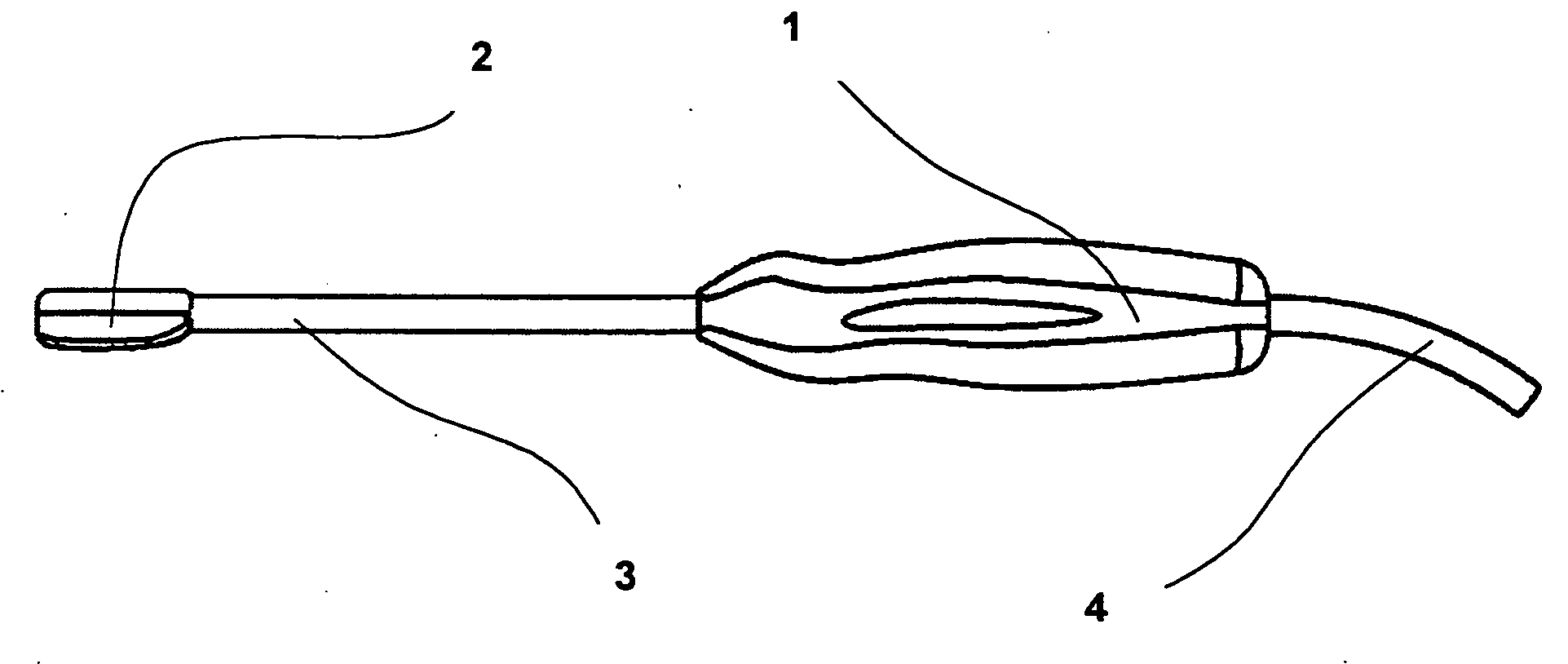
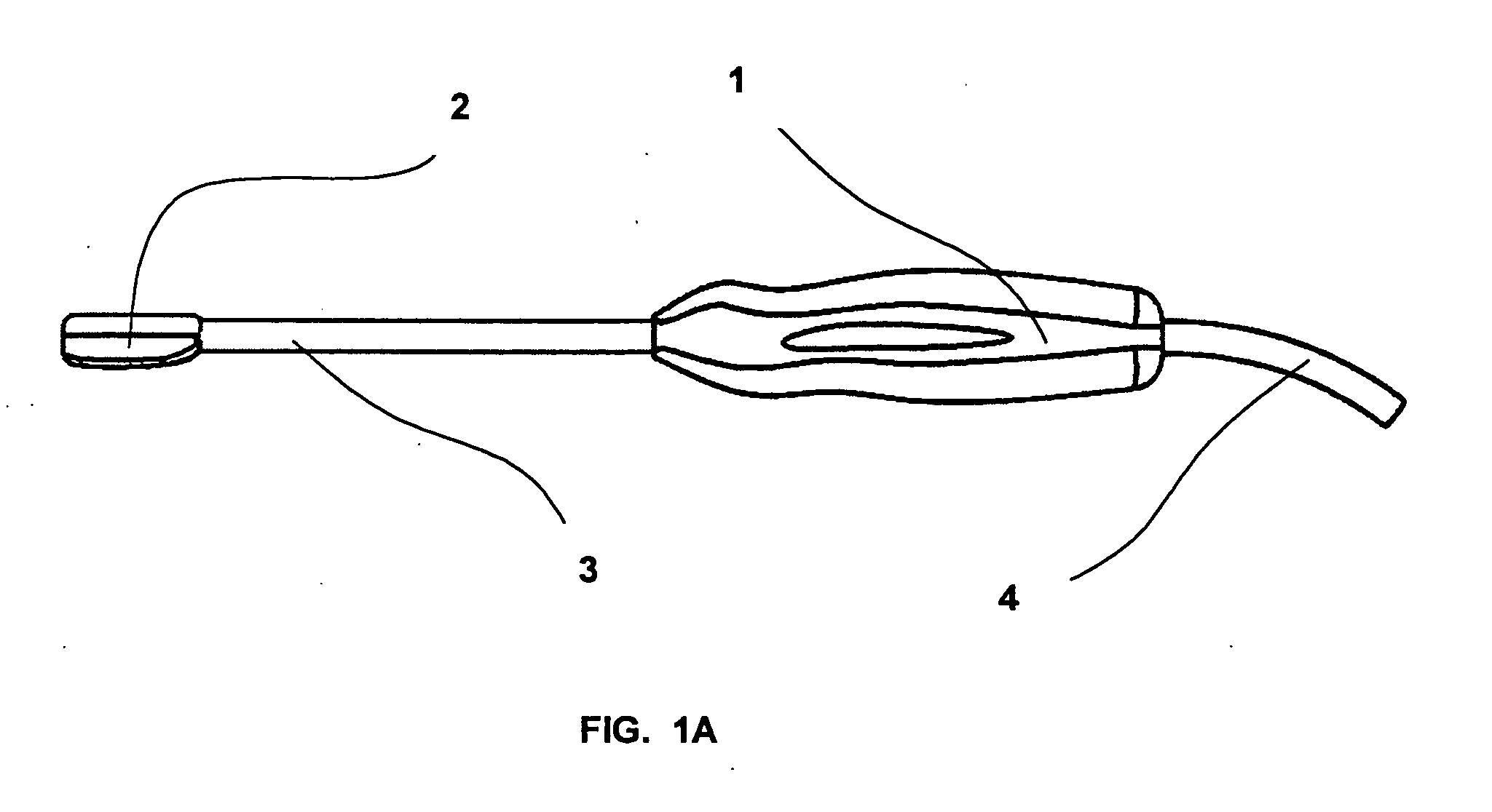
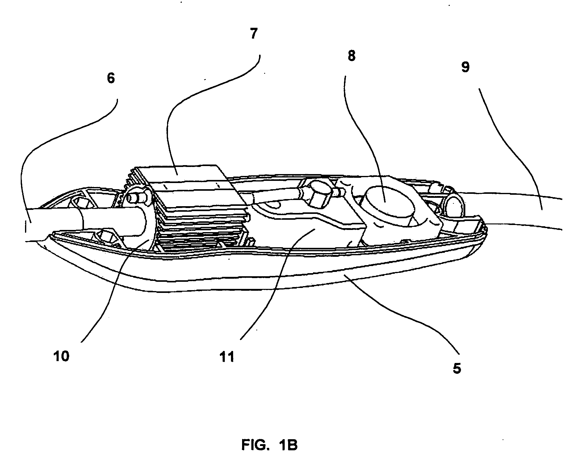
[0019] The present invention describes both an apparatus and a method, using high intensity ultrasound, which may be used alone or in combination with a chemotherapy agent, to treat benign and malignant tumor cells. The method comprises the use of (1) HIFU, to treat the major portion of the tumor (e.g. the primary tumor and a suitable margin surrounding the tumor) without surgical resection by inducing coagulative necrosis of the tumor cells by elevated temperature, and (2) sonoporation, though which the uptake of a locally-delivered chemotherapy agent into tumor cells is enhanced by the use of ultrasound to treat those tumor cells not destroyed by direct HIFU therapy. The present invention permits the direct destruction of tissue (ablation) of the tumor and its margin as well as the indirect destruction of tumor cells lying outside the margin, all of which can be accomplished by one or more ultrasound apparatuses used during therapy.

Problems solved by technology

There is no improvement in mortality from radiation treatments to the breast.
In addition to the large block of time, travel and expense, radiation therapy has significant short and long term side effects, including fatigue; damage to the skin including blistering, peeling and color changes; fullness; stiffness and swelling of the breast.
There may be permanent size, color and texture changes to the breast and long term damage to the heart and lungs with associated morbidity and mortality from radiation therapy.
No significant advances have been made during the past 25 years in the treatment of GBMs and most patients die within three months without therapy.
Tumors of the head and neck are often visible and somewhat accessible to the surgeon; however, the surgical removal of these tumors results in significant morbidity and reduction in the quality of life.
However, many of these drugs are toxic when given systemically.

Method used


Image

  • Applications of HIFU and chemotherapy
  • Applications of HIFU and chemotherapy
  • Applications of HIFU and chemotherapy

Examples


Embodiment Construction

[0024] The details of the present invention will be discussed with reference to particular embodiments and the accompanying drawings which are intended to represent the present invention by way of example only.

[0025] The present invention comprises a surgical probe which emits ultrasound to uniformly cauterize (or destroy by heat) tumor tissue directly, when the ultrasound is delivered at high intensities, and indirectly, through enhanced cell uptake of a chemotherapy agent, when the ultrasound is delivered at low intensities. The invention also comprises the approach of applying high intensity ultrasound with or without sonoporation with a local chemotherapeutic drug to the region surrounding a surgical procedure, such as a breast lumpectomy or brain tumor, thereby potentially eliminating the need for “second lumpectomies” due to tumor cells in the surgical margin, potentially eliminating post lumpectomy radiation treatment, and reducing or eliminating remaining tumors in the marg...


PUM


Abstract

A method, using high intensity ultrasound, which may also be combined with a chemotherapy agent, that can result the direct destruction of tumor cells and in the reduction or elimination of local reoccurrence of cancer after removal of cancerous tissue, such as a surgical breast lumpectomy or surgical excision of a brain tumor. The method comprises either (1) the treatment of the tumor directly with High Intensity Focused Ultrasound, or (2) the treatment of the margins of the tissue surrounding the surgical cavity or void with ablative continuous wave high intensity ultrasound and a combination of a locally delivered chemotherapy agent and high intensity ultrasound, termed sonoporation. The present invention permits both the direct destruction (ablation) of tumor tissue as well as the destruction of tissue around the surgical margin and may include the enhanced local cellular uptake of locally injected chemotherapeutic drugs, all of which can be accomplished by a therapeutic ultrasound device used during surgery.

Description

[0001] This application claims the benefit of U.S. Provisional Patent Application No. 60 / 726,254 filed Oct. 13, 2005 the entire disclosure of which is hereby incorporated by reference.FIELD OF THE INVENTION [0002] The present invention describes an apparatus and a method for treating a tumor as well as a margin of tissue surrounding the tumor with both High Intensity Focused Ultrasound (HIFU) and chemotherapy. The therapy provided combines the ability of HIFU to induce coagulative necrosis with the synergistic effects of ultrasound and chemotherapy, generally described as sonoporation. This approach is intended to treat the tumor and / or a margin surrounding the tumor to prevent the local recurrence of cancer due to cancer cells remaining in adjacent or nearby tissue. BACKGROUND OF THE INVENTION [0003] Improved imaging technology and adoption of earlier and repeated mammography result in many breast cancers being diagnosed when tumors are one centimeter diameter or smaller, or are co...

Claims


Application Information

Patent Timeline
IPC IPC(8): A61B18/04A61N1/30
CPCA61B8/4438A61B2017/00482A61B2018/00005A61K41/0047A61N7/02A61N7/022A61N2007/0078
Inventor LARSON, EUGENE A.KAMINSKI, PERRY W.
Owner UST INC
Learn More