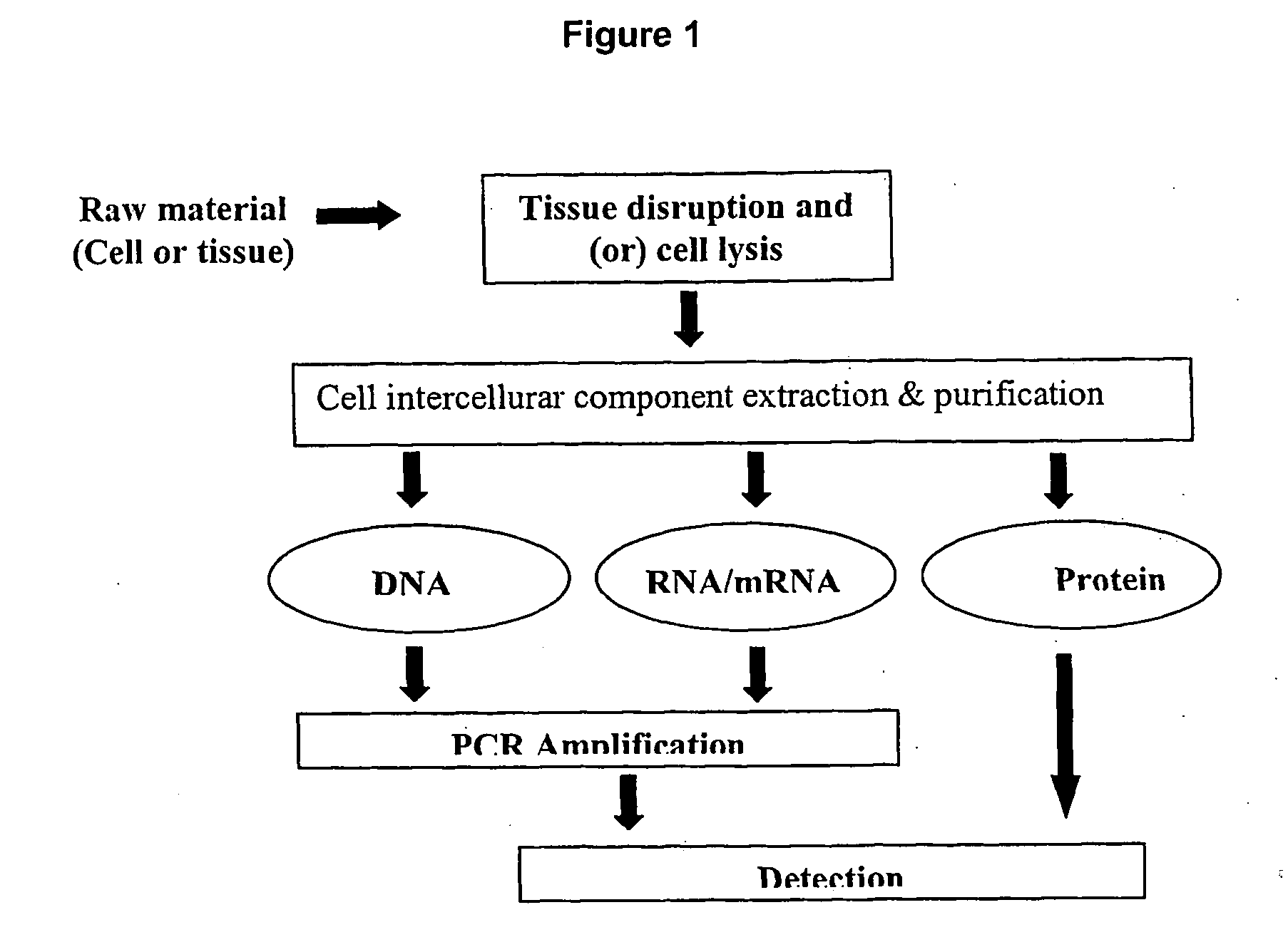
Method and system for cell and/or nucleic acid molecules isolation
- Summary
- Abstract
- Description
- Claims
- Application Information
AI Technical Summary
Benefits of technology
Problems solved by technology
Method used
Image

Examples
example 1
[0194] Trypsins and collagenases were used as exemplary models of certain embodiments of the invention. The process set forth herein are however applicable to other types of tissues, including human tissues, plant tissues, adipose tissues, and the like.
[0195] Trypsin-EDTA digestion of rat liver was carried out as follows: freshly harvested tissue was cut into 2 mm3 sample sizes, followed by washing twice in 500 μl iced Phosphate Buffered Saline (PBS). Trypsin-EDTA solution was added to the tissue sample, which was incubated in a shaking water bath at 37° C. for 30 min, and triturated from time to time until no further tissue disruption was observed. A similar procedure was followed using collagenase, except that: 1) incubation time was increased to 90 min and shaking was not necessary; and 2) gentle flicking of the sample was applied instead of trituration after incubation. The cell suspension obtained using these procedures yielded a homogenous solution that could be used for down...
example 2
[0204] Trypsin and collagenases were used as exemplary models of this embodiment. As an example, Trypsin-EDTA digestion of rat liver was carried out as follows: freshly harvested tissue was cut into 8 mm3 (10 mg in weight) sample size, followed by washing twice in 500 μl iced Phosphate Buffered Saline (PBS). Trypsin-EDTA solution was added to the tissue sample, which was incubated in a shaking water bath at 37° C. for 30 min, pipette the solution until no further tissue disruption was observed. A similar procedure was followed using collagenase except that: 1) incubation time was increased to 90 min and shaking force was not necessary; and 2) gentle flicking was applied instead of triturating after incubation.
[0205] By our experiment, 0.01% to 0.15% of trypsin concentration was found to be preferable in terms of cell yield. Other concentrations could be used, however, by adjusting the time of exposure to the protease. Table 4 is the optimized experimental trypsin concentration for ...
example 3
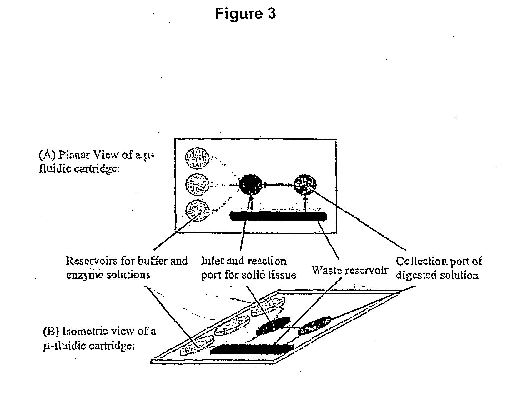
[0207] The process of the tissue disruption device including the following steps:
[0208] 100 μl of protease [0.05-0.15% (wt / vol) for Trypsin and 100-300 unit / ml for collagenases] solution is first injected into the incubation chamber and preheated to 37° C. Fresh or frozen mammal tissue (up to 10 mg) is then put into the chamber and sealed. The tissue sample is incubated inside the chamber for about 15 minutes so that it becomes softened by the enzymolysis of the protease solution.
[0209] Once the incubation time is over, the softened tissue and the solution are passed through the disruption channel for tissue disruption with the help of a micropump, which is connected, to the inlet and outlet of the device (Refer to FIG. 12, Components 18&19). Shear force generated in the disruption components breaks the softened tissue into smaller size. These smaller pieces of tissue will, then be softened with the enzymolysis of the protease reagent. As the dimension of the disruption components...
PUM
| Property | Measurement | Unit |
|---|---|---|
| Volume | aaaaa | aaaaa |
| Volume | aaaaa | aaaaa |
| Volume | aaaaa | aaaaa |
Abstract
Description
Claims
Application Information
 - Generate Ideas
- Intellectual Property
- Life Sciences
- Materials
- Tech Scout
- Unparalleled Data Quality
- Higher Quality Content
- 60% Fewer Hallucinations
Browse by: Latest US Patents, China's latest patents, Technical Efficacy Thesaurus, Application Domain, Technology Topic, Popular Technical Reports.
© 2025 PatSnap. All rights reserved.Legal|Privacy policy|Modern Slavery Act Transparency Statement|Sitemap|About US| Contact US: help@patsnap.com