Tissue marker for multimodality radiographic imaging

a multi-modality radiographic imaging and tissue marker technology, applied in the field of tissue markers, can solve the problems of difficult identification and differentiation of small signal voids produced by some conventional markers from naturally occurring dark artifacts, and the difficulty of manifoldization, so as to achieve good visualization characteristics and increase the effect of signal intensity

Active Publication Date: 2007-05-17
BREAST MED
View PDF86 Cites 29 Cited by
  • Summary
  • Abstract
  • Description
  • Claims
  • Application Information

AI Technical Summary

Benefits of technology

[0009] Various embodiments may provide certain advantages. For instance, a contrast agent selected to produce an increase in signal intensity in an MR imaging modality may produce good visualization characteristics without also producing an excessive artifact and without interfering with MR spectroscopy. Production of an increase in signal intensity in an MR imaging modality may be particularly beneficial in certain contexts, such as, for example, imaging of breast tissue, which is heterogeneous.

Problems solved by technology

This manifestation can be particularly problematic in some contexts.
For example, heterogeneous breast tissue produces many dark artifacts under MR imaging, thereby rendering small signal voids produced by some conventional markers difficult to identify and distinguish from naturally occurring dark artifacts.
In addition, some markers produce large susceptibility artifacts under MR imaging, thereby distorting images in both MRI and spectroscopic modalities.
Some markers incorporate an external gel that may produce a positive or bright signal, but such gels are not permanent.
Some other markers contain collagen or polylactic acid, which may interfere with magnetic resonance spectroscopy.

Method used


Image

  • Tissue marker for multimodality radiographic imaging
  • Tissue marker for multimodality radiographic imaging
  • Tissue marker for multimodality radiographic imaging

Examples


Embodiment Construction

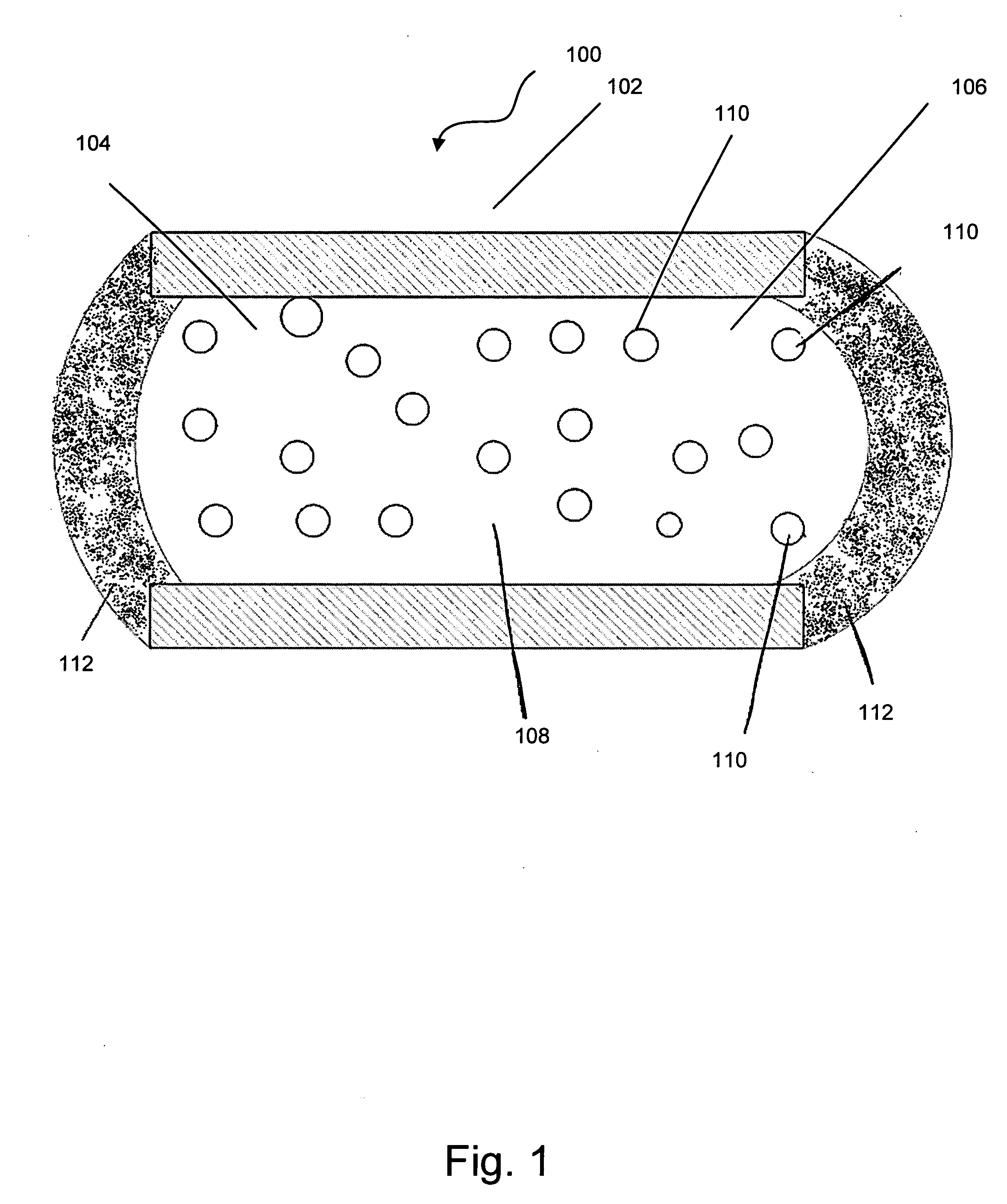
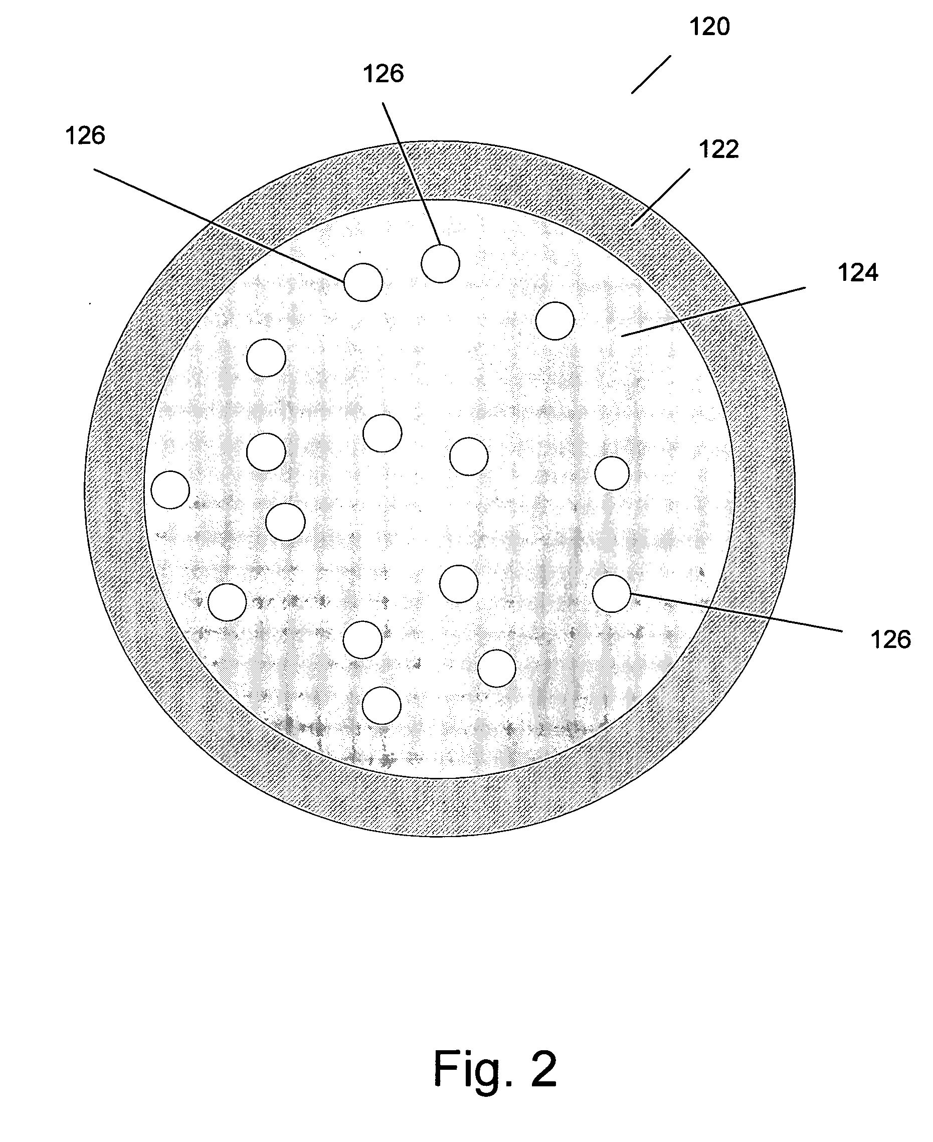
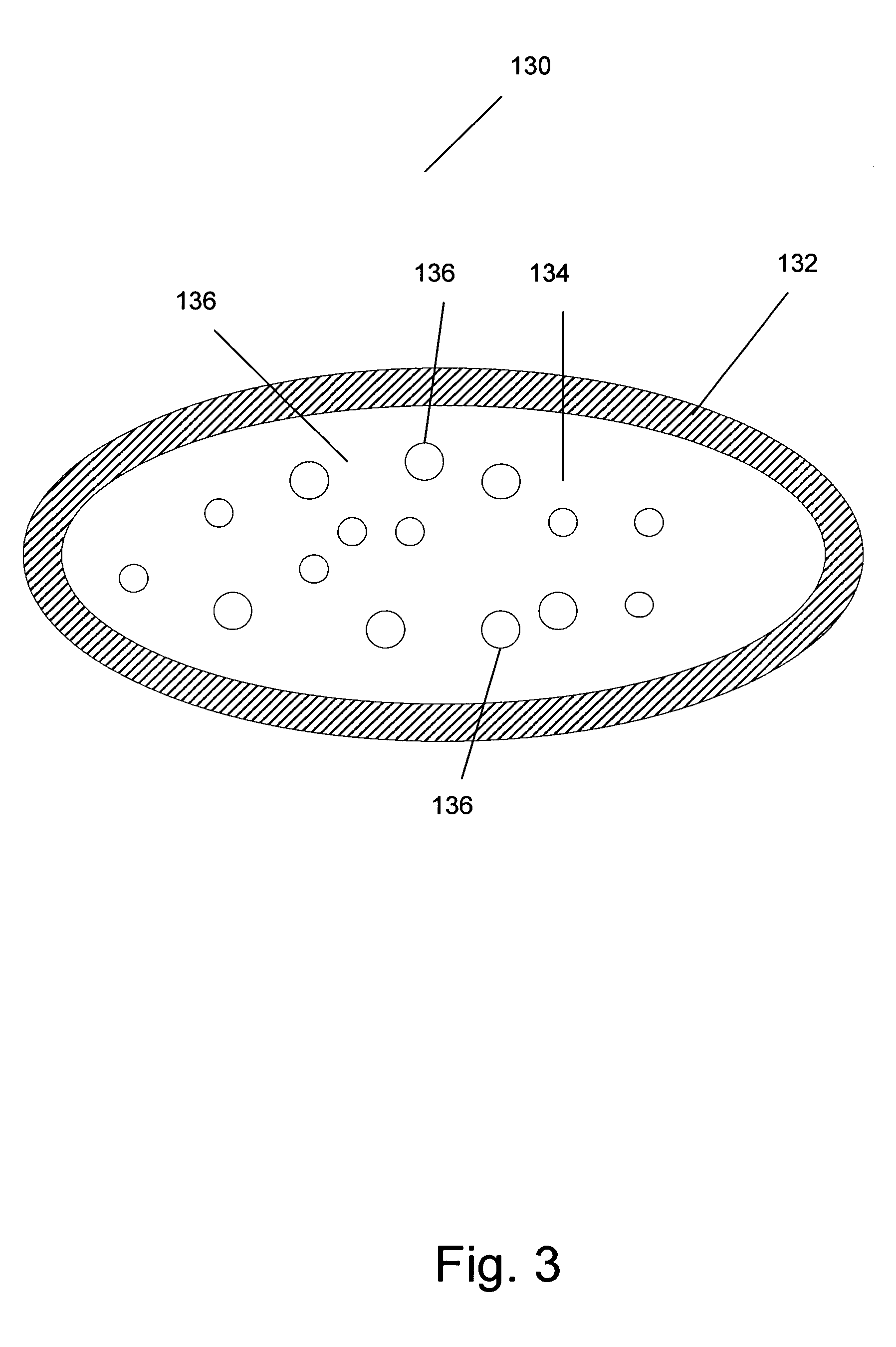
[0017] According to various embodiments, an implantable tissue marker incorporates a contrast agent sealed within a chamber in a container formed from a solid material. The contrast agent is selected to produce an increase in signal intensity under magnetic resonance imaging (MRI). An additional contrast agent may also be sealed within the chamber to provide visibility under another imaging modality, such as computed tomographic (CT) imaging or ultrasound imaging.

[0018] In this way, certain advantages may be realized. For instance, a contrast agent selected to produce an increase in signal intensity in an MR imaging modality may produce good visualization characteristics without also producing an excessive artifact and without interfering with MR spectroscopy. Producing an increase in signal intensity in an MR imaging modality may be particularly beneficial in certain contexts, such as, for example, imaging of breast tissue. Most conventional markers appear as a signal void in MR i...


PUM

PropertyMeasurementUnit
lengthaaaaaaaaaa
outer diameteraaaaaaaaaa
magnetic resonanceaaaaaaaaaa

Abstract

An implantable tissue marker incorporates a contrast agent sealed within a chamber in a container formed from a solid material. The contrast agent is selected to produce a change, such as an increase, in signal intensity under magnetic resonance imaging (MRI). An additional contrast agent may also be sealed within the chamber to provide visibility under another imaging modality, such as computed tomographic (CT) imaging or ultrasound imaging.

Description

TECHNICAL BACKGROUND [0001] The disclosure relates generally to tissue markers. More particularly, the disclosure relates to implantable tissue markers for use in magnetic resonance imaging. BACKGROUND [0002] Certain medical conditions, including breast cancer, are increasingly being diagnosed and treated using minimally invasive medical techniques. Such techniques typically involve the use of clinical imaging methods that allow the physician to visualize interior portions of a patient's body without the need to make excessive incisions. Imaging can be performed using any of variety of modalities, including, for example, X-rays, computed tomographic (CT) X-ray imaging, portal film imaging devices, electronic portal imaging devices, electrical impedance tomography (EIT), magnetic resonance (MR) imaging, or MRI, magnetic source imaging (MSI), magnetic resonance spectroscopy (MRS), magnetic resonance mammography (MRM), magnetic resonance angiography (MRA), magnetoelectro-encephalograph...

Claims


Application Information

Patent Timeline
Patent Type & Authority Applications(United States)
IPC IPC(8): A61K51/00A61K49/04
CPCA61B19/54Y10T29/49982A61B2019/5425A61B2019/5454A61B2019/547A61B2019/5495A61K49/0002A61K49/0447A61K49/18A61B8/481A61B17/3468A61K49/223A61M31/005G01R33/4814G01R33/5601Y10T29/49888Y10T29/4998A61B2019/5408A61B90/39A61B2090/3908A61B2090/3925A61B2090/3954A61B2090/3958A61B2090/3966A61B2090/397A61B2090/3995A61M31/002G01R33/4812H01F41/02H01F41/0253Y10T29/49075
Inventor BOLAN, PATRICK J.GARWOOD, MICHAELNELSON, MICHAEL T.HALPERN, DANIEL A.
Owner BREAST MED
Why Eureka
Try Eureka