Stem cells within gel microenvironments

a microenvironment and stem cell technology, applied in the field of stem cell cell biology of stem cells and their differentiation, delivery and use in regenerative medicine, can solve the problems of limiting the usefulness of repairing bone in situ, inability to control the induction and maintenance of cell differentiation,

Inactive Publication Date: 2007-05-24
RENESSELAER POLYTECHNIC INST
View PDF18 Cites 121 Cited by
  • Summary
  • Abstract
  • Description
  • Claims
  • Application Information

AI Technical Summary

Problems solved by technology

However, the chief factor limiting stem cell-based therapies is the inability to control the induction and maintenance of cell differentiation.
However, many of the current methods used to induce desired differentiation of hMSC in vitro involve treatments that are difficult to implement for use as in vivo therapies.
While these media supplements clearly induce osteoblast differentiation under some conditions, they may suppress bone growth in vivo (Ng et al., 2002), which may limit their usefulness for repairing bone in situ.

Method used


Image

  • Stem cells within gel microenvironments
  • Stem cells within gel microenvironments
  • Stem cells within gel microenvironments

Examples


example 1

Collagen-Agarose Beads

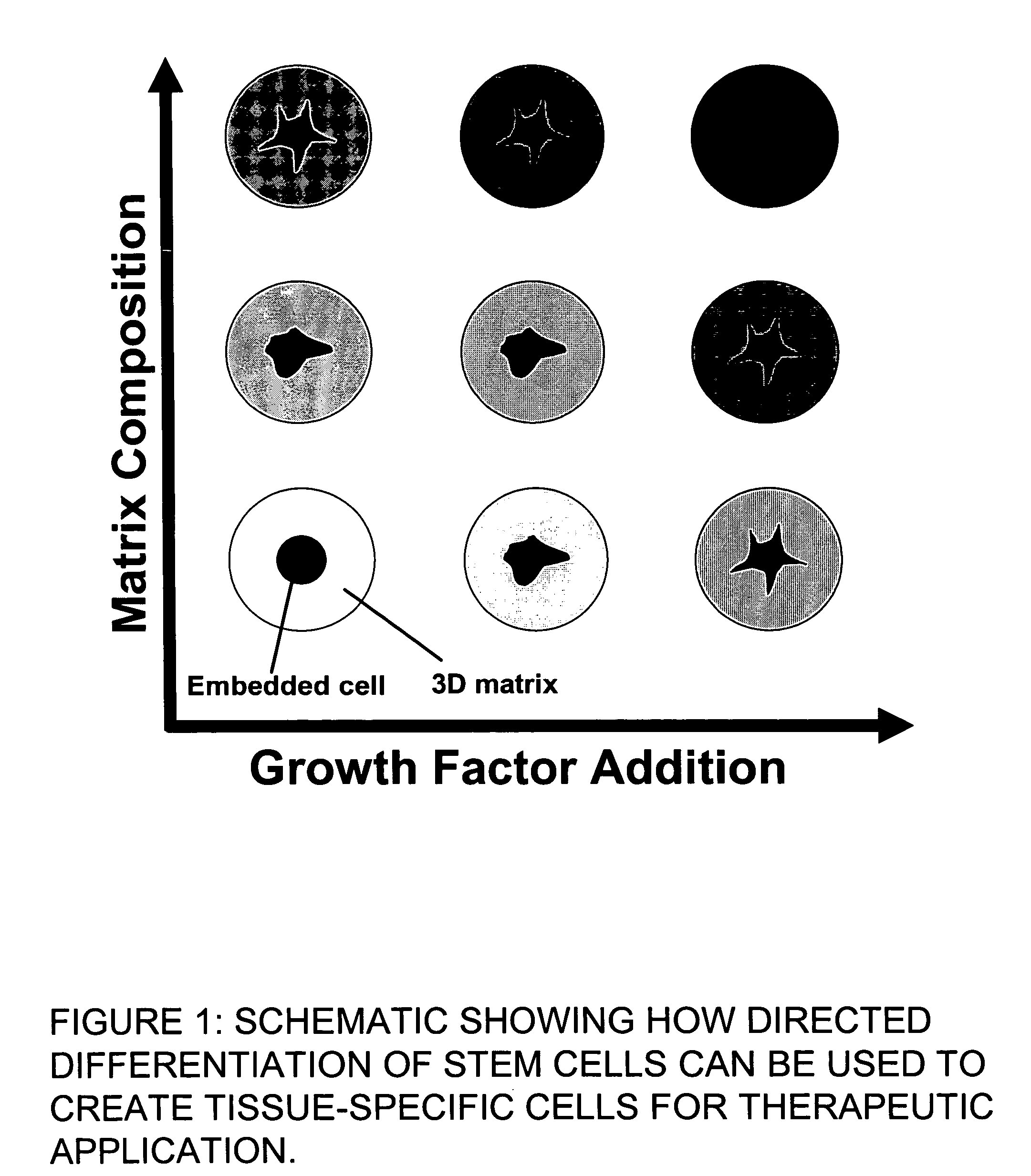
[0030] In one embodiment of the present invention, hMSC were directly embedded into the matrix of 3D microbeads consisting of varying amounts of agarose and collagen Type I. The inventors examined factors that influence the bead production process, as well as the effects of varying matrix composition on hMSC viability and morphology. Collagen Type I was used because of its established involvement in influencing the phenotype of hMSCs, in particular towards the osteoblastic lineage. Agarose was used as an inert filler material to provide structural integrity to the beads and facilitate bead harvesting. By varying the ratio of agarose to collagen, the effect of ECM on hMSC function could be examined.

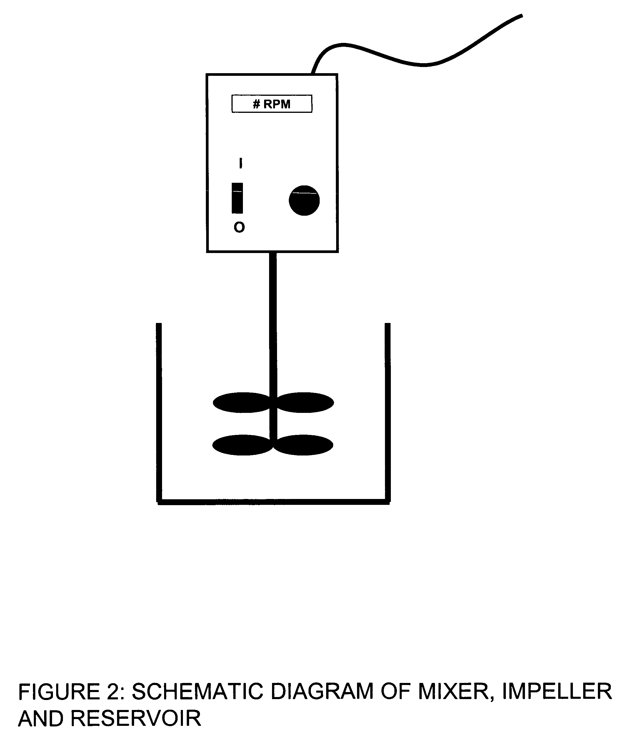
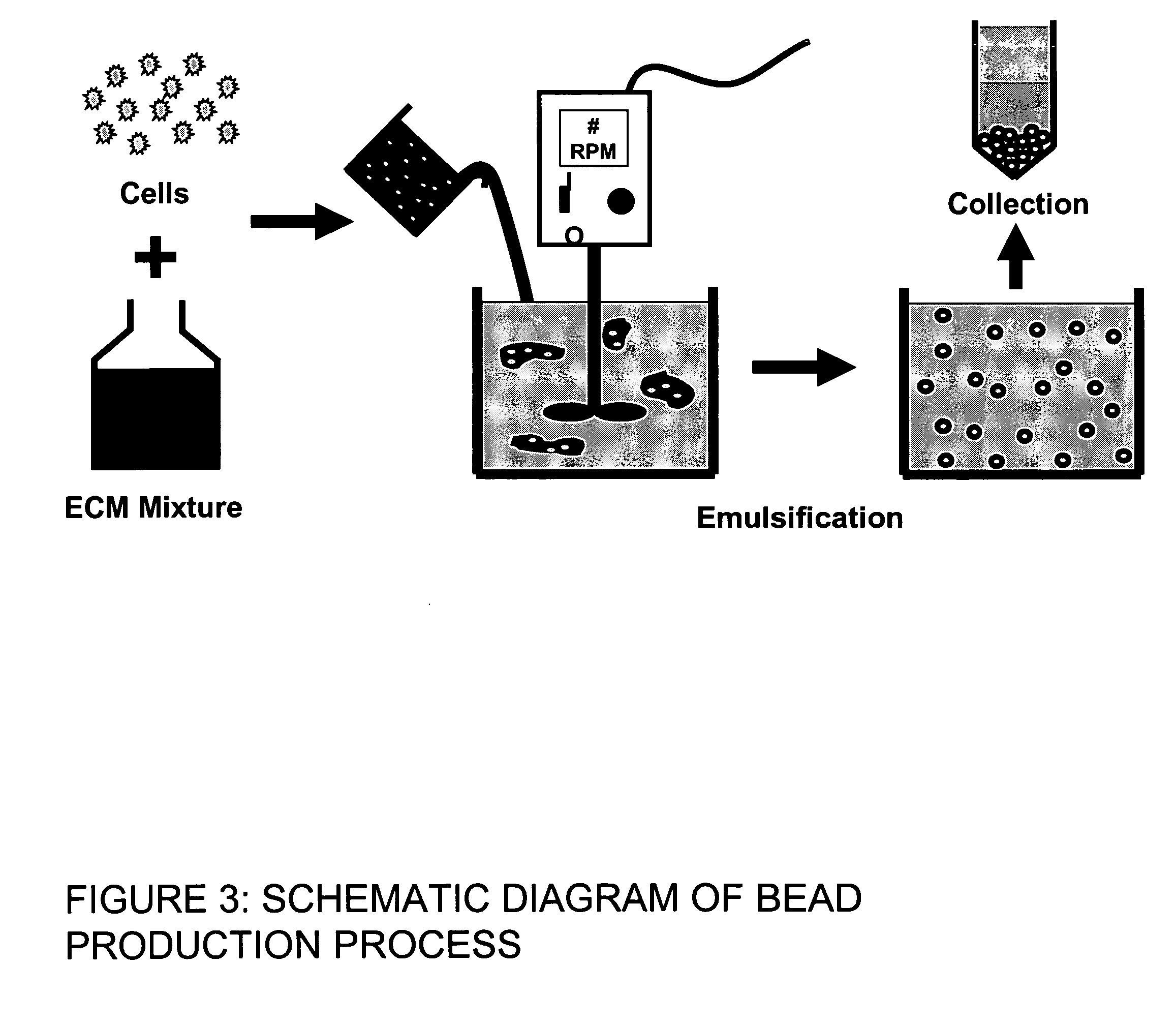
[0031] The bead production system is shown schematically in FIGS. 2 and 3. Cells were prepared for encapsulation through detachment from tissue culture flasks using trypsin-EDTA. The cells were counted and resuspended in a mixture of 5× DMEM, FBS, 0.1 M NaOH, 4.0 mg...

example 2

Gelatin Beads

[0039] Using a process analogous to that described in Example 1, gelatin beads can be made. In this case, a warm solution of gelatin and cells is emulsified and the emulsion is cooled to form gel beads with embedded cells. Because gelatin will re-melt when warmed to body temperature, gelatin beads can be stabilized using cross-linking to prevent remelting. Genipin or other agents that crosslink proteins (including transglutaminases and ribosylation) can be used for this purpose. FIG. 8(A) shows gelatin beads that have been stabilized with genipin cross-linking.

example 3

Collagen-Gelatin Beads

[0040] Using a process analogous to that described in Example 1, collagen-gelatin beads can be made. In this case, a warm solution of collagen, gelatin and cells is emulsified and the emulsion is cooled to form gel beads with embedded cells. Because gelatin will re-melt when warmed to body temperature, collagen-gelatin beads can be stabilized using cross-linking to prevent remelting. Genipin or other agents that crosslink proteins (including transglutaminases, ribosylation) can be used for this purpose. FIG. 8(B) shows collagen-gelatin beads that have been stabilized with genipin cross-linking. Alternately, the gelatin can be allowed to remelt after bead collection, thereby producing pure collagen beads (see Example 5, below).


PUM

PropertyMeasurementUnit
sizeaaaaaaaaaa
sizeaaaaaaaaaa
timeaaaaaaaaaa

Abstract

This invention provides a system to embed stem cells within three-dimensional (3D) hydrogel microenvironments consisting of naturally derived proteins, proteoglycans and / or polysaccharides. Pure matrices or combinations of materials can be used. The method involves suspending stem cells in solutions of the matrix components of interest, emulsifying these solutions in a hydrophobic phase, triggering gelation of the matrix components by changing the environmental conditions, and collection of the resulting hydrogel beads. The unique bead format of this invention has the advantage of allowing the use of small amounts of rare matrix proteins. Bead preparations can be concentrated into a paste for use as a cell delivery vehicle to damaged tissues, either directly after encapsulation or after a period of culture to promote stem cell differentiation. Defined 3D microenvironments can guide stem cell differentiation, and the resulting beads can be used directly as a cell delivery vehicle in various tissue repair applications.

Description

CROSS-REFERENCE TO RELATED APPLICATION [0001] This application claims benefit under 35 U.S.C. § 119(e) of U.S. provisional patent application No. 60 / 738,172, filed Nov. 11, 2005, the disclosure of which is herein incorporated in its entirety by reference.TECHNICAL FIELD [0002] The present invention relates generally to the field of cell biology of stem cells and their differentiation, delivery and use in regenerative medicine. More specifically, it describes methods of embedding stem cells within three-dimensional (3D) microenvironments, thereby guiding their differentiation to a desired phenotype. BACKGROUND OF THE INVENTION [0003] Stem cells are progenitor cells that have the ability to divide to form additional stem cells, as well as to differentiate into tissue-specific cells. The ability to self renew and differentiate into numerous cell types makes the use of stem cells to repair or replace damaged tissues a promising approach to many clinical problems. However, the chief fact...

Claims


Application Information

Patent Timeline
Patent Type & Authority Applications(United States)
IPC IPC(8): A61K35/12C12N5/08C12N5/06C12N5/0775
CPCA61K9/0019A61K9/5036A61K9/5052A61K9/5057A61K2035/126C12N2533/52C12N2533/54C12N2533/70C12N2533/72C12N5/0663
Inventor STEGEMANN, JAN P.LORD, EVAN J.
Owner RENESSELAER POLYTECHNIC INST
Why Eureka
Try Eureka