Fusion proteins comprising bacteriophage coat protein and asingle-chain T cell receptor

a technology of bacteriophage coat protein and asingle which is applied in the field of fusion proteins comprising bacteriophage coat protein and a single-chain t cell receptor, can solve the problems of reducing tcr yield, negatively affecting tcr stability and functionality, and difficult to isolate tcrs in significant quantities, so as to enhance the solubility and functionality of sctcr fusion proteins, the effect of enhancing

Inactive Publication Date: 2007-05-24
ALTOR BIOSCIENCE CORP
View PDF3 Cites 5 Cited by
  • Summary
  • Abstract
  • Description
  • Claims
  • Application Information

AI Technical Summary

Benefits of technology

[0014] The present invention relates to scTCR fusion proteins that include a scTCR molecule covalently linked (ie. fused) to a bacteriophage coat protein (e.g., gene III or gene VIII protein). The bacteriophage coat protein unexpectedly increases solubility of the scTCR fusion proteins, thereby substantially enhancing yield and functionality thereof. The scTCR fusion proteins are fully soluble and functional, and can be isolated in significant quantities without performing difficult solubilization, cleaving or re-folding steps. Solubility and functionality of the scTCR fusion proteins are further enhanced by providing slow host cell induction conditions, as well as optimizing vector sequences (ribosome binding, leader, and promoter sequences) which affect fusion protein expression. The scTCR fusion proteins can be produced in a variety of formats including bacteriophage display libraries to screen for binding molecules which specifically bind the scTCR fusion proteins.

Problems solved by technology

In general, TCRs have been difficult to isolate in significant quantities.
These steps substantially reduce TCR yields and negatively impact TCR stability and functionality.
However, only small amounts of soluble and functional TCR are usually produced by these methods.
However, many scTCR isolation methods have been reported to produce insoluble and improperly folded molecules.
The methods generally produce scTCRs in low yields.
However, the scTCR fusion proteins must be cleaved from the cell surface and are often improperly folded, thereby reducing yields.
However, the scTCR fusion proteins usually require difficult protein refolding steps to obtain significant amounts of the fusion protein.

Method used


Image

  • Fusion proteins comprising bacteriophage coat protein and asingle-chain T cell receptor
  • Fusion proteins comprising bacteriophage coat protein and asingle-chain T cell receptor
  • Fusion proteins comprising bacteriophage coat protein and asingle-chain T cell receptor

Examples


example 1

Construction of Soluble scTCR Fusion Proteins Protein

[0140] The DNA sequence of the murine DO11.10 cell TCR has been reported (Kappler, J. et al. PNAS (1994) 91 8462). The TCR recognizes and binds a chicken ovalbumin peptide spanning amino acids 323-339 (ie. OVA) in the context of an I-Ad MHC class II molecule. DNA encoding the TCR was prepared generally along the lines of the method disclosed in Kappler and Marrack, supra.

[0141] Briefly, mRNA from 1×10⁶ DO11.10 cells was isolated using oligo-dT coated magnetic beads in accordance with the manufacturer's instructions (Dynal). TCR α chain cDNA was made by incubating a mixture containing the C-α specific “back” primer, KC113 (SEQ ID No. 6) along with the DO11.10 mRNA. Subsequently, standard amounts of nucleotides and reverse transcriptase were added to the mixture to form cDNA. The β chain cDNA was made in a similar manner with the exception that the “back” primer KC111 (SEQ ID NO.4) was used instead of the KC113 primer. Alpha chain...

example 2

Vectors for Expressing scTCR Fusion Proteins

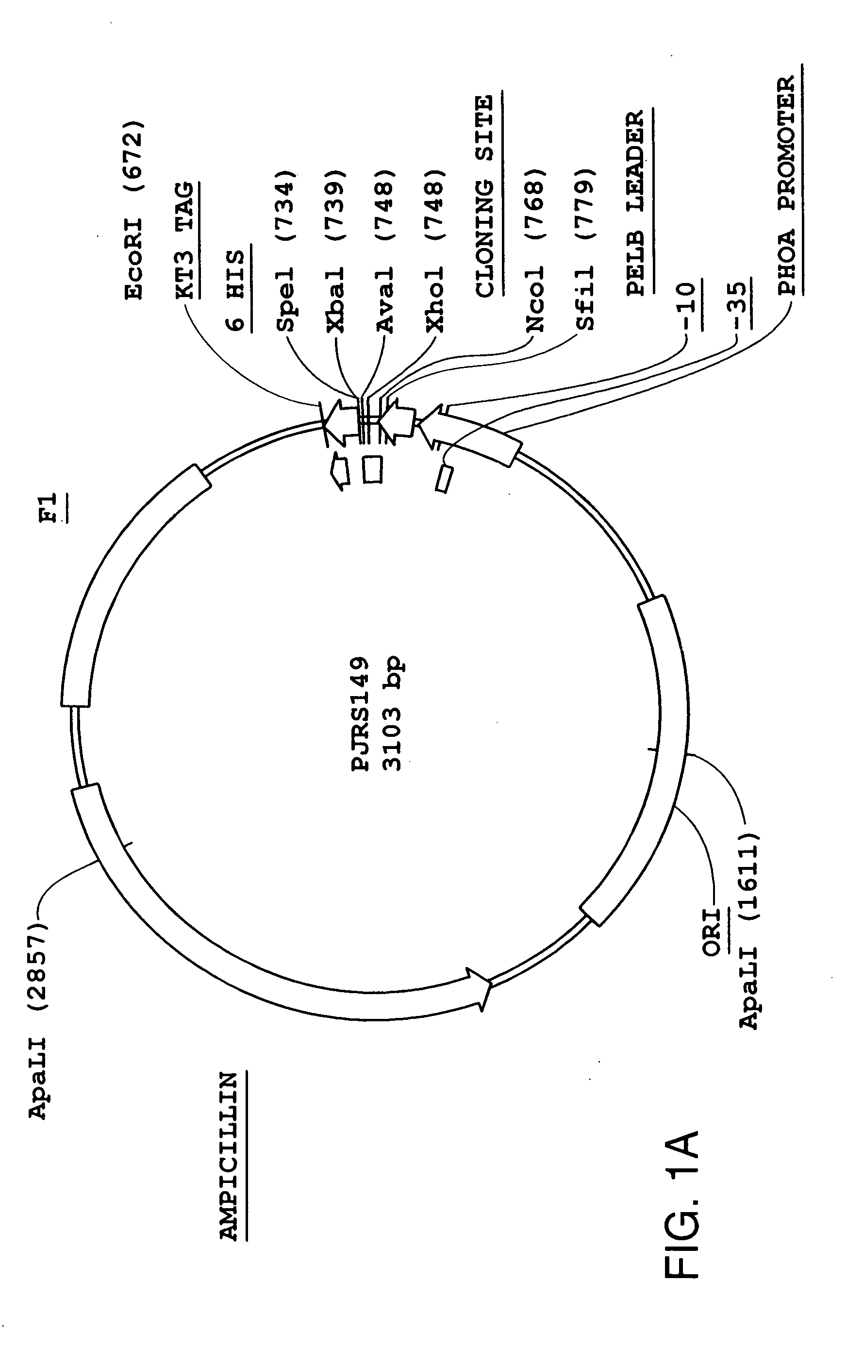
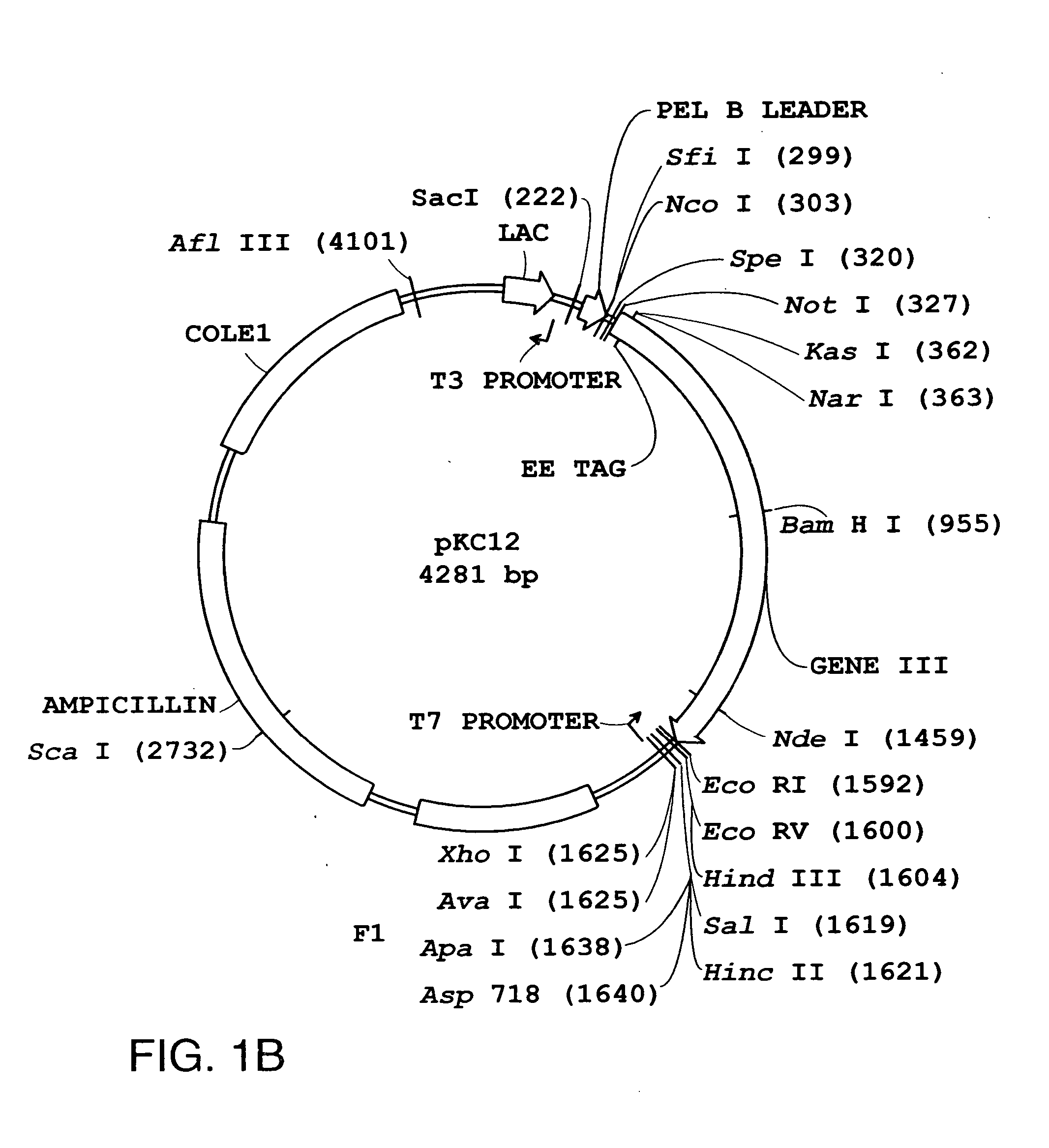
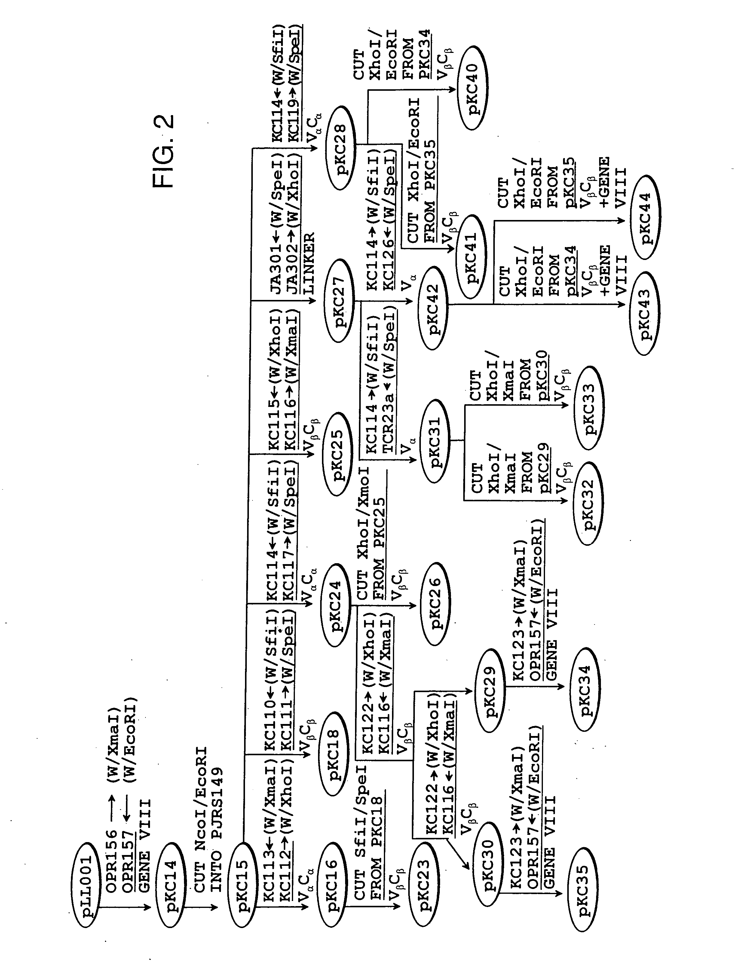
[0143] The scTCR DNA provided in Example 1 was inserted into vectors bearing a strong (phoA) or weak (lacZ) bacterial promoter. The vectors used to express the scTCR were the pJRS149 and pKC12 DNA vectors illustrated in FIGS. 1A and 1B, respectively. These vectors allow fusion of the scTCR to a bacteriophage coat protein. The construction of DNA vectors encoding fusion proteins is schematically outlined in FIG. 2 and as follows.

[0144] A. DNA Vector pJRS149 [0145] The pJRS149 DNA vector is a phagemid with a pBluScript™ (Invitrogen) backbone. The vector was used to produce soluble scTCR fusion proteins for use in bacteriophage display experiments described in Examples 14 to 18 which follows.

[0146] B. DNA Vector pKC12 [0147] DNA encoding gene III was cloned into the pKC12 vector illustrated in FIG. 1B.

[0148] C. DNA Vectors pKC14, and Vector pKC15 [0149] The gene VIII DNA sequence was PCR amplified from fd tet bacteriophage (ATCC No.37000)...

example 3

Cloning DNA Encoding Fusion Proteins into DNA Vector pEN2

[0160] Expression of the soluble scTCR produced in Example 1 was increased by subcloning into the pEN2 vector illustrated in FIG. 5. The pEN2 vector includes a phoA promoter, a gene 10 ribosomal binding site, and a modified pel B leader. Soluble scTCR inserts in a pEN2 vector format are schematically illustrated in FIG. 6A and FIG. 6B and described as follows. In FIG. 6A and 6B, the DNA vectors were constructed from the pEN2 vector.

[0161] A. Construction of DNA Vector pKC60 [0162] The pKC60 vector was made by introducing a 1204 bp SfiI-EcoRI fragment into pEN2. The SfiI-EcoRI fragment consisted of the V-α and V-β, C-β domains. The fragment was made by amplifying pKC51 DNA as template and using primers KC114 (“front” SEQ ID NO: 7 ) and JWTCR208 (“back” SEQ ID NO:75). The JWTCR209 primer included a EE-tag and a XmaI site 3′ of the C-β domain. The addition of an XmaI site facilitated cloning of the gene III and gene VIII genes....


PUM

PropertyMeasurementUnit
induction timeaaaaaaaaaa
OD/mlaaaaaaaaaa
OD/mlaaaaaaaaaa

Abstract

The present invention relates to novel fusion proteins comprising a bacteriophage coat protein and a single-chain T cell receptor and uses of such complexes. In one aspect, the invention relates to soluble fusion protein comprising a bacteriophage coat protein covalently linked to a single-chain T cell receptor which comprises a V-alpha chain covalently linked to a V-beta chain by a peptide linker sequence. The soluble fusion proteins of the invention are useful for a variety of applications including: 1) making a bacteriophage library for displaying single-chain T cell receptors for use in screens for identification and isolation of ligands that bind single-chain T cell receptors, and 2) methods for isolating soluble and fully functional single-chain T cell receptors from the fusion proteins.

Description

RELATED APPLICATIONS [0001] This application is a continuation of U.S. application Ser. No. 08 / 813,781 filed Mar. 7, 1997, pending. The entire contents which is expressly incorporated herein by reference.1. FIELD OF THE INVENTION [0002] The present invention relates to fusion proteins comprising bacteriophage coat protein and a single-chain T cell receptor, as well as methods of making, and using such fusion proteins. The fusion proteins are useful for a variety of applications including making bacteriophage display libraries to screen binding molecules in vitro. 2. BACKGROUND [0003] A T cell response is modulated by antigen binding to a T cell receptor (TCR). One type of TCR is a membrane bound heterodimer consisting of an α and β chain configured to resemble an immunoglobin variable (V) and constant (C) region. The TCR α chain includes a covalently linked V-α and C-α chain, whereas the β chain includes a V-β chain covalently linked to a C-β chain. The V-α and V-β chains form a poc...

Claims


Application Information

Patent Timeline
Patent Type & Authority Applications(United States)
IPC IPC(8): A61K39/12C07H21/04C12P21/04C07K14/005C07K14/705C12N15/86C07K14/01C07K14/725C12N7/01C12N15/10C40B40/02
CPCC07K14/005C07K14/7051C07K2319/00C12N15/1037C12N2795/10022C40B40/02
Inventor WEIDANZ, JON A.CARD, KIMBERLYN F.WONG, HING C.
Owner ALTOR BIOSCIENCE CORP
Why Eureka
Try Eureka