Novel translocation assay

a translocation assay and translocation technology, applied in the field of in vitro translocation assay, can solve the problems of limiting the transport of substrates (both naturally occurring substrates and small molecules) into and/or outside the cell, nephrogenic diabetes insipidus, and inconvenient methods, so as to increase the level of mutant membrane transport protein, reduce the rate of recycling or transporter internalization

Inactive Publication Date: 2007-06-21
GARVAN INST OF MEDICAL RES
View PDF3 Cites 47 Cited by
  • Summary
  • Abstract
  • Description
  • Claims
  • Application Information

AI Technical Summary

Benefits of technology

[0052] For example, the present inventors have developed an assay useful for determining the level of GLUT4 translocation in a cell. The assay uses a GLUT4 protein that is labeled with a tag or marker that facilitates detection of the GLUT4. Preferably, the tag or marker is located within an extracellular domain of the GLUT4 protein. The location of the tag or marker facilitates detection of the GLUT4 protein at the plasma membrane of an intact cell. By determining the level of tagged / marked GLUT4 protein at the plasma membrane of a cell relative to the level of tagged / marked GLUT4 in the cell, the level of GLUT4 translocation is determined.
[0067] For instance, the reduced rate of recycling or transporter internalization of the mutant membrane transport protein increases the level of the mutant membrane transport protein at the plasma membrane of a cell compared to the level of a wild-type form of the membrane transport protein.

Problems solved by technology

However, as the membrane-localized form is capable of transport activity, the amount of any membrane transport protein present in the plasma membrane limits the transport of substrates (both naturally-occurring substrates and small molecules) into and / or outside of the cell.
Additionally, deficiency of the water channel protein aquaporin 2 hinders its translocation to the apical surface of the cell abolishing reabsorption of water from the collecting duct and resulting in nephrogenic diabetes insipidus.
These methods are imprecise, as any redundancy in the transport process of interest, e.g. if a cell expresses multiple proteins that transport the same molecule, may mask or reduce the effect of a mutation of one of the constituents (i.e. transport proteins) of the process.
These assays are both laborious and subject to inter-assay variability, and furthermore, are only semi-quantitative.
Accordingly, the quantitative nature of these assays is limited.
Furthermore, these assays are not readily adapted to high-throughput analysis, for example, for screening compounds that modulate translocation of a membrane transport protein.

Method used


Image

  • Novel translocation assay
  • Novel translocation assay
  • Novel translocation assay

Examples


example 1

Generation and Expression of a labeled GLUT4 Protein

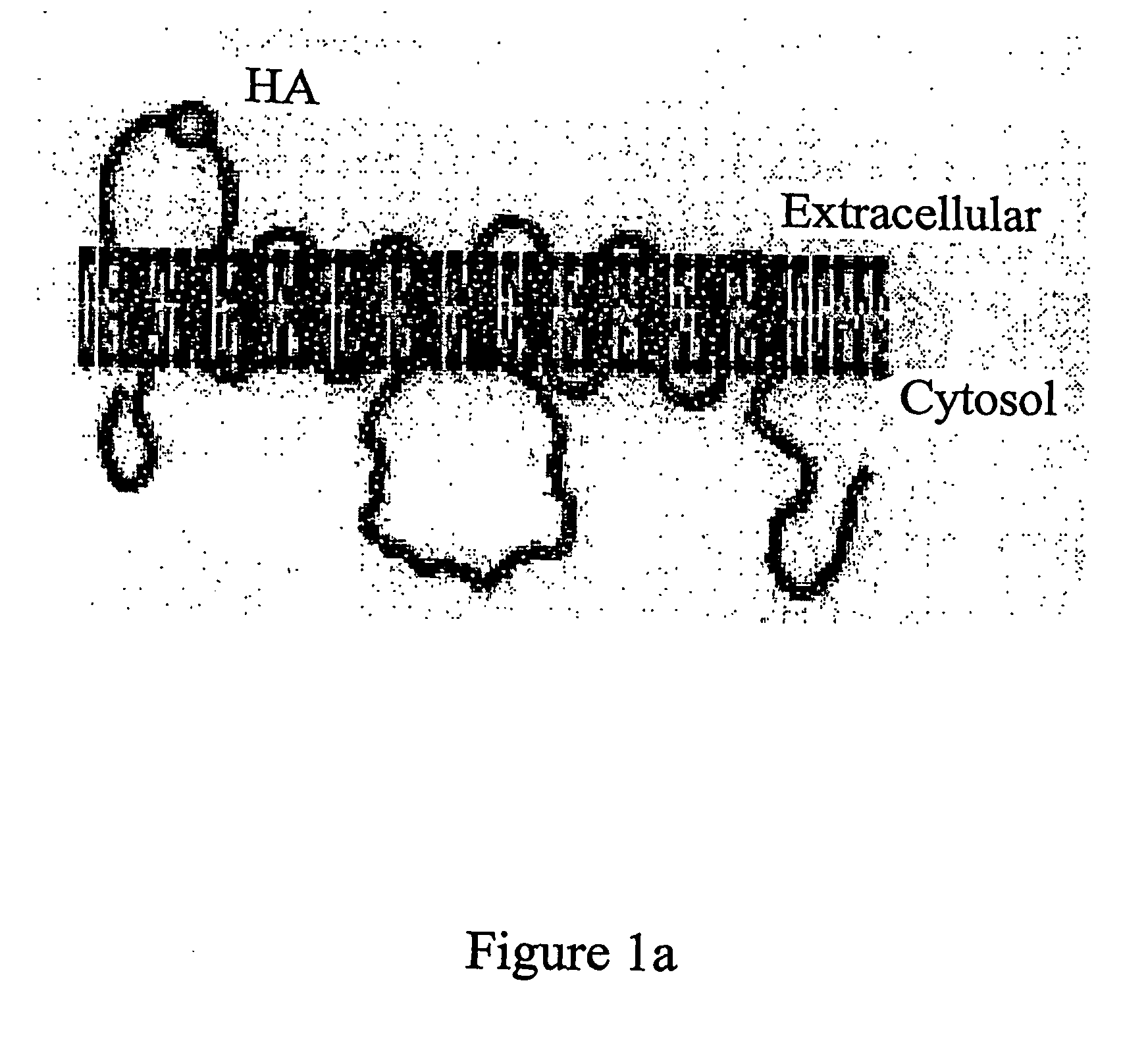
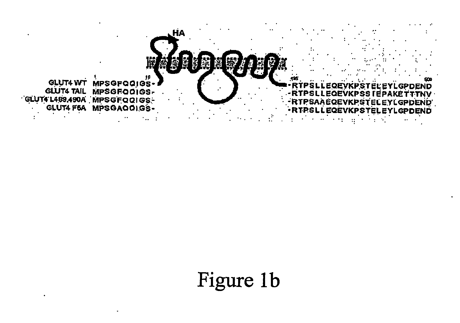
[0569] A HA-tagged GLUT4 protein was produced essentially as described in Quon et al., Proc. Natl. Acad. Sci USA 94: 5587-5591, 1994. Essentially, the cDNA encoding GLUT4 was digested with SauI and a double stranded oligonucleotide was inserted by ligation. The double stranded oligonucleotide was formed by hybridizing two oligonucleotides one comprising the sequence TGAGATCGATTATCCTTATGATGTTCCTGATTATGG (SEQ ID NO: 63) and the other TCA GCA TAA TCA GGA ACA TCA TAA GGA TAA TCG ATC (SEQ ID NO: 64). The inserted nucleic acid encodes a HA tag between amino acids 67 and 68 in the first exofacial loop of GLUT4 (SEQ ID NO: 4). This gene construct was inserted into the vector pBABE (Pear et al. Proc. Natl Acad Sci. U.S.A. 90: 8392-8396 1993). The polypeptide encoded by this protein is shown schematically in FIG. 1A.

[0570] Additional gene constructs were generated comprising a nucleic acid encoding mutant forms of GLUT4 (these constructs e...

example 2

Generation of an Assay to Determine the Localization of GLUT4
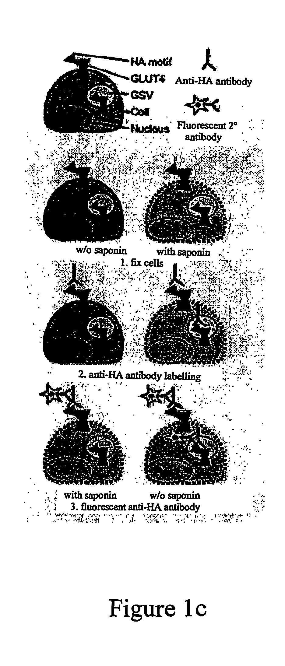
[0577] 2.1 Methods

[0578] Retrovirally-transduced fibroblasts expressing HA-tagged GLUT 4 or a mutant therof were differentiated into adipocytes essentially as described above. These adipocytes were then subcultured for 30 hours. Insulin was then added at different time points, after which the cells were fixed in 3% formaldehyde. After washing and quenching with 50 mM glycine, cells were incubated for 20 min with 5% normal swine serum (NSS) in the absence or presence of 0.1% saponin to analyse the level of GLUT4 at the plasma membrane (PM) or the total cellular GLUT4 content, respectively. Cells were incubated for 60 min with a saturating concentration of either an antibody directed against the HA tag or a control non-relevant antibody (mouse IgG MOPC21) in PBS containing 2% NSS. After extensive washing, the cells were incubated for 20 min with 5% NSS in the presence or absence of 0.1% saponin to permeabilize all cells. C...

example 3

Insulin Induced Translocation of GLUT4 in 3T3-L1 Fibroblasts and Adipocytes

[0581] In fibroblasts, insulin induced the translocation of wild-type GLUT4 and each of the GLUT4 mutants to the PM (FIG. 3). The maximum level of surface GLUT4 was reached after 6 min of insulin stimulation, representing a 5-fold increase above that observed in non-stimulated cells, followed by a rapid reduction. The PM level of the GLUT4 F5A mutant was slightly higher than that of the other GLUT4 molecules in insulin-stimulated fibroblasts. In adipocytes we observed an ˜8-fold increase in cell surface GLUT4 levels in response to insulin stimulation. Neither wild-type GLUT4 nor any of the GLUT4 mutants showed an overshoot as was observed in fibroblasts. The GLUT4 TAIL mutant showed translocation characteristics similar to those of GLUT4 WT, although cell surface levels in both the absence and presence of insulin were increased by approximately 5%, in accordance with previous studies (Shewan et al., Mol. Bio...


PUM

PropertyMeasurementUnit
pHaaaaaaaaaa
densityaaaaaaaaaa
concentrationsaaaaaaaaaa

Abstract

The present invention relates to a novel in vitro assay for determining the level of a protein, in particular, a membrane transport protein that is located at the plasma membrane of a cell compared to the level of the protein in the cell. The process of the invention is also useful for determining the level of recycling of a membrane transport protein. The present invention additionally provides a process for identifying an agent that modulates the translocation of a protein, in particular, a membrane transport protein, to the plasma membrane and, as a consequence, the activity of that protein.

Description

FIELD OF THE INVENTION [0001] The present invention relates to a novel in vitro assay for determining the level of a protein, in particular, a membrane transport protein that is located at the plasma membrane of a cell compared to the level of the protein in the cell. In one embodiment, the present invention provides a method for identifying an agent that modulates the translocation of a protein, in particular, a membrane transport protein, to the plasma membrane and, as a consequence, the activity of that protein. BACKGROUND OF THE INVENTION [0002] General [0003] This specification contains nucleotide and amino acid sequence information prepared using Patent in Version 3.1, presented herein after the claims. Each nucleotide sequence is identified in the sequence listing by the numeric indicator <210> followed by the sequence identifier (e.g. <210>1, <210>2, <210>3, etc). The length and type of sequence (DNA, protein (PRT), etc), and source organism for each ...

Claims


Application Information

Patent Timeline
Patent Type & Authority Applications(United States)
IPC IPC(8): G01N33/567G01N33/50G01N33/68
CPCG01N33/5035G01N33/6872G01N2500/10
Inventor JAMES, DAVID
Owner GARVAN INST OF MEDICAL RES
Why Eureka
Try Eureka