Fabrication of vascularized tissue using microfabricated two-dimensional molds

a two-dimensional mold and vascularization technology, applied in the field of organ transplantation and reconstructive surgery, can solve the problems of limiting the size of newly formed tissue, countless patients suffer, and critical organ shortage,

Inactive Publication Date: 2007-06-28
THE GENERAL HOSPITAL CORP +1
View PDF9 Cites 17 Cited by
  • Summary
  • Abstract
  • Description
  • Claims
  • Application Information

AI Technical Summary

Benefits of technology

[0012] A method and materials to create complex vascularized living tissue in three dimensions from a two-dimension microfabricated mold has been developed. The method involved creating a two dimensional surface having a branching structure etched into the surface. The pattern begins with one or more large channels which serially branch into a large array of charmels as small as individual capillaries, then converge to one or more large channels. The etched surface serves a template within a mold formed with the etched surface for the circulation of an individual tissue or organ. Living vascular cells are then seeded onto the mold, where they form living vascular channels based on the pattern etched in the mold. Once formed and sustained by their own matrix, the top of the mold is removed. The organ or tissue specific cells are then added to the etched surface, where they attach and proliferate to form a thin, vascularized sheet of tissue. The tissue can then be gently lifted from the mold using techniques such as fluid flow and other supporting material, as necessary. The tissue can then be systematically folded and compacted into a three-dimensional vascularized structure. This structure can then be implanted into animals or patients by directly connecting the blood vessels to flow into and out of the device. Immediate perfusion of oxygenated blood occurs, which allows survival and function of the entire living mass.

Problems solved by technology

However, the shortage of organs needed for transplantation has become critical and continues to worsen.
Therefore, countless patients suffer as a result.
However, the relatively long time course for angiogenesis limits the size of the newly formed tissue.
However, it falls short for thick, complex tissues such as large vital organs, including liver, kidney, and heart.
However, this approach is essentially a two-dimensional technology and it has not been apparent that it might be adapted to the generation of thick, three-dimensional tissues.
However, this technique is limited by the characteristics and chemistry of the particular polymers.
Also, it severely limits the types of tissue to be fabricated.
Polymer walls do not allow the plasma exchange that is needed at the alveolar capillary wall of the lung.

Method used


Image

  • Fabrication of vascularized tissue using microfabricated two-dimensional molds
  • Fabrication of vascularized tissue using microfabricated two-dimensional molds
  • Fabrication of vascularized tissue using microfabricated two-dimensional molds

Examples


example 1

Micromachining of Template to Tissue Engineer Branched Vascularized Channels for Liver Fabrication.

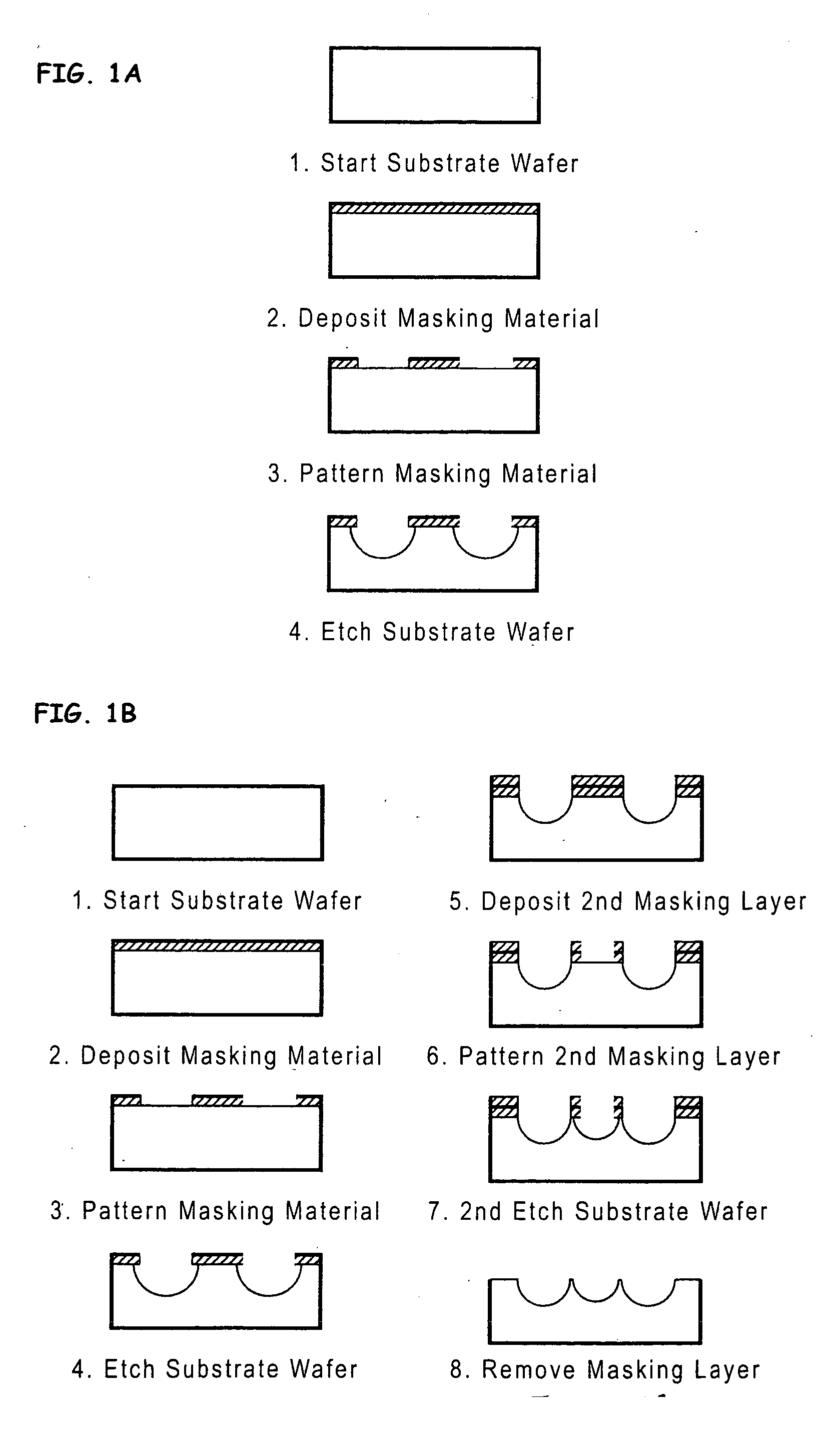
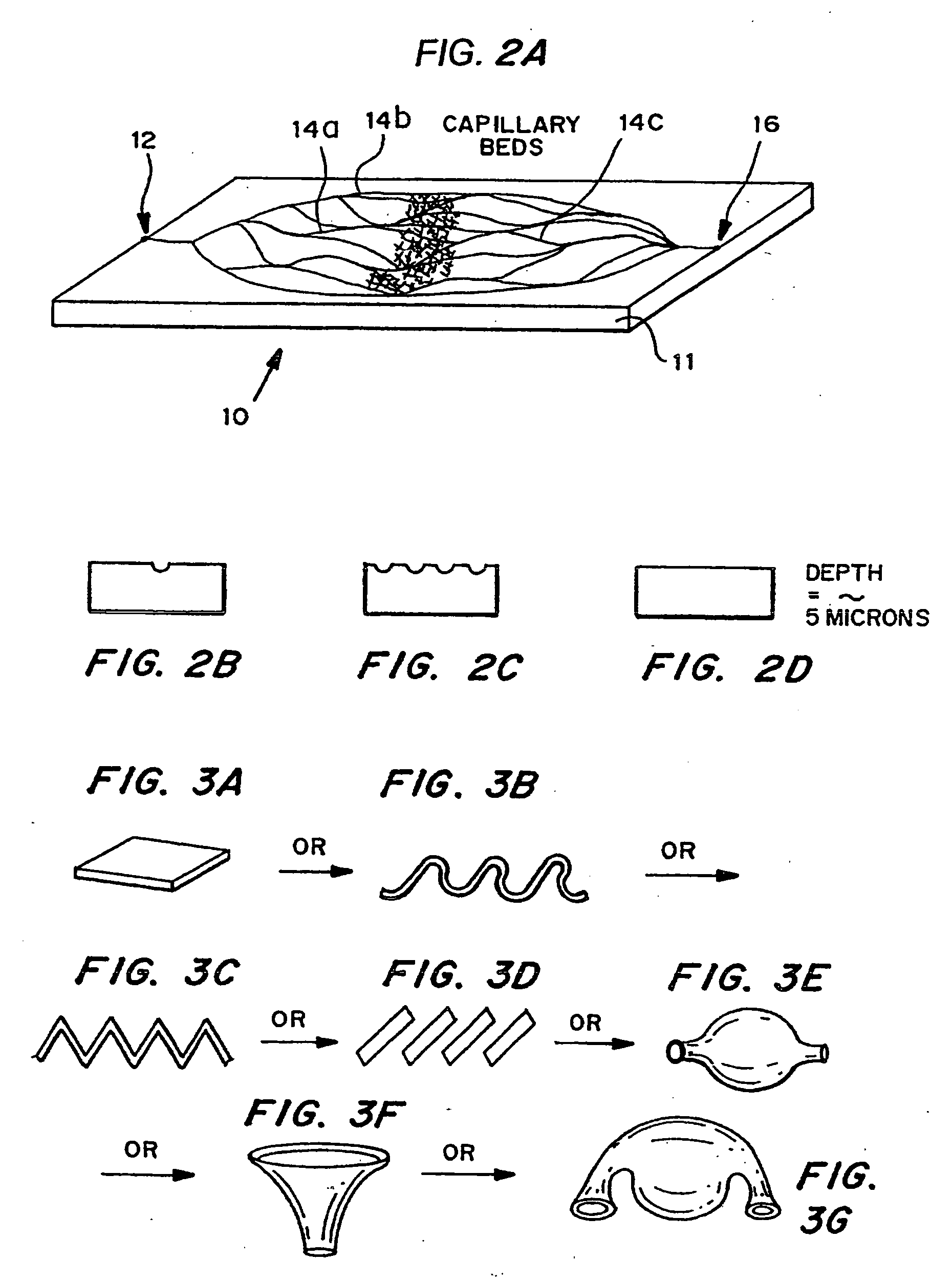
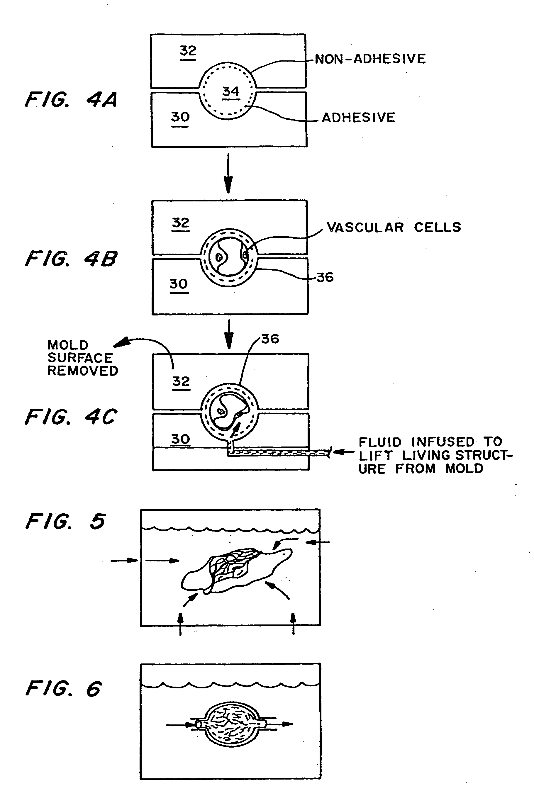
[0086] Micromachining technologies were used on silicon and pyrex surfaces to generate complete vascular systems that may be integrated with engineered tissue before implantation. Trench patterns reminiscent of branched architecture of vascular and capillary networks were etched using standard photolithographic techniques onto silicon and pyrex surfaces to serve as templates. Hepatocytes and endothelial cells were cultured and subsequently lifted as single-cell monolayers from these two dimensional molds. Both cell types were viable and proliferative on these surfaces. In addition, hepatocytes maintained albumin production. The lifted monolayers were then folded into compact three-dimensional tissues. The goal is to lift these branched vascular networks from two dimensional templates so that they can be combined with layers of parenchymal tissue, such as hepatocytes, to form three di...


PUM

PropertyMeasurementUnit
lengthaaaaaaaaaa
lengthaaaaaaaaaa
diameteraaaaaaaaaa

Abstract

A method and materials to create complex vascularized living tissue in three dimensions from a two-dimension microfabricated mold has been developed. The method involved creating a two dimensional surface having a branching structure etched into the surface. The pattern begins with one or more large channels which serially branch into a large array of channels as small as individual capillaries, then converge to one or more large channels. The etched surface serves a template within a mold formed with the etched surface for the circulation of an individual tissue or organ. Living vascular cells are then seeded onto the mold, where they form living vascular channels based on the pattern etched in the mold. Once formed and sustained by their own matrix, the top of the mold is removed. The organ or tissue specific cells are then added to the etched surface, where they attach and proliferate to form a thin, vascularized sheet of tissue. The tissue can then be gently lifted from the mold using techniques such as fluid flow and other supporting material, as necessary. The tissue can then be systematically folded and compacted into a three-dimensional vascularized structure. This structure can then be implanted into animals or patients by directly connecting the blood vessels to flow into and out of the device. Immediate perfusion of oxygenated blood occurs, which allows survival and function of the entire living mass.

Description

[0001] This application claims priority ot U.S. Ser. No. 60 / 131,930 filed Apr. 30, 1999, and U.S. Ser. No. 60 / 165,329 filed Nov. 12, 1999.[0002] The United States government has certain rights in this invention by virtue of grant number DAMD 17-99-2-9001 from the Department of Defense.BACKGROUND OF THE INVENTION [0003] The present invention generally relates to the fields of organ transplantation and reconstructive surgery, and to the new field of Tissue Engineering. It more specifically is a new method and materials for generating tissues requiring a blood vessel supply and other complex components such as a nerve supply, drainage system and lymphatic system. [0004] Organ transplantation, as currently practices, has become a major lifesaving therapy for patients afflicted with disease which destroy vital organs including the heart, liver, lungs, kidney and intestine. However, the shortage of organs needed for transplantation has become critical and continues to worsen. Likewise, ev...

Claims


Application Information

Patent Timeline
Patent Type & Authority Applications(United States)
IPC IPC(8): A61K35/14A61K9/70A61F2/02A61F2/04C12M3/00A61F2/06A61F2/28A61K35/12A61K35/44A61L27/38A61M1/10C12N5/071
CPCA61K35/12A61K35/44A61L27/3604A61L27/38C12N5/0671C12N5/0697C12N2502/14C12N2502/28C12N2535/10B33Y70/00A61L27/3895A61L27/3886
Inventor VACANTI, JOSEPH P.BORENSTEIN, JEFFREY T.PIEN, HOMERCUNNINGHAM, BRIAN T.
Owner THE GENERAL HOSPITAL CORP
Why Eureka
Try Eureka