Methods and systems for segmentation and surface matching

a technology of surface matching and segmentation, applied in the field of methods and systems for verifying anatomical features of patients undergoing radiation therapy, can solve the problems of cancerous tissue extending beyond the boundaries of original treatment, original segmentation, contoured image not accurately representing the lesion being treated, etc., to facilitate rapid and accurate treatment position adjustment

Active Publication Date: 2007-07-19
ELEKTA AB
View PDF73 Cites 89 Cited by
  • Summary
  • Abstract
  • Description
  • Claims
  • Application Information

AI Technical Summary

Benefits of technology

[0009] In the radiotherapy application, for example, adjustments to the treatment parameters and / or patient positioning can be made based on the newly segmented volume image, and thus the invention facilitates rapid and accurate treatment position adjustment just prior to treatment delivery while including important clinical and / or practical concerns not addressed by other conventional methods.

Problems solved by technology

Changes such as lesion movement, growth, organ shifting, or other morphisms can cause healthy tissue to become subject to potentially harmful radiation, and cancerous tissue to extend beyond the boundaries of the original treatment plan.
Because organs can change location and / or shape from the time of planning to the delivery of the initial fraction, as well as from fraction to fraction, the original segmented, contoured image may no longer accurately represent the lesion being treated.
As a result, the treatment plan may no longer be optimal.
However, because of the time constraints imposed during the individual fractions, and the lack of a trained physician during the fractions, it may not be possible to generate an updated segmented or contoured image of the lesion.

Method used


Image

  • Methods and systems for segmentation and surface matching
  • Methods and systems for segmentation and surface matching
  • Methods and systems for segmentation and surface matching

Examples


Embodiment Construction

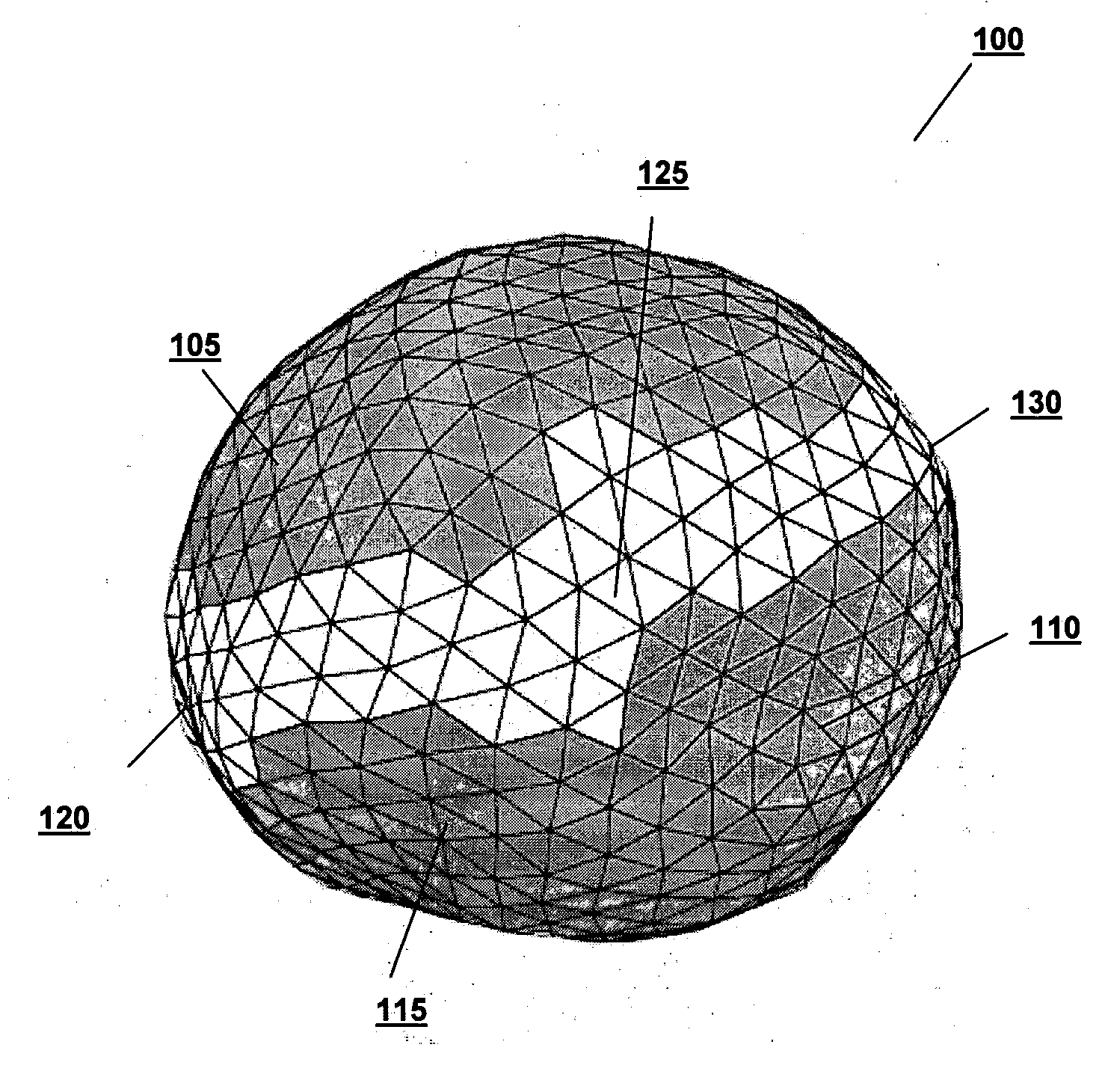
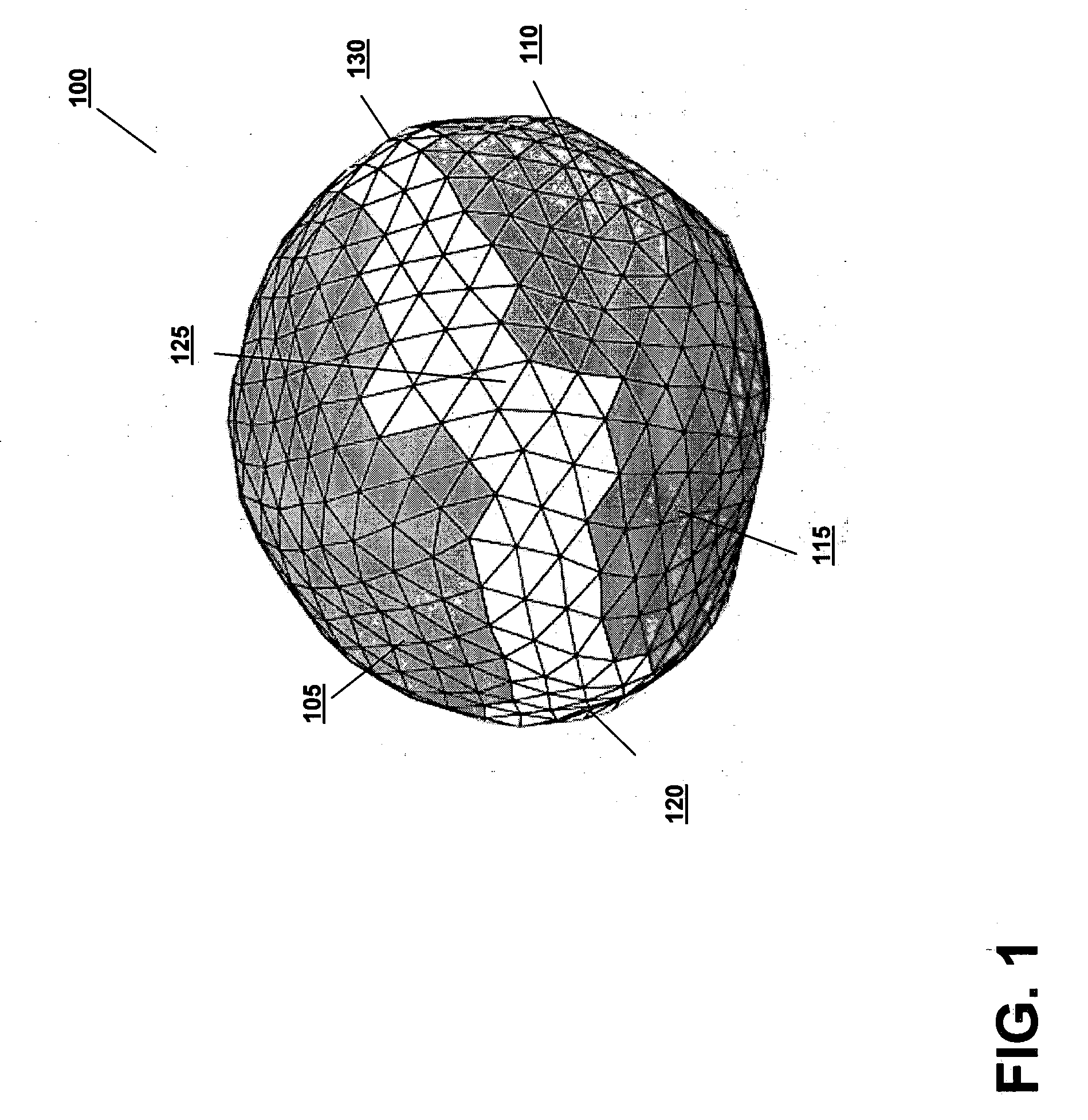
[0025] Referring to FIG. 1, an image 100 of a lesion is obtained during a treatment planning stage. The image can be an individual two-dimensional image, multiple two-dimensional sectional images or slices that collectively represent a volume and may be combined into a composite image, or three-dimensional images rendered programmatically or manually (or a combination). The image can be obtained using devices such as CT scanners, ultrasound devices, MRI devices, PET scanners, and x-ray devices, as well as other imaging modalities commonly used throughout the art. The structure of interest may be a tumor or lesion independent of other organs, a cancerous gland (such as the prostate) requiring treatment, a non-cancerous organ of interest, or any identifying landmark within the image. The image 100 may be, for instance, used by an oncologist, physician, or radiation technician to determine the location and shape of the lesion to be treated and to determine the parameters of the radiati...


PUM


Abstract

A contoured surface map of a lesion within a patient is obtained by shifting a reference surface to an estimated location in operational images. The process can be repeated to minimize errors, and the contoured surface map can then be segmented.

Description

TECHNICAL FIELD [0001] This invention relates to methods and systems for verifying anatomical features of a patient undergoing radiation therapy and, more particularly, to methods and systems for using reference images to identify surface elements of anatomical elements imaged at a later time. BACKGROUND INFORMATION [0002] Radiation-emitting devices are used for the treatment of cancerous tumors within patients. The primary goal of treating cancerous tumors with radiation therapy is the complete eradication of the cancerous cells, while the secondary goal is to avoid, to the maximum possible extent, damaging healthy tissue and organs in the vicinity of the tumor. Typically, a radiation therapy device includes a gantry that can be rotated around a horizontal axis of rotation during the delivery of a therapeutic treatment. A particle linear accelerator (“LINAC”) is located within the gantry, and generates a high-energy radiation beam of therapy, such as an electron beam or photon (x-r...

Claims


Application Information

Patent Timeline
Patent Type & Authority Applications(United States)
IPC IPC(8): A61B5/05
CPCG06T2207/30096G06T2200/04G06T2207/20116G06T2207/20104G06T7/0089G06T7/0083G06T2207/10081G06T7/149G06T7/12
Inventor LATHUILIERE, FABIENNEAUDET, VERONIQUEFALCO, TONYLACHAINE, MARTIN
Owner ELEKTA AB
Why Eureka
Try Eureka