

Methods and apparatuses for diagnosing AML and MDS
a technology of aml and mds, applied in the field of methods, systems and equipment for diagnosing aml and mds, can solve the problems of laborious and time-consuming
- Summary
- Abstract
- Description
- Claims
- Application Information
AI Technical Summary
Benefits of technology
Problems solved by technology
Method used
Image

Examples
example 1
Isolation of RNA and Preparation of Labeled Microarray Targets
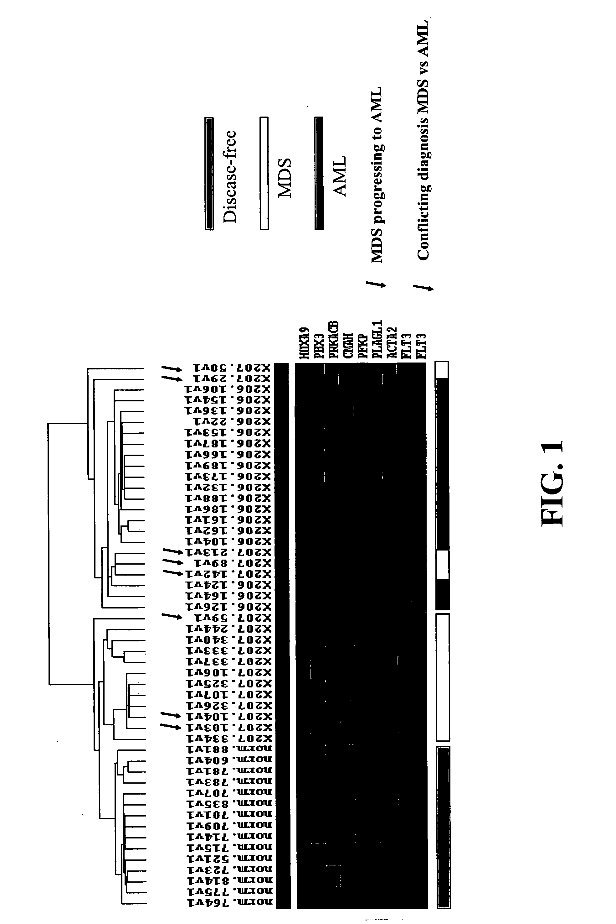
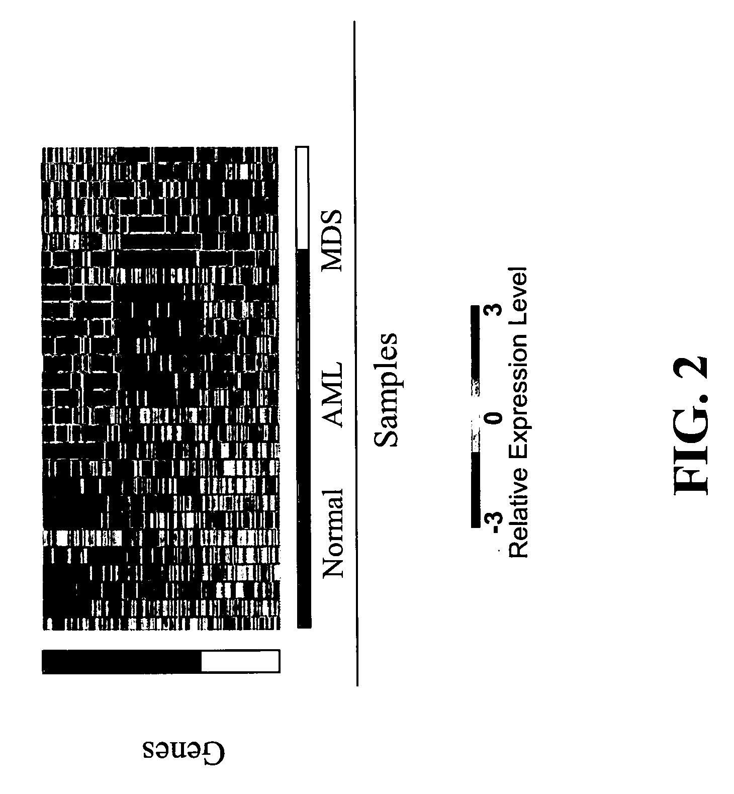
[0111] BMMCs were isolated from bone marrow aspirates taken from 15 disease-free volunteers, 17 patients with MDS, and 18 patients with AML. Informed consents for the pharmacogenomic portions of these clinical studies were received and the project was approved by the local Institutional Review Boards at the participating clinical sites. MDS patients were primarily of Caucasian descent and had a mean age of 66 years (range of 52-84 years). AML patients were exclusively of Caucasian descent and had a mean age of 45 years (range of 19-65 years). Disease-free volunteers were exclusively of Caucasian descent with a mean age of 23 years (range of 18-32 years).
[0112] At screening, bone marrow aspirates from each patient were obtained for pharmacogenomic assessment and histopathologically examined by two independent pathologists. Each bone marrow sample was examined initially by an on-site pathologist and secondly by a single c...
example 2
Hybridization to Affymetrix Microarrays and Detection of Fluorescence
[0114] 2 μg total RNA is converted to cDNA by priming with an oligo-dT primer containing a T7 DNA polymerase promoter at the 5′ end. The cDNA is used as the template for in vitro transcription using a T7 DNA polymerase kit (Ambion, Woodlands, Tex.) and biotinylated CTP and UTP (Enzo). Labeled cRNA can be fragmented in 40 mM Tris-acetate pH 8.0, 100 mM KOAc, 30 mM MgOAc for 35 minutes at 94° C. in a final volume of 40 μl.
[0115] Individual diseased and disease-free samples are hybridized to HgU95Av2 or HG-U95A genechips (Affymetrix). No samples are pooled. 10 μg of labeled target can be diluted in 1×MES buffer with 100 μg / ml herring sperm DNA and 50 μg / ml acetylated BSA. To normalize arrays to each other and to estimate the sensitivity of the oligonucleotide arrays, in vitro synthesized transcripts of 11 bacterial genes can be included in each hybridization reaction as described in Hill et al., SCIENCE, 290: 809-81...
example 3
Gene Expression Data Analysis
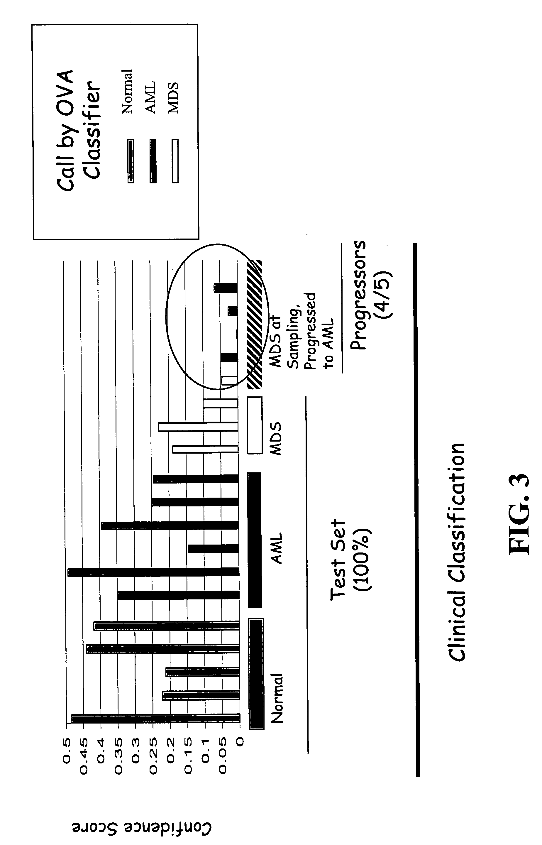
[0118] Data analysis and absent / present call determination was performed on raw fluorescent intensity values using GENECHIP 3.2 software (Affymetrix). The “average difference” values for each transcript were normalized to “frequency” values using the scaled frequency normalization method in which the average differences for 11 control cRNAs with known abundance spiked into each hybridization solution were used to generate a global calibration curve. See Hill et al., GENOME BIOL., 2(12):research0055.1-0055.13 (2001), which is incorporated herein by reference. This calibration was then used to convert average difference values for all transcripts to frequency estimates, stated in units of parts per million ranging from 1:300,000 (3 parts per million (ppm)) to 1:1000 (1000 ppm).
[0119] GENECHIP 3.2 software uses algorithms to calculate the likelihood as to whether a gene is “absent” or “present” as well as a specific hybridization intensity value or “avera...
PUM
 Abstract
Description
Claims
Application Information