Induction of Myocardial Cell From Mammalian Bone Marrow Cell or Cord Blood-Derived Cell and Fat Tissue

a technology of which is applied in the field of induction of myocardial cells from mammalian bone marrow cells or cord blood-derived cells and fat tissues, can solve the problems of ethical problems, inability to apply es cells to actual medical practice, and inability to ethically prepare es cells for each patient, etc., to achieve high safety and rare life-threatening risks for patients

Inactive Publication Date: 2007-09-13
JAPAN SCI & TECH CORP
View PDF2 Cites 47 Cited by
  • Summary
  • Abstract
  • Description
  • Claims
  • Application Information

AI Technical Summary

Benefits of technology

[0015] According to the present invention, myocardial cells can be easily obtained from mammalian bone marrow cells or cord blood-derived cells and fat tissues. The resulting myocardial cells are highly safe because they have not been subjected to genetic engineering, and they exhibit gene expression patterns or phenotypic traits peculiar to myocardial cells. Thus, the myocardial precursor cells and/or myocardial cells obtained by the method of the present invention can be applied to the evaluation of drugs for the regeneration of the heart or drugs affec...

Problems solved by technology

However, preparation of ES cells for each patient is ethically problematic, and myocardial cells differentiated from the randomly prepared ES cells cause immunological rejection.
Thus, ES cells cannot be applied to actual medical practice at present.
The myocardial cells prepared via such method have been subjected...

Method used


Image

  • Induction of Myocardial Cell From Mammalian Bone Marrow Cell or Cord Blood-Derived Cell and Fat Tissue
  • Induction of Myocardial Cell From Mammalian Bone Marrow Cell or Cord Blood-Derived Cell and Fat Tissue
  • Induction of Myocardial Cell From Mammalian Bone Marrow Cell or Cord Blood-Derived Cell and Fat Tissue

Examples


example 1

Differentiation of Fat Tissues into Myocardial Cells

[0050] Fat tissues (about 1.5 ml) were removed from the cervical or abdominal region of a mouse or rat, the tissues were sliced using ophthalmic surgery scissors, and the sliced tissues were immersed in 1 ml of dispase solution at 37° C. for 15 minutes to loosen the cells. The cells were then filtered through a 40-micron nylon mesh filter, sowed at a cell density of 1×10⁶ cells / ml, and then subjected to two-dimensional culture on a 24-well culture dish (diameter: about 1.3 cm) in 5% CO2 at 37° C. using a DMEM medium containing 10% FCS.

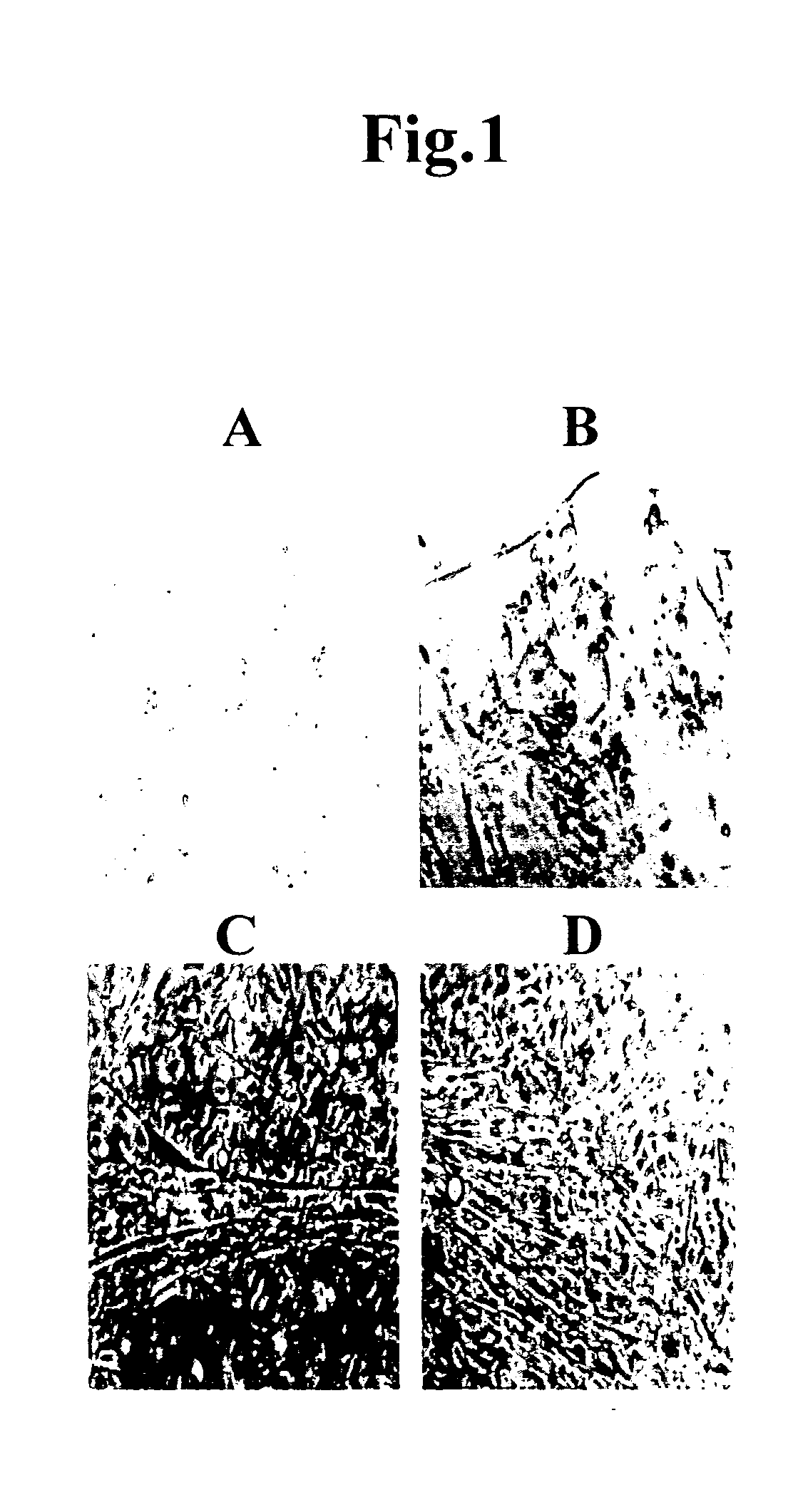
[0051]FIG. 1 shows the results of culturing mouse fat cells. Beating myocardial cell-like cells were observed 3 days after the initiation of culture, and proliferation of spherical myocardial precursor / stem cell-like cells was initiated simultaneously therewith. Myocardial cells can be identified by abundant mitochondria, ANP granules, and Z bands with the use of a stereoscopic microscope, and by mo...

example 2

Immunostaining

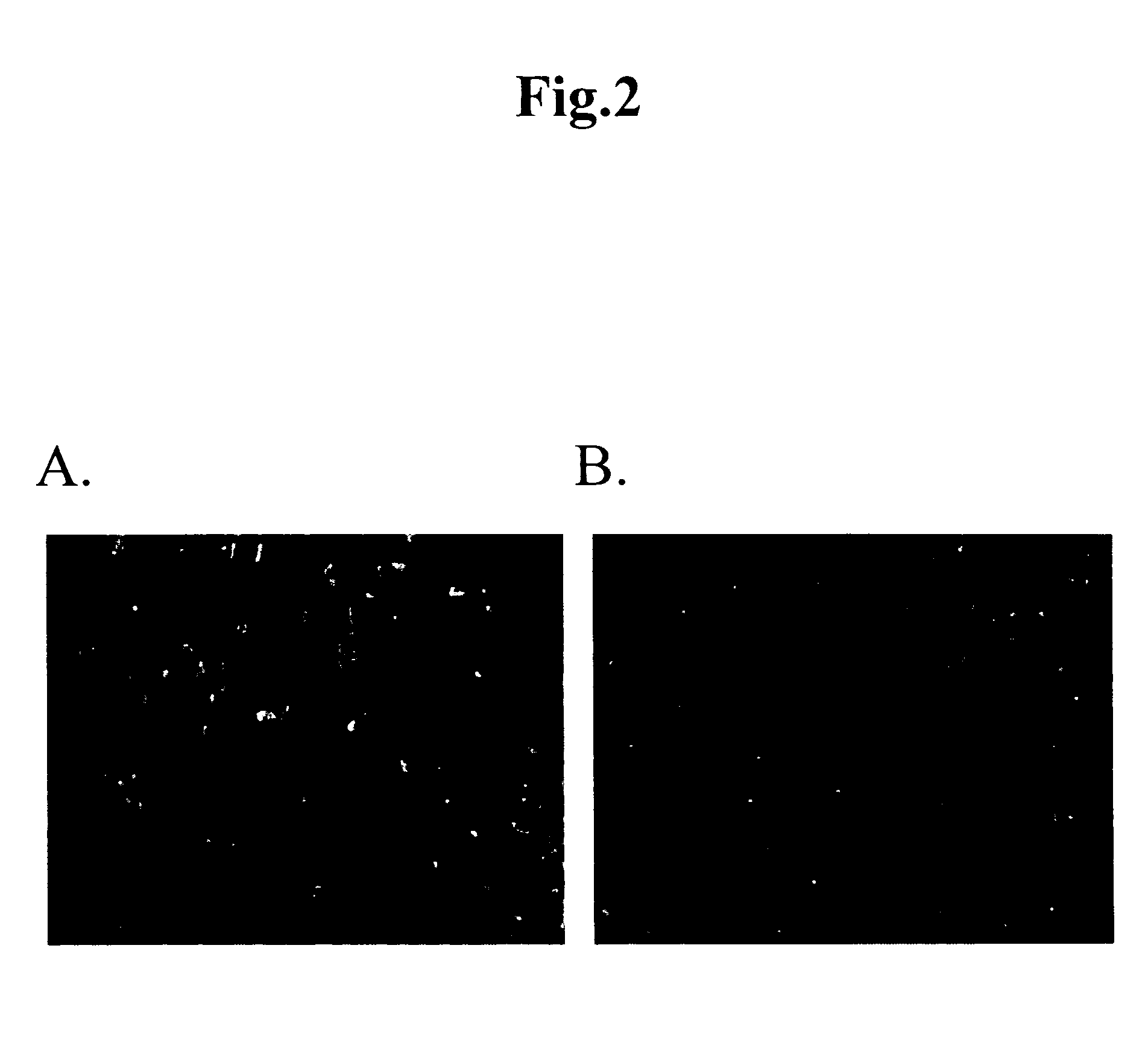
[0052] In order to confirm that the cells obtained in Example 1 had the characteristics of myocardial cells, immunostaining was carried out using a fluorescence-labeled anti-sarcomeric actin (α-Sarcomeric muscular Actin (Sr-1)) antibody (Dako) or anti-cardiac actin (MBL). Sarcomeric actin and cardiac actin are proteins that exhibit expression patterns peculiar to myocardial cells.

[0053] Immunostaining was carried out by culturing mouse fat tissue-derived cells in the same manner as in Example 1 for 14 days and then adding 1 μg / ml of antibody thereto. The results are shown in FIG. 2. As is apparent from FIG. 2, the cultured cells were labeled by the green-fluorescence, which indicates that they were sarcomeric actin- and cardiac actin-positive cells.

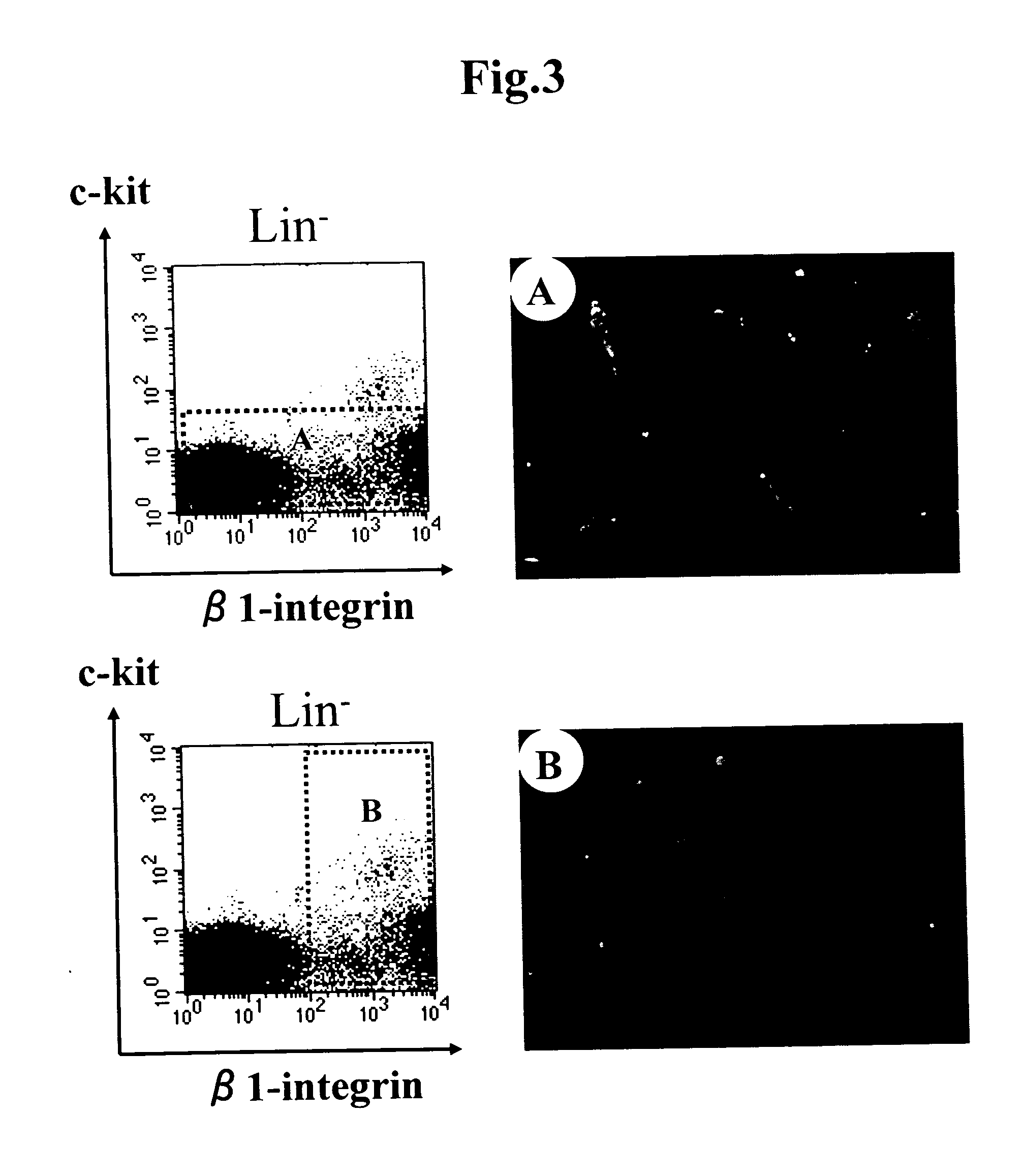
[0054] The fat tissues were allowed to disperse in the same manner as in Example 1, staining was carried out using the Lin antibody (a mixture of CD4, CD8, Gr-1, Mac-1, and TER119 (Pharmingen) that can recognize mature bl...

example 3

Gene Expression Analysis

[0055] In order to confirm that the cells obtained in Example 1 are myocardial cells, gene expression analysis was carried out via RT-PCR. At the outset, mouse fat tissue-derived cells were cultured in the same manner as in Example 1 for 14 days, total RNA was extracted using the RNeasy Mini Kit (Qiagen), and the extracted total RNA was reversely transcribed into cDNA using the PCR kit (Clontech). Subsequently, RT-PCR was carried out with the Advantage polymerase Mix (Clontech) and using the following PCR primers for detecting α,β-MHC, α-skeletal A, α-cardiac A, MLC-2a,2v, and BNP.

α-MHC-S(SEQ ID NO: 1)5′-tgt ctg ctc tcc acc ggg aaa atc t-3′α-MHC-AS(SEQ ID NO: 2)5′-cat ggc caa ttc ttg act ccc atg a-3′β-MHC-S(SEQ ID NO: 3)5′-aac cca ccc aag ttc gac aag atc g-3′β-MHC-AS(SEQ ID NO: 4)5′-cca act ttc ctg ttg ccc caa aat g-3′α-skeletal A-S(SEQ ID NO: 5)5′-gga gat tgt gcg cga cat caa aga g-3′α-skeletal A-AS(SEQ ID NO: 6)5′-tgg tga tcc aca tct gct gga agg t-3′α-car...


PUM

PropertyMeasurementUnit
Timeaaaaaaaaaa
Ratioaaaaaaaaaa

Abstract

This invention provides a method for differentiating mammalian bone marrow cells or cord blood-derived cells into myocardial precursor cells and/or myocardial cells by culturing said bone marrow cells or cord blood-derived cells with cells isolated from mammalian fat tissues or a culture supernatant thereof.

Description

TECHNICAL FIELD [0001] The present invention relates to a technique of inducing mammalian bone marrow cells or cord blood-derived cells and fat tissues to differentiate into myocardial cells. BACKGROUND ART [0002] Myocardial cells stop their proliferation upon the maturation of a host. Accordingly, restoration of myocardial cells could not be expected in a heart that has been once afflicted with myocardial infarction, and the heart was considered to be a nonregenerative organ. In recent years, however, the existence of myocardial precursor / stem cells was discovered in the heart, and division thereof or differentiation thereof into myocardial cells was occasionally observed (Beltrami A. P., et al., “Adult Cardiac Stem Cells Are Multipotent and Support Myocardial Regeneration,” Cell, Vol. 114, pp. 763-776, 2003). The development of a technique for inducing ectopic cells, which can be differentiated into myocardial cells, to differentiate into myocardial cells enables the treatment of ...

Claims


Application Information

Patent Timeline
IPC IPC(8): C12N5/08C12Q1/00A61L27/00A61L27/36A61L27/38C12N5/07C12N5/077C12N15/09C12Q1/02
CPCA61L27/3826A61L27/383A61L27/3839C12Q1/02C12N5/0657C12N2502/1305C12N2506/21A61L27/3895A61L27/3834C12N5/0652C12N5/0662C12N5/0647A61K35/34
Inventor TAKAKURA, NOBUYUKIYAMADA, YOSHIHIRO
Owner JAPAN SCI & TECH CORP
Why Eureka
Try Eureka