Vascular mimic for drug and device evaluation

a technology of mimicry and drug, applied in the field of tissue engineered vascular grafts, can solve the problems of no other available research or other problems

Inactive Publication Date: 2007-10-18
UNIV ARIZONA
View PDF0 Cites 27 Cited by
  • Summary
  • Abstract
  • Description
  • Claims
  • Application Information

AI Technical Summary

Benefits of technology

[0007] The present invention is based, in part, on the inventors' discovery that specifically designed tissue engineered vascular grafts (TEVGs) can be effectively used as model blood vessel mimics (BVMs) for the testing and evaluation of various treatment modalities including, but not necessarily limited to, therapeutic agents and an array of therapeutic intravascular devices. Accordingly, in one aspect, the present invention provides tissue engineered vascular grafts and / or BVMs for use in an in vitro model system comprising a tubular polymeric structure, wherein the luminal surface of the tubular structure comprises at least one layer of cells. The TEVGs of the present invention may comprise cells of any mammalian cell type. Additionally, the cells used in the TEVGs and BVMs of the present invention may include neoplastic and genetically modified cells. The polymeric graft scaffold structure used in the present invention may be comprised of either degradable and non-degradable polymers. In a particular embodiment, the polymeric graft scaffold structure comprises ePTFE.

Problems solved by technology

These constantly evolving drugs and devices possess great potential, but need to be evaluated before clinical applications are possible.
However, none of these publications, nor any other available research, describes the creation of a tissue engineered, three dimensional blood vessel mimic in vitro model system which can be used to evaluate intravascular devices such as stents as well as therapeutic agents.

Method used


Image

  • Vascular mimic for drug and device evaluation
  • Vascular mimic for drug and device evaluation
  • Vascular mimic for drug and device evaluation

Examples


example 1

[0071] Materials and Methods

[0072] Stent surface modifications were evaluated in an in vitro blood vessel mimic model system. BVMs were created by pressure-sodding human microvessel endothelial cells onto the lumen of serum-conditioned 3 mm I.D. expanded polytetrafluoroethylene vascular grafts (C.R. Bard, Inc). Following cell sodding, BVMs were cultivated under flow in an in vitro environment in order to establish the cellular lining. After 1 week, stents were deployed into the BVM systems (see FIG. 4) via a catheter and introducer port.

[0073] Following deployment of the stents, flow was continued for 1 week, at which point stented vessels were taken out of the system and fixed in 10% formalin. Vessels were cut longitudinally into three sections. Stent surface analysis was performed using scanning electron microscopy (SEM) to assess cell coverage and cell morphology. In addition, bisbenzimide (BBI) staining of cell nuclei provided information regarding endothelialization of the de...

example 2

[0077] Materials and Methods

[0078] Expanded polytetrafluoroethylene (ePTFE) of 3 mm and 4 mm inner diameter was cut into 4.5 cm lengths, steam sterilized, and denucleated. ePTFE grafts were conditioned with proteins by capping the grafts and forcing a serum dilution through the pores for 1 hour.

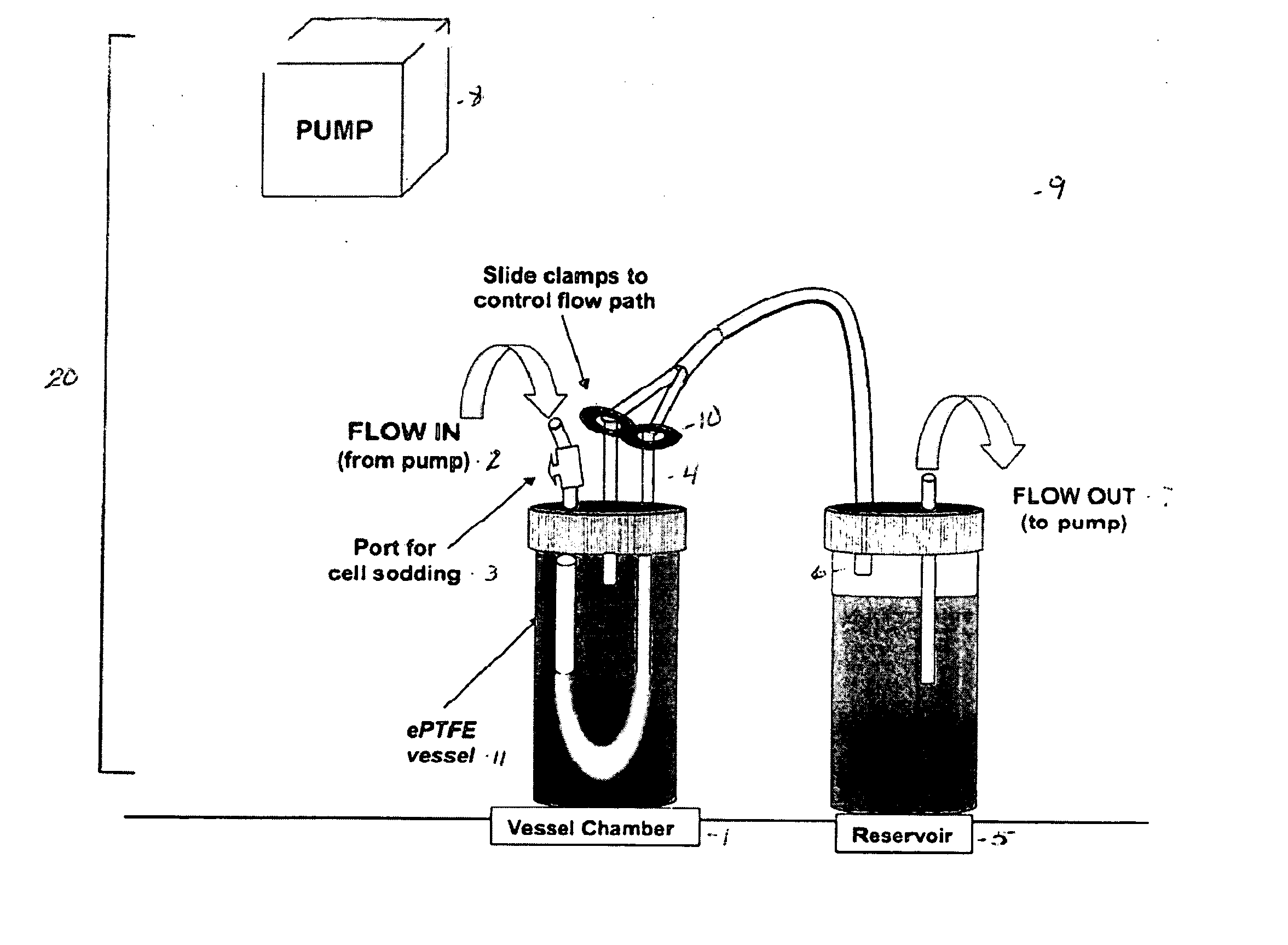
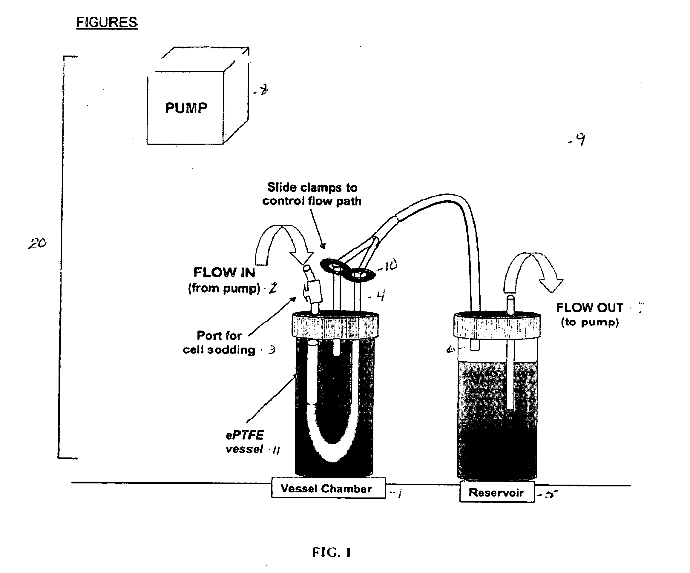
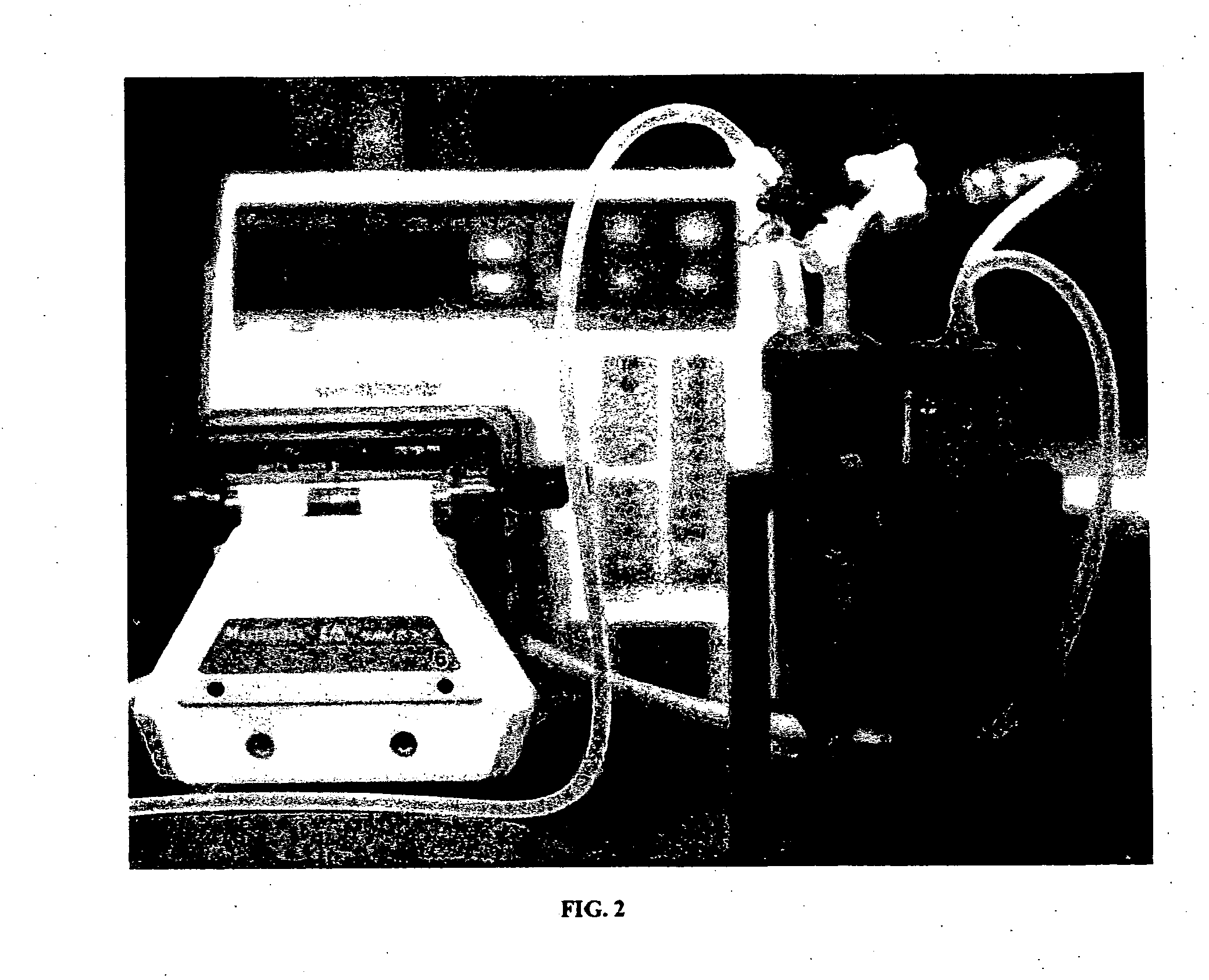
[0079] Conditioned grafts were placed in a bioreactor system, as shown in FIG. 2, and pressure-sodded with human microvessel endothelial cells (HMVECs), isolated from human liposuction fat. Transmural pressure was maintained for 1 hour to facilitate cell deposition.

[0080] Bioreactors were placed in a 37° C. incubator, and media was circulated luminally through each system at 15 mL / min for 10 days to allow for the establishment of a cellular lining. Developments of a cellular lining was verified with scanning electron microscopy (SEM) and hematoxytin and eosin (H&E) staining.

[0081] After 10 days of BVM development, flow was temporarily stopped to allow for stent deployment. Bare metal sten...

example 3

[0089] Materials and Methods

[0090] The in vitro bioreactor system was developed with two chambers placed in series with tubing, and flow established with a Watson-Marlow peristaltic pump. The pump allowed flow regulation through the graft at rates ranging from 4-200 mL / min. The system permitted pressure-sodding of cells, stent deployment, and solute injection to take place internally to maintain sterility. 4 cm lengths of 3 mm I.D. expanded polytetrafluoroethylene (ePTFE, C.R. Bard, Inc.) vascular grafts were denucleated, treated with bovine serum, and inserted into the bioreactor.

[0091] Once each graft / bioreactor system was prepared, endothelial cells were freshly isolated through collagenase digestion of rat epididymal fat pads, and the cells were immediately pressure-sodded onto the grafts. Williams S K, Rose D G, Jarrell B E. Microvascular Endothelial Cell Sodding of ePTFE vascular grafts. Frontiers in Bioscience 2004; 9:1412-1421. Flow was increased to 6 mL / min and the constr...


PUM

PropertyMeasurementUnit
pressureaaaaaaaaaa
pressureaaaaaaaaaa
pressureaaaaaaaaaa

Abstract

The present invention provides tissue engineered vascular grafts (TEVGs) and Blood Vessel Mimics (BVMs) and methods for using TEVGs as BVMs in in vitro model systems for the evaluation of intravascular devices and drugs. The present invention additionally relates to devices and methods for preparing TEVGs, BVMs and BVM model systems.

Description

[0001] This application claims priority to U.S. Patent Application Ser. No. 60 / 763,125, filed Jan. 27, 2006, and U.S. patent application Ser. No. 11 / 314,281, filed Dec. 22, 2005, both of which are incorporated herein by reference.FIELD OF THE INVENTION [0002] The present invention relates to tissue engineered vascular grafts (TEVGs) and Blood Vessel Mimics (BVMs) and methods for using TEVGs as BVMs in in vitro model systems for the evaluation of intravascular devices and drugs. The present invention additionally relates to devices and methods for preparing TEVGs, BVMs and BVM model systems. BACKGROUND OF THE INVENTION [0003] Vascular and cardiac treatments continue to evolve and change as research advances and new technologies are developed. Stent technology, for example, is constantly changing as new modifications and coating technologies become available. Drug treatments are likewise constantly evolving. Stents can be drug-eluting, protein or polymer coated, or modified via numero...

Claims


Application Information

Patent Timeline
Patent Type & Authority Applications(United States)
IPC IPC(8): C12Q1/02C12M1/00A01N1/00C12N5/02
CPCC12N5/0691G01N33/5082C12N2533/30C12N2503/00
Inventor WILLIAMS, STUART K.O'HALLORAN CARDINAL, KRISTEN
Owner UNIV ARIZONA
Why Eureka
Try Eureka