
Method of differentiating stem cells into cells of the endoderm and pancreatic lineage

Inactive Publication Date: 2007-11-08
WISCONSIN ALUMNI RES FOUND
View PDF2 Cites 151 Cited by
  • Summary
  • Abstract
  • Description
  • Claims
  • Application Information

AI Technical Summary

Benefits of technology

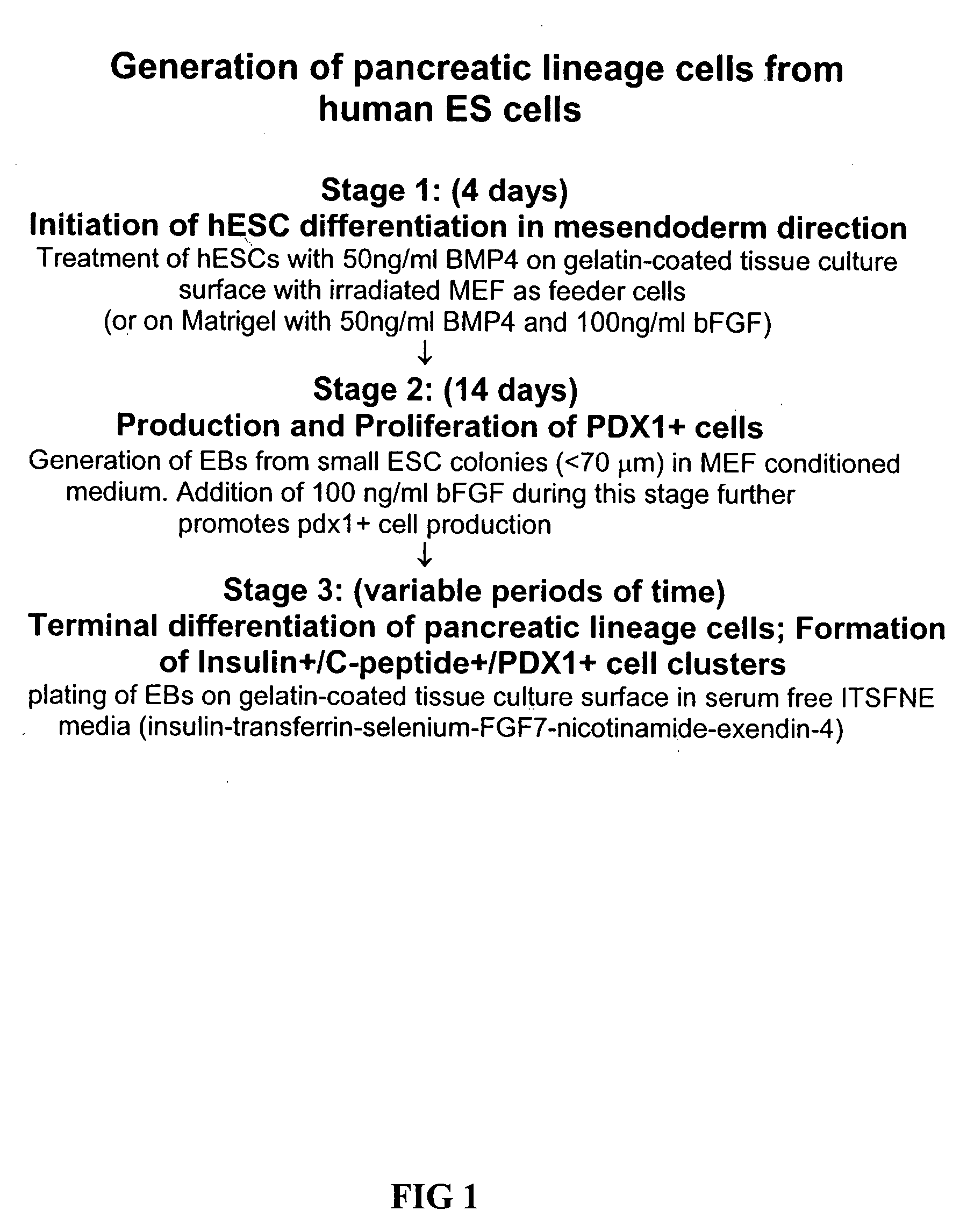
[0010] The present invention is broadly summarized as novel methods for direct in vitro differentiation of human pluripotent stem cells to cells of the endoderm and pancreatic lineage. The method involves culturing the stem cells with an effective amount of a bone morphogenetic protein to induce differentiation in the direction of mesendoderm. These mesendoderm cells are further cultured to form embryoid bodies (EBs), which under defined conditions terminally differentiate to cells of to the pancreatic lineage. By utilizing defined components that promote pancreatic islet differentiation from human pluripotent stem cells, the methods of the invention provide a simple, reproducible culture protocol enabling large-scale production of pancreatic cell types for research or therapeutic uses.

Problems solved by technology

However, the lack of available organs or islet cells has restricted this therapy only to very selected patients.
The amount of islet cells which can be harvested from human cadavers is extremely limited.
However, the experimental methods would be impractical to adopt into a high-throughput culture protocol and the nature of the molecular signals was not revealed by this study.
Yet, the state of the art is that reproducible, highly efficient differentiation to pancreatic precursors and islet cells is not routinely achievable.
The previous techniques reported for culturing human ESCs into cells of the pancreatic lineage, while suitable for laboratory scale investigations, can not be readily scaled up to reliably and consistently produce large numbers of pancreatic cell types for research or for therapeutic uses.

Method used


Image

  • Method of differentiating stem cells into cells of the endoderm and pancreatic lineage
  • Method of differentiating stem cells into cells of the endoderm and pancreatic lineage
  • Method of differentiating stem cells into cells of the endoderm and pancreatic lineage

Examples


example 1

Cell Culture and Differentiation

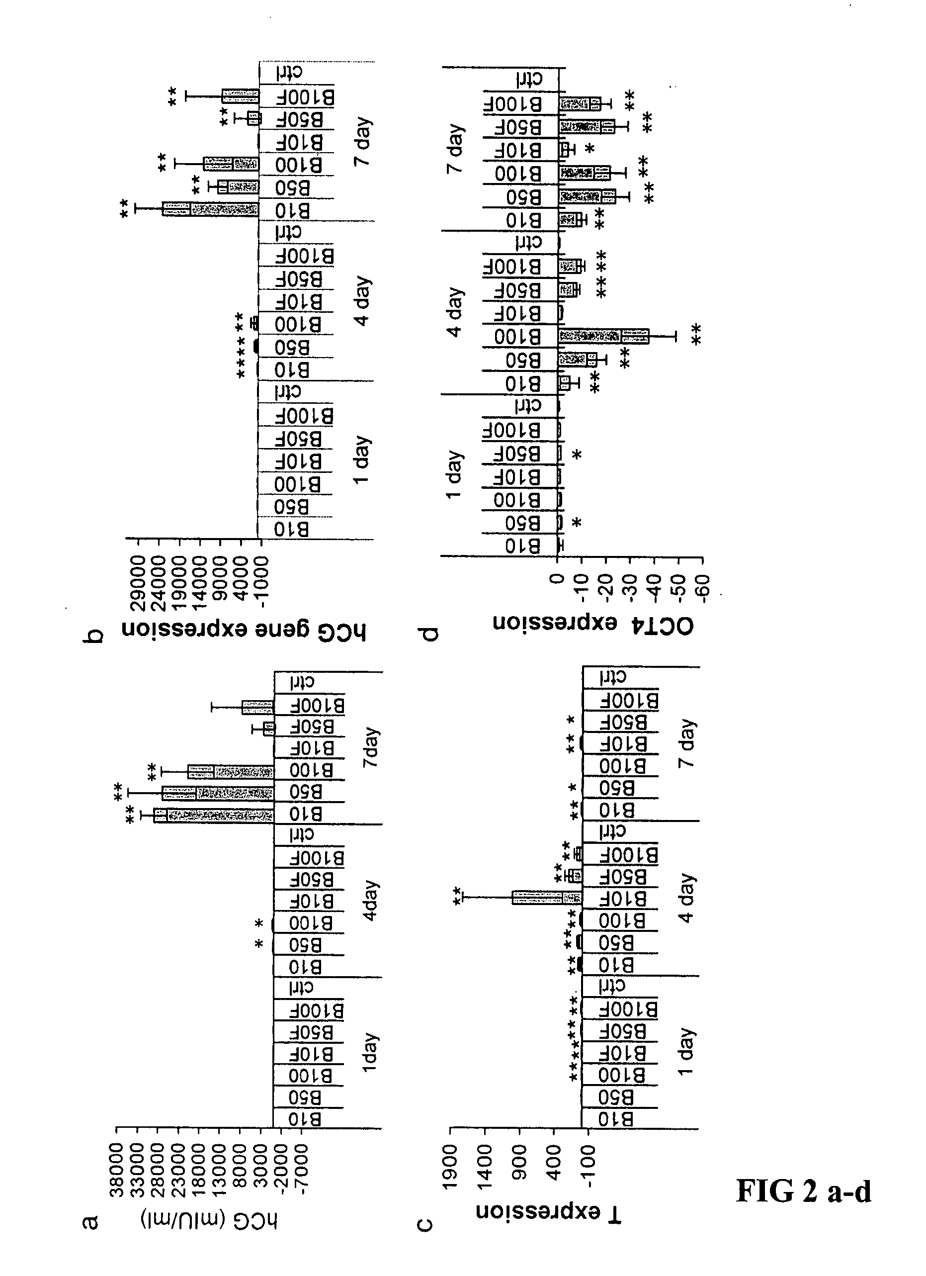
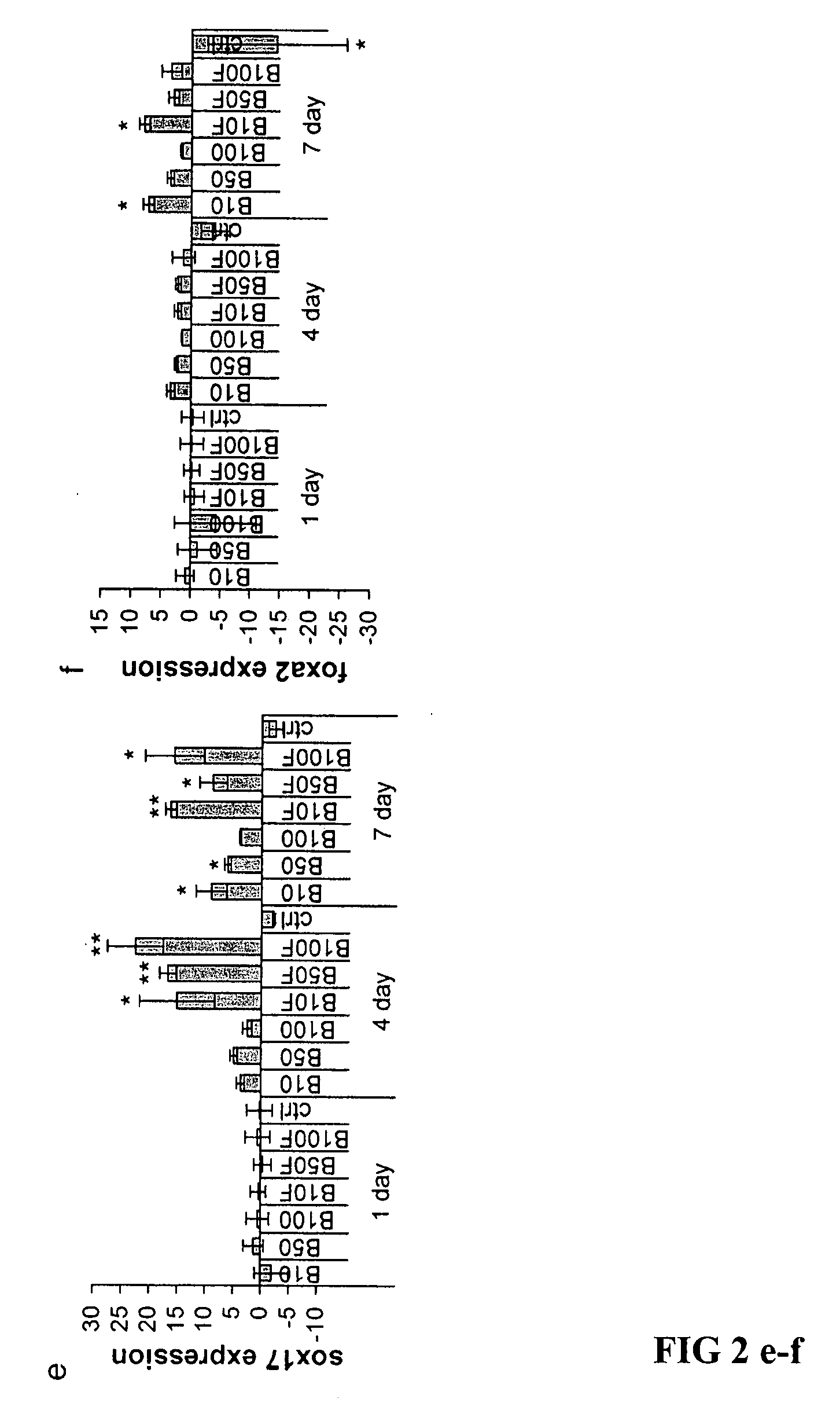
[0052] The general differentiation method is illustrated in simplistic fashion in FIG. 1. NIH approved HESC lines, H1 (WA01) and H9 (WA09) were used between passage 24 to 40. Media for undifferentiated ESCs was comprised of 80% DMEM / F12 and 20% Knockout serum replacement supplemented with 1 mM L-glutamine, 1% nonessential amino acids, 0.1 mM 2-mercaptoethanol and 4 ng / ml bFGF (all from Invitrogen). hESCs were cultured in 6-well plates on a feeder layer of irradiated mouse embryonic fibroblasts (MEFs) in either ESC media (control group), ESC media plus 50 ng / ml BMP4 (BMP4 group; R&D systems), or ESC media plus 50 ng / ml BMP4 plus 300 ng / ml noggin (noggin group; R&D systems) for 4 days.

[0053] In experiments with Matrigel™, hESCs were grown on growth-factor depleted Matrigel™ (BD Biosciences) instead of MEF and culture media was MEF-conditioned media (CM, control group), CM plus 50 ng / ml BMP4 or CM plus 50 ng / ml BMP4 plus 100 ng / ml bFGF. Colonies were t...

example 2

Quantitative PCR and RT-PCR

[0054] Total cellular RNA was extracted with TriZol (Invitrogen). cDNA was synthesized from 1 μg total RNA using a SuperScript First-Strand Synthesis kit (Invitrogen). Quantitative real time RT-PCR (Q-PCR) was performed using Assays-on-demand agents (Applied Biosystems) on an ABI PRISM 7700 Sequence Detection System (Applied Biosystems) for the following transcripts: foxa2, sox17, brachyury, ngn3, pdx1, insulin, glucagon, glut2 and an endogenous control, β-actin. Q-PCR was performed according to equipment manufacturer's instructions. Relative quantification was carried out using the comparative cycle threshold (CT) methods recommended by the supplier. Fold change was calculated as: 2−ΔΔCT Mean ΔΔCT values from Q-PCR analyses were compared using the unpaired, two-tailed Student's t-test. P values <0.05 were considered significant.

[0055] For non-quantitative RT-PCR, oligonucleotide primer pairs were generated against human transcripts using Genebank sequen...

example 3

Immunofluorescence Staining

[0056] Immunofluorescence staining of coverslips was carried out as previously described (Kahan, B. W. et al., Pancreatic precursors and differentiated islet cell types from murine embryonic stem cells: an in vitro model to study islet differentiation. Diabetes 52, 2016-2024 (2003)). The following primary antibodies were used at the listed dilutions: PDX1 rabbit anti-mouse serum 1:4000 (gift of C. Wright); insulin mouse monoclonal 10 μg / ml (ATCC No: HB124); glucagon mouse monoclonal 1:2000 (Sigma); somatostatin mouse monoclonal 1:2000 (Novonordisk); Ki-67 mouse monoclonal 1:25 (BD Pharmingen); C-peptide rat monoclonal 1:3000 (BCBC 1921); Brachyury goat anti-human 1:20 (R&D); OCT4 goat anti mouse 1:100 (Santa Cruz); Sox17 goat anti-human 1:40 (R&D); FOXA2 rabbit anti-rat1:4000 (Gift of R. Costa). Secondary antibodies (Goat anti-mouse IgG Alexa Fluor 488, 1:2000; Goat anti-rabbit Alexa Fluor 568, 1:4000; Goat anti-rat Alexa Fluor 488, 1:2000; Goat anti-rabb...


PUM


Abstract

Methods are described to more efficiently produce cells of the endoderm and pancreatic lineage from mammalian pluripotent stem cells. These methods provide a simple, reproducible culture protocol using defined media components to enable consistent, large-scale production of pancreatic cell types for research or therapeutic uses.

Description

CROSS-REFERENCE TO RELATED APPLICATIONS [0001] This application claims the benefit of U.S. Provisional Application No. 60 / 796,662 filed May 2, 2006. This application is incorporated herein by reference in its entirety.STATEMENT REGARDING FEDERALLY SPONSORED RESEARCH OR DEVELOPMENT [0002] This disclosure was made with United States government support awarded by the following agency: NIH DK042502. The United States has certain rights in this disclosure.BACKGROUND OF THE INVENTION [0003] Type I diabetes is an autoimmune disease of humans caused by destruction of pancreatic islet beta cells. At present the disease is irreversible, although its symptoms are controlled by the administration of exogenous insulin. Type I diabetes is one of the most common autoimmune diseases in human populations and is a major public health concern. [0004] It has previously been found that transplantation of a whole pancreas or of isolated islet cells is an effective treatment for Type I diabetes to restore...

Claims


Application Information

Patent Timeline
IPC IPC(8): C12N5/08C12N5/071
CPCC12N5/0676C12N2500/25C12N2500/38C12N2506/02C12N2501/117C12N2501/155C12N2501/335C12N2501/115C12N5/06C12N5/0606C12N5/0678
Inventor ODORICO, JONXU, XIAOFANG
Owner WISCONSIN ALUMNI RES FOUND
Learn More