
Biomarkers for use in the diagnosis and treatment of colorectal cancer

a colorectal cancer and biomarker technology, applied in the field of large intestinal diseases, can solve the problems of increasing the risk of developing early adenomas with low-grade dysplasia, low diagnostic yield compared to other methods, and limited reliable detection methods, especially for early detection of the diseas

Inactive Publication Date: 2008-01-24
MIRACULINS
View PDF0 Cites 81 Cited by
  • Summary
  • Abstract
  • Description
  • Claims
  • Application Information

AI Technical Summary

Benefits of technology

[0018]The present invention relates to methods for a differential diagnosis of colorectal cancer or a non-malignant disease of the large intestine by detecting one or more differentially expressed biomolecules within a test sample of a given subject, comparing results with samples from healthy subjects, subjects having a precancerous lesion of the large intestine, subjects with non-malignant disease of the large intestine, subjects with localized colorectal cancer, subjects with metastasised colorectal cancer, and / or subjects with an acute or a chronic inflammation of the large intestines, wherein a comparison allows for a differential diagnosis of a subject as healthy, having a precancerous lesion of the large intestines, having non-malignant disease of the large intestine, having a localized colorectal cancer, having a metastasised colorectal cancer, or having an acute or chronic inflammation of the large intestine.

Problems solved by technology

A mutation in the gene encoding the APC (Adenomatous Polyposis Coli) protein leads to the disruption of its biological activity and subsequently increases the risk of developing early adenomas with low-grade dysplasia from the normal mucosa of the colon.
Despite the present knowledge of the molecular mechanisms leading to the development of CRC, reliable detection methods, particularly for the early detection of the disease, are somewhat limited.
Despite its effectiveness as a screening method, a major disadvantage to this test is its low diagnostic yield compared to other methods, as well as its high false-positive rate (Galiatsatos & Foulkes, 2006).
Unfortunately, FS is only able to detect 50% of adenomas and the level of patient discomfort is compromised (Hendon & DiPalma, 2005).
Both the BE and DCBE are also cost effective and safe, but their sensitivity is low and they lack therapeutic capability (Hendon & DiPalma, 2005; Huang et al., 2005).
The disadvantages to this technique are multiple and include high costs, the use of conscious sedation thereby increasing patient recovery time following the procedure, the need for highly trained personnel, and higher complication rates as compared to other screening methods (Huang et al., 2005).
Currently, the disadvantages of this screening tool involves poor sensitivity for polyp detection at less than 5 mm and a relatively high false-positive rate, which may result in an unnecessary follow-up colonoscopy (Huang et al., 2005).
Moreover, its radiation dose may pose a long-term risk for screened individuals (Prokop, 2005).
Its current limitations are lack of dada from screening populations and the need to confine and determine how many and which markers are necessary, as well as the necessary expenses to execute the test (about $500-$800 per test) (Huang et al., 2005).
Despite the availability of screening methods for the detection of CRC, no one method is able to detect CRC within its early stages.
Most patients exhibit symptoms such as rectal bleeding, pain, abdominal distension or weight loss only after the disease is in its advanced stages, leaving little therapeutic options available.
However, the utilization of CRC screening methods remains low.
Some of the major problems from the public include a fear of being hurt by the techniques used, particularly the colonoscopy, as well as an unawareness of the necessity for screening for the disease without symptoms (Hendon & DiPalma, 2005).

Method used


Image

  • Biomarkers for use in the diagnosis and treatment of colorectal cancer
  • Biomarkers for use in the diagnosis and treatment of colorectal cancer
  • Biomarkers for use in the diagnosis and treatment of colorectal cancer

Examples


example 1

Detection of Serum Biomarkers

Biomarker Discovery

[0250]A total of 136 serum samples were collected from patients recruited through the Departments of Gastroenterology and Surgery of the Universities of Erlangen and Magdeburg (both in Germany), and maintained by the European Tumor Sample Institute gGmbH (Hennigsdorf, Germany).

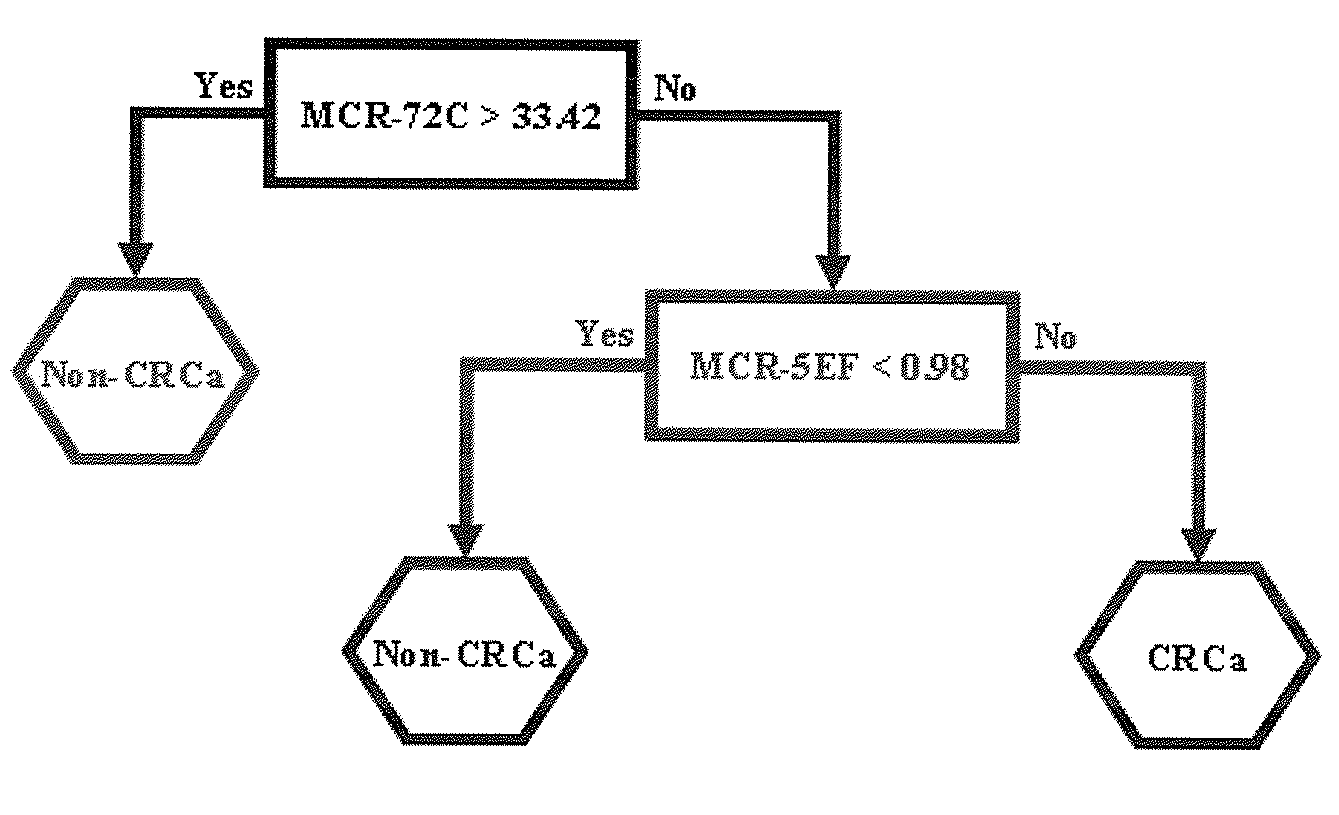
Sample groups include colorectal cancer (68 patients), benign (45 patients) and controls (23 patients) (Table 1).

[0251]

TABLE 1Summary of the distribution of samples for the discovery ofbiomarkers for colorectal cancer.GenderMaleFemaleSiteEMDTotalEMDTotalCRCa5313632932Benign0181802727Healthy09901414CRCa: Colorectal CancerMD: Magdeburg.E: Erlangen.

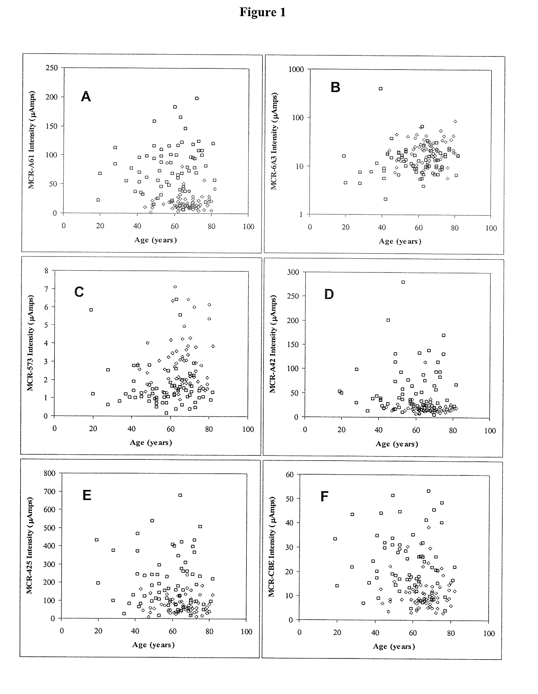
[0252]To determine if there was a significant bias between patient genders or patient ages in different sample groups (CRCa vs. benign disease vs. control; or CRCa vs. non-CRCa), χ2 contingency table analyses was performed. Patient age was categorized as either less than 55 years, 56 to 65 years, 66 to 75 years, over 75 y...

example 2

Validation of Serum Biomarkers for Colorectal Cancer Diagnosis

[0260]A total of 371 serum samples were collected. Of the 371 samples, 165 serum samples were obtained from ETSI (European Tumour Sample Institute, Hennigsdorf, Germany), and 206 were obtained from FCCC (Fox Chase Cancer Centre, Philadelphia, Pa.). Samples obtained from both sites included three different groups of subjects. Group A sera were drawn from 146 colorectal cancer patients. Diagnosis was made based on endoscopy, ultrasonic testing, and / or other means of colorectal cancer detection, and was confirmed by post-surgical histological evaluation.

[0261]Group B consisted of sera drawn from 104 patients with non-malignant (“benign”) disease symptoms of the large intestine (for example, benign polyps, adenoma, inflammation, diverticulitis). Sera were collected following colorectal endoscopy to confirm the absence of colorectal cancer.

[0262]Group C sera were drawn from 121 healthy patients who were not suffering from a di...

example 5

Purification and Identification of Biomarker M1

[0281]Biomarker M1 was purified from healthy blood donor serum. 4800 μl serum was mixed with 4800 μl denaturing buffer (7M urea, 2M thiourea, 1% DTT and 0.02% Triton®-X 100), incubated on ice for 10 min and diluted 1:10 in SAX binding buffer (0.1M Tris-HCl, 0.02% Triton®-X 100, pH8.5) to a final volume of 96 mL.

[0282]The chromatographic steps were performed (i) at 4° C. by using the Äkta system (Amersham Biosciences, Uppsala, Sweden) or (ii) at 10° C. by using the Vision Workstation (Applied Biosystems, Foster City, Calif., USA). The anion-exchange chromatography of the diluted serum was performed on a HiTrap Q FF (5 ml, Amersham Biosciences) column with 0.1M Tris-HCl (pH 8.5), 0.02% Triton®-X 100, 0.25 M urea, 0.08% DTT and a linear NaCl gradient from 0 to 2 M over 50 ml for elution of the proteins (two runs in parallel).

[0283]All fractions were analyzed by MALDI-TOF. 2011 of a fraction was concentrated and desalted using ZipTipμ-C18 (...


PUM

PropertyMeasurementUnit
temperatureaaaaaaaaaa
massaaaaaaaaaa
mass spectrometryaaaaaaaaaa

Abstract

The present invention relates to the field of the diagnosis of large intestine diseases. More particularly, embodiments of the invention provide a method for differential diagnosis of colorectal cancer from a non-malignant disease of the large intestine, and from a healthy large intestine.

Description

CROSS-REFERENCE TO RELATED APPLICATIONS[0001]This application claims the benefit of priority under 35 U.S.C. §119(e) of U.S. Provisional Application No. 60 / 820,134, filed Jul. 24, 2006, U.S. Provisional Application No. 60 / 866,769, filed Nov. 21, 2006, and U.S. Provisional Application No. 60 / 940,317, filed May 25, 2007, the entire disclosures of which are hereby incorporated by reference.FIELD OF THE INVENTION[0002]The present invention relates to the field of the diagnosis of large intestinal diseases (including colon and rectum). More particularly, embodiments of the invention provide a method for differential diagnosis of colorectal cancer from a non-malignant disease of the large intestine, and from a healthy large intestine.BACKGROUND[0003]Colorectal cancer (CRC) is the number three leading type of cancer, and the second leading cancer for estimated cancer deaths in the United States (Huang et al., 2005). In 2005, it was estimated that 149,250 new cases of CRC would be diagnosed...

Claims


Application Information

Patent Timeline
Patent Type & Authority Applications(United States)
IPC IPC(8): C40B30/04C40B40/04
CPCA61K38/4833G01N27/447G01N2030/027G01N33/57419G01N33/6848G01N30/7233
Inventor STEDRONSKY, KATRINBARKER, DOUGLASZHANG, YILAN
Owner MIRACULINS
Learn More