

Multi-Dimensional Image Reconstruction and Analysis for Expert-System Diagnosis
a multi-dimensional image and expert system technology, applied in tomography, applications, instruments, etc., can solve the problems of inability to distinguish radio-isotopes, inability to provide spectral information for pet imaging, and bias in estimated kinetic parameters, so as to improve detection efficiency and quantitative accuracy of measurement of radiopharmaceutical sources.
- Summary
- Abstract
- Description
- Claims
- Application Information
AI Technical Summary
Benefits of technology
Problems solved by technology
Method used
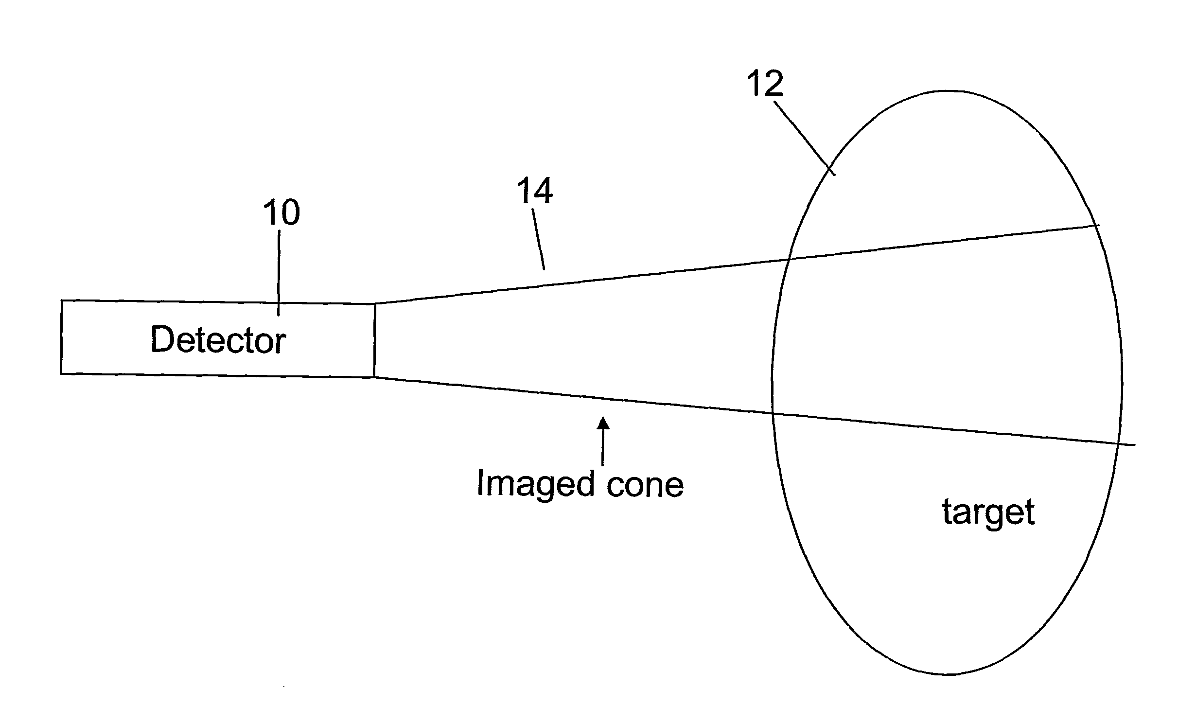
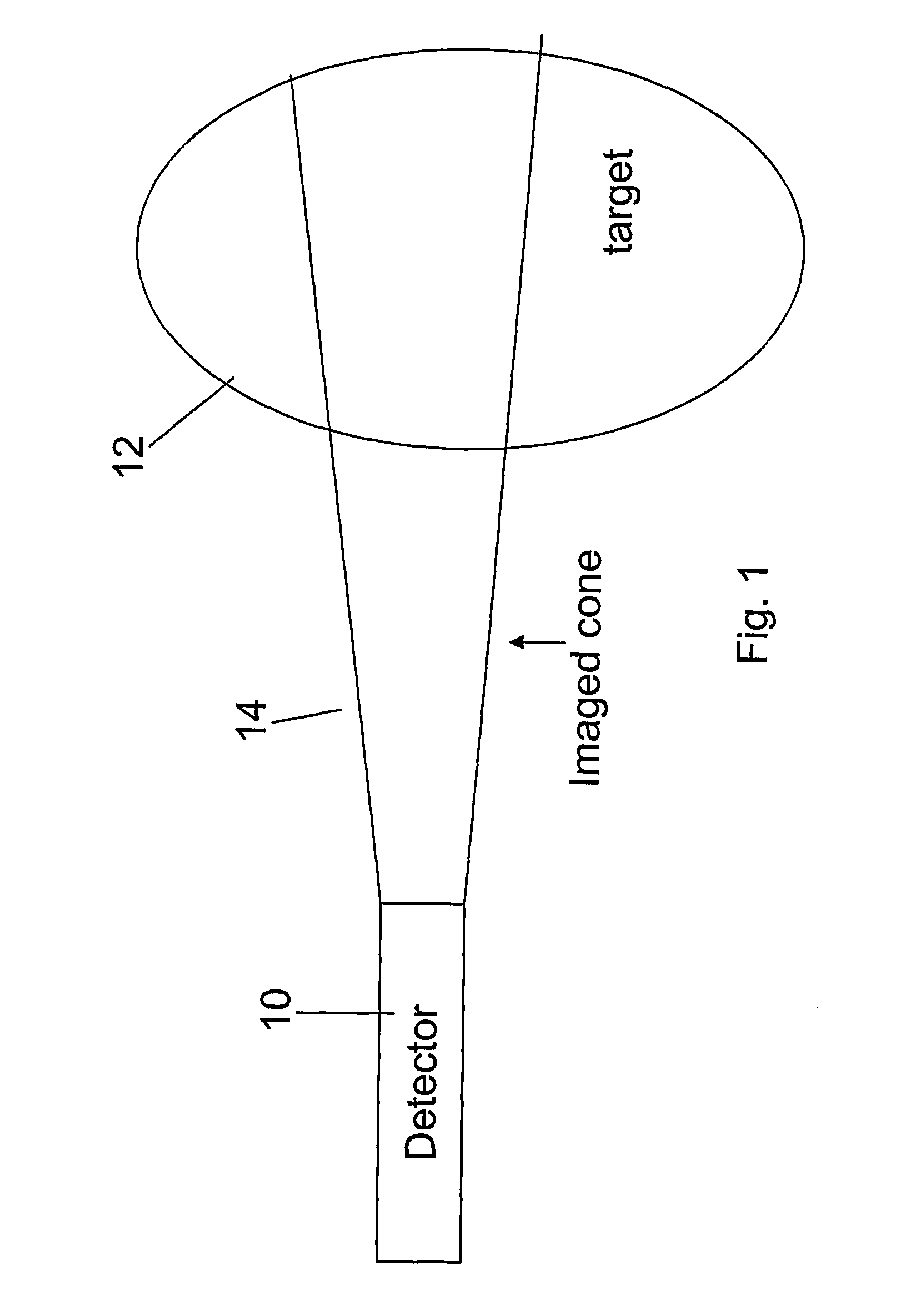
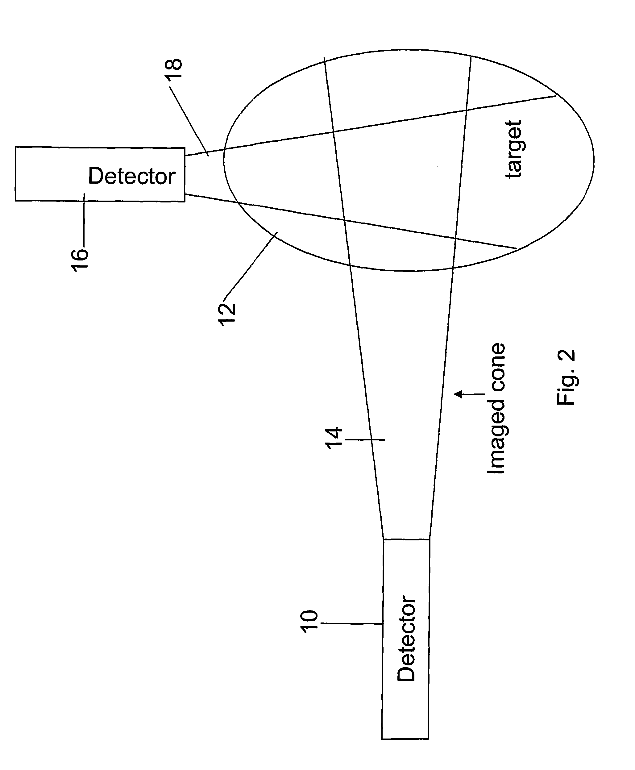
Image

Examples
example 1
[0483] In accordance with an embodiment of the present invention, a cardiac vulnerable-plaque and myocardial-perfusion protocol is employed, for two purposes:
i. identify plaques that may be released into the blood stream; and
ii. evaluate the extent of perfusion defect, if any, caused by the plaque.
[0484] The cardiac vulnerable-plaque and myocardial-perfusion protocol is carried out as follows: A patient is injected with up to about 5 mCi of Annexin radiopharmaceutical labeled with 111-In. After about 24 hours, the patient is injected with up to about 5 mCi of AccuTec radiopharmaceutical labeled with Tc-99m. After the AccuTec injection, the patient is subjected to a pharmacological stress, for example, by the administration of adenosine. At peak vasodialation, the patient is injected with up to about 1 mCi Tl-201 thallous chloride. Immediately following the Tl-201 thallous chloride injection, dynamic imaging is carried out for up to 30 minutes.
[0485] The AccuTec binds to activa...
example 2
[0497] The effect of acetazolamide (Diamox) used for treatment of disorders such as epilepsy by controlling fluid secretion may be tested by using Tc-99m-HMPAO or Tc-99m-Ceretec for quantitative measurements of regional cerebral blood flow (rCBF). Patients will be administered Diamox and after sufficient time for the drug to take effect, the patient will be injected with up to 30 mCi of HMPAO or Ceretec. Immediately following the injection, the patient will be imaged for 30 minutes with a dynamic imaging protocol. The time of maximal rCBF increase is estimated to be at 15 minute post injection 1 g of Diamox. The rCBF increases from 58±8 at rest to 73±5 ml / 100 g / min. In normals with relatively low rCBF values at rest, Diamox increases the reserve capacity much more than in normals with high rCBF values before provocation. It can be expected that this concept of measuring rCBF at rest and the reserve capacity will increase the sensitivity of distinguishing patients with reversible cer...
example 3
[0498] This example relates to FIGS. 10H-10J.
[0499] Tc-99m-MAG3 renography can differentiate the severity of renal angiopathy in patients with Type 2 diabetes through the depletion of renal vasoreactivity after an intravenous injection of acetazolamide (ACZ). Patients will be administered ACZ and after sufficient time for the drug to take effect, the patient will be injected with up to 30 mCi of MAG3Tc99m. Immediately following the injection, the patient will be imaged for 30 minutes with a dynamic imaging protocol to determine the renal perfusion flow. Values of effective renal plasma flow (ERPF) significantly differ between the baseline and the ACZ, both for the normal group and for hypertension group, both having p<0.01 However, for the diabetes group values are distinctly different from the other two groups, with p<0.13.
PUM
 Abstract
Description
Claims
Application Information