
Promotion of Cell Migration and Axon Regeneration in the Cns

a technology of axon regeneration and cell migration, applied in the field of promoting cell migration and axon regeneration in the cns, can solve the problems of poor prognosis for improvement and recovery, interruption of axon pathways, and limited repair ability of adult mammalian cns progenitor cells, etc., and achieve the effect of enhancing the migration of neural cells

Inactive Publication Date: 2008-04-24
MEMORIAL SLOAN KETTERING CANCER CENT
View PDF0 Cites 10 Cited by
  • Summary
  • Abstract
  • Description
  • Claims
  • Application Information

AI Technical Summary

Benefits of technology

[0010] The present invention relates broadly to ministrations to certain portions of the central nervous system that provide enhanced migration of neural cells, or their extensions, from an originating region to a second region in which, or at which, a need for such cells and / or their extensions has been identified.

Problems solved by technology

Adult mammalian CNS progenitor cells, however, manifest only a limited ability to repair damage, such as may arise from various pathologies or traumas.
There are numerous CNS pathologies or diseases for which treatments are limited, and that offer poor prognosis for improvement and recovery.
The CNS environment interferes with axon regeneration, and the glial scar that forms at the lesion site provides a molecular barrier to regeneration.
Traumatic injury to the CNS can lead to interruption of axon pathways, such as the corticospinal tract, formation of scar tissue at the site of the injury, and consequent loss of transmission of nerve impulses to distal synapses.
At present there is no effective therapy to improve the prognosis for patients suffering such traumas.

Method used


Image

  • Promotion of Cell Migration and Axon Regeneration in the Cns
  • Promotion of Cell Migration and Axon Regeneration in the Cns
  • Promotion of Cell Migration and Axon Regeneration in the Cns

Examples


example 1

PSA Expression in COS-7 Cells after Transfection with the GFP-PST Plasmid

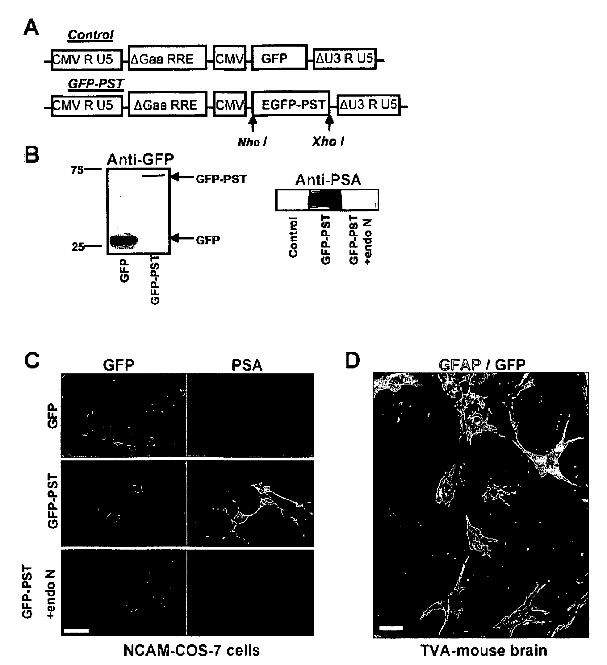
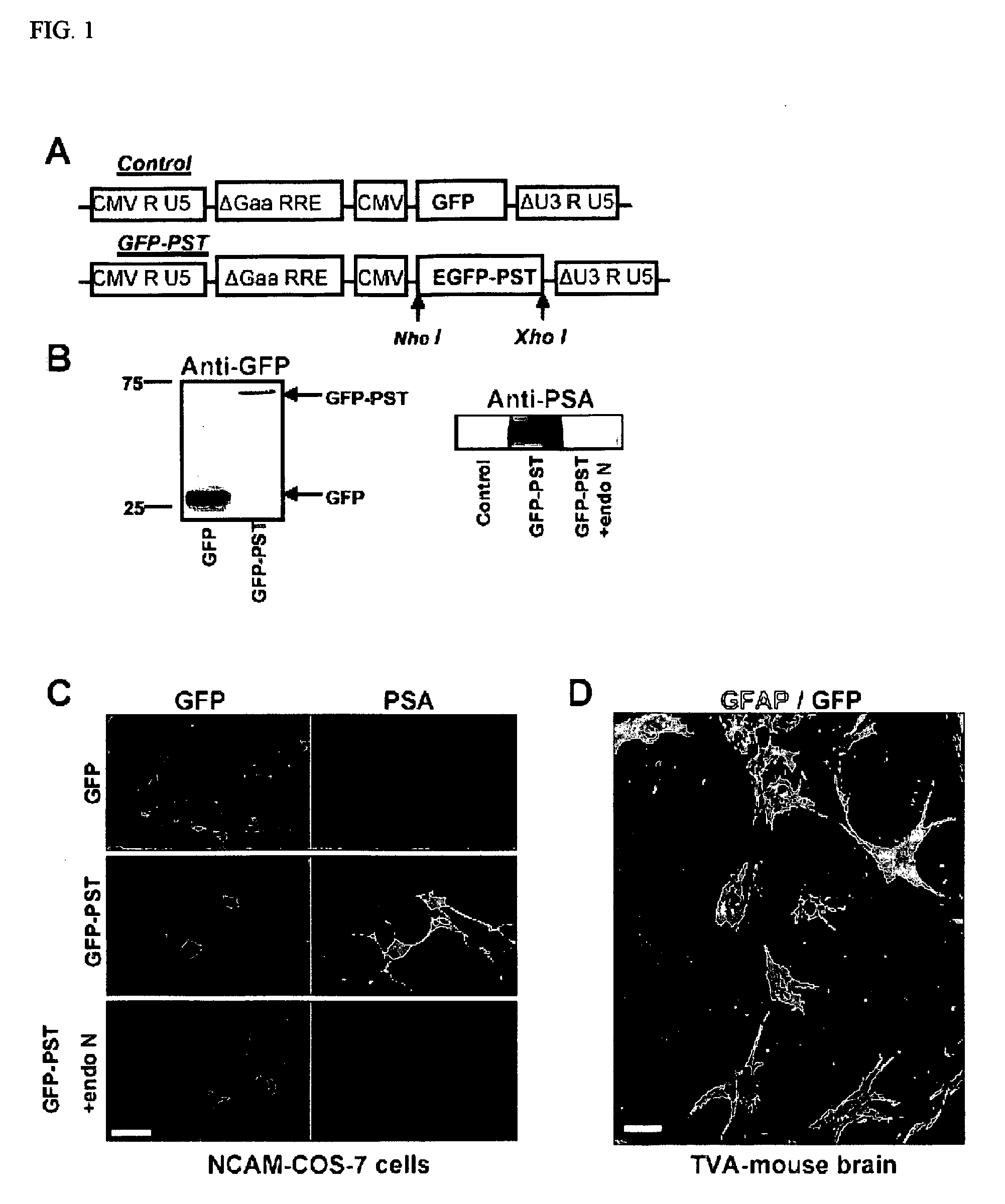
[0268] The GFP-PST fusion protein was inserted into the pCS-CG plasmid (FIG. 1A), and tested by co-transfection into COS-7 cells along with the NCAM plasmid. The Western blot shows a GFP-positive band at the expected molecular weight of 27 kDa in an extract of COS-7 cells transfected with GFP (control; FIG. 1B, left blot, left lane). Cells transfected with GFP-PST fusion protein yielded a band at 69 kDa (FIG. 1B, left blot, right lane), which is the expected molecular weight for the fusion protein. On the right (FIG. 1B, right blot), a Western blot specific for PSA shows a band at molecular weights over 150 kDa in NCAM / GFP-PST transfected COS-7 cells (center lane). These cells normally lack NCAM-PSA and are unable to express PSA after co-transfection with NCAM DNA only (control, left lane). The PSA band disappeared when the lysate was treated for 30 min with the PSA-specific endoneuraminidase N(PST+endo N, rig...

example 2

Viral-Infection and PST Expression in Astrocytes of Adult GFAP-TVA transgenic mice

[0270] GFAP-TVA transgenic animals express the TVA receptor exclusively in astrocytes, which allows selective infection of these cells by HIV (ALSV-A) lentiviral vectors. Accordingly, when brain slices from these animals were incubated with lentivirus carrying either the GFP-PST or the GFP construct, GFP expression (green) was restricted to only to astrocytes (red; GFAP positive) (FIG. 1D). After 6 days in culture, PSA appeared on the surface of the GFP-PST-infected astrocytes (data not shown).

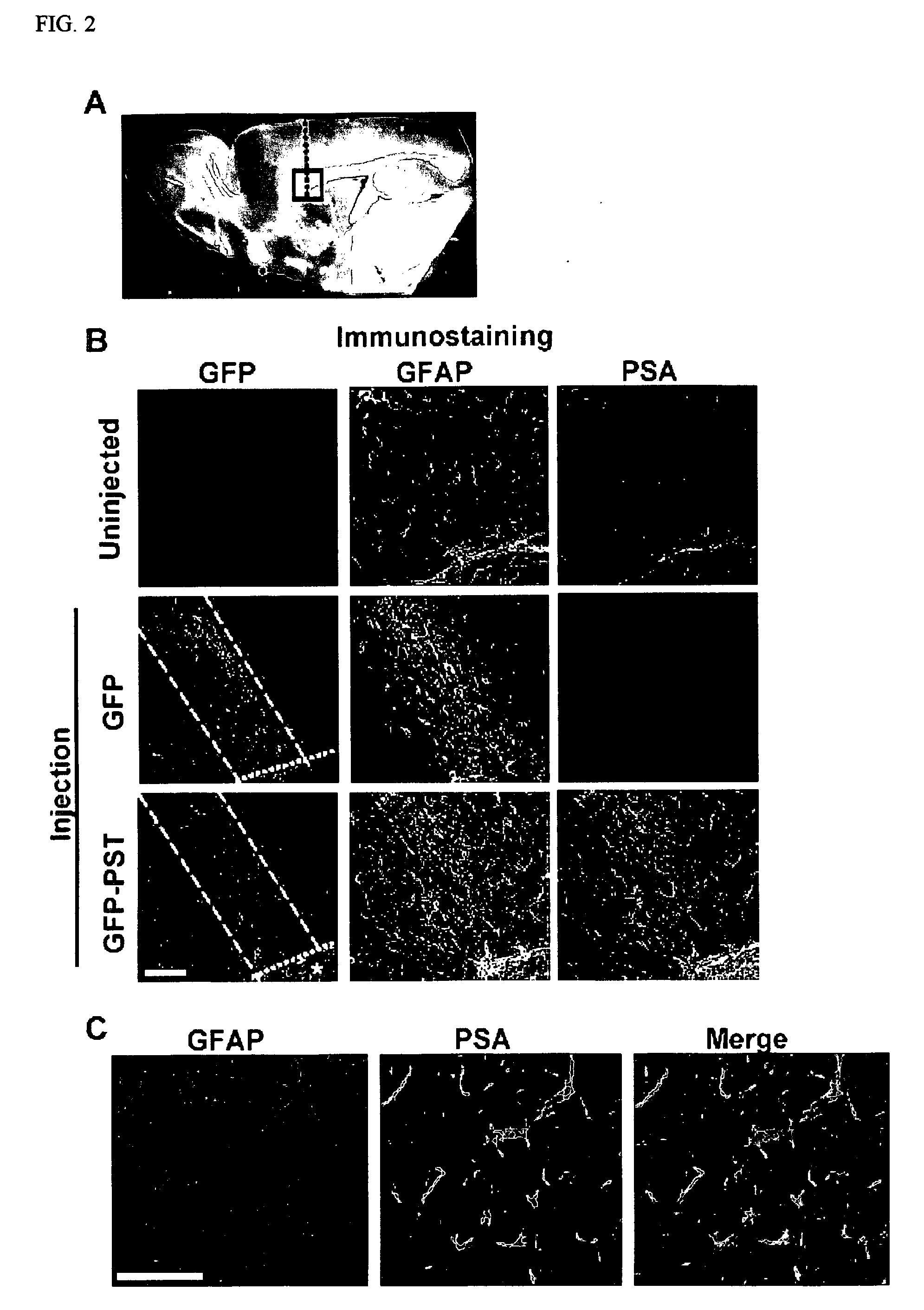
[0271] After the in vitro studies, virus encoding GFP or GFP-PST was injected in vivo in the transgenic mice along a needle track extending through the cortex and corpus callosum (CC) to the SVZ (FIG. 2A). Immunohistological examination of the injected regions after 30 days revealed that the virus had selectively infected GFAP-expressing astrocytes (FIG. 2B), which have been reported to be found at lesioned are...

example 3

PSA Expression Enhances SVZ Progenitor Migration into the Corpus Callosum

[0272] To determine whether ectopic PSA expression would influence the migration of SVZ cells, experiments similar to those in Example 2 were carried out using the GFAP-TVA transgenic mice. The progenitor cells were pre-labeled by repetitive BrdU injections to identify cells undergoing DNA replication and cell proliferation, and were visualized by co-staining for BrdU and nestin (FIG. 3). Injection of the control GFP virus alone resulted in deviation of a small but significant number of SVZ progenitors into the CC as compared to an uninjected animal. (The induced expression of PSA in the control studies involves a needle track lesion, which can increase the presence of SVZ precursors in white matter (Goings et al., 2004)). Injection of the PST construct, on the other hand, increased the migration of progenitor cells into the CC by more than 4 times (FIGS. 3A and 3B; p<0.001). The injections did not induce glia...


PUM

PropertyMeasurementUnit
molecular weightaaaaaaaaaa
thickaaaaaaaaaa
dissociation constantaaaaaaaaaa

Abstract

The present invention relates broadly to methods for stimulating neural progenitor cell migration to certain regions of the nervous system where the neural progenitor cell naturally would not migrate. These methods have wide interest in fields related to the development of therapeutic approaches for addressing a broad range of neurodegenerative and demyelinating pathologies of the central nervous system. In order to accomplish these various outcomes, the invention provides modalities for introducing into a cell of the CNS a substance that promotes polysialylation of a protein component of the cell. This process underlies various methods of the invention, such as a method of polysialylating a protein of the cell, a method of promoting migration of a neural progenitor cell from a first region of a brain to a second region of the brain, or a method of promoting a neural progenitor cell originating in a first region of a brain to differentiate in a second region of the brain. The methods described above provide the basis for various therapeutic methods disclosed in the invention. In one example a method is disclosed of inhibiting the development of, treating, or ameliorating a neurological pathology in a subject, wherein the method includes introducing into brain cells, located in a path starting in a location close to where neural progenitor cells are located and ending in an area of the CNS where it is desirable that the neural progenitor cells migrate to, of the subject a substance that promotes polysialyltransferase activity that polysialylates a protein of the cells. In a second example a method is disclosed of inhibiting the development of, treating, or ameliorating a neurological pathology in a subject, including administering to the subject a substance that promotes polysialyltransferase activity in an amount effective to treat the pathology.

Description

FIELD OF THE INVENTION [0001] The present invention relates generally to methods and compositions that provide enhanced migration of neural cells, or their extensions, from an originating region to a second region in which, or at which, a need for such cells and / or their extensions has been identified. BACKGROUND OF THE INVENTION [0002] Central nervous system (CNS) progenitor cells are located in discrete regions of the adult mammalian CNS including the subventricular zone (SVZ) of the lateral ventricle and the dentate gyrus in the hippocampus. New neural progenitors are continuously born in the adult mammalian brain, where they migrate to replenish particular cell populations. Adult mammalian CNS progenitor cells, however, manifest only a limited ability to repair damage, such as may arise from various pathologies or traumas. [0003] At early stages of CNS cell fate determination, progenitors express the polysialylated form of the neural cell adhesion molecule (PSA-NCAM). PSA is a l...

Claims


Application Information

Patent Timeline
Patent Type & Authority Applications(United States)
IPC IPC(8): A61K39/00A61P43/00C07K16/00C12N15/00
CPCA01K67/027C12N2830/008A01K2217/05A01K2227/105A01K2267/0356A01K2267/0393A61K47/4823A61K47/48384A61K48/00A61K49/0008C12N9/1081C12N15/8509C12N15/86C12N2740/16043A01K67/0275A61K47/61A61K47/6803A61P43/00
Inventor RUTISHAUSER, URSPETRIDIS, ATHANASIOS K.MAAROUF, ABDERRAHMAN EL
Owner MEMORIAL SLOAN KETTERING CANCER CENT
Learn More