Mitochondriotropic Phospholipid Vesicles

a phospholipid vesicles and mitochondria technology, applied in the field of mitochondrial phospholipid vesicles, can solve the problems of not being addressed, familial deafness and some cases of alzheimer's disease, and achieve the effect of protecting mitochondria and slowing down the natural aging process

Inactive Publication Date: 2008-04-24
NORTHEASTERN UNIV
View PDF14 Cites 12 Cited by
  • Summary
  • Abstract
  • Description
  • Claims
  • Application Information

AI Technical Summary

Benefits of technology

[0012] The delivery of both small drug molecules and large macromolecules to and into mitochondria provides the basis for a large variety of cytoprotective and cytotoxic therapies. For example, the delivery of therapeutic DNA and RNA such as antisense oligonucleotides, ribozymes, plasmid DNA expressing mitochondrial encoded genes as well as wild-type mtDNA can provide the basis for treatment of mitochondrial DNA diseases. The delivery of antioxidants can protect mitochondria from oxidative stress caused by a variety of insults, perhaps even contributing to slowing down t

Problems solved by technology

Certain deleterious base substitutions can cause familial deafness and some cases of Alzheimer's disease and Parkinson's disease.
Both studies, however, were done w

Method used


Image

  • Mitochondriotropic Phospholipid Vesicles
  • Mitochondriotropic Phospholipid Vesicles
  • Mitochondriotropic Phospholipid Vesicles

Examples


example i

Synthesis and Characterization of the Bromide Salt Containing Stearyl-Triphenylphosphonium Cation as a Hydrophobized Amphiphilic Delocalized Cation According to the Invention

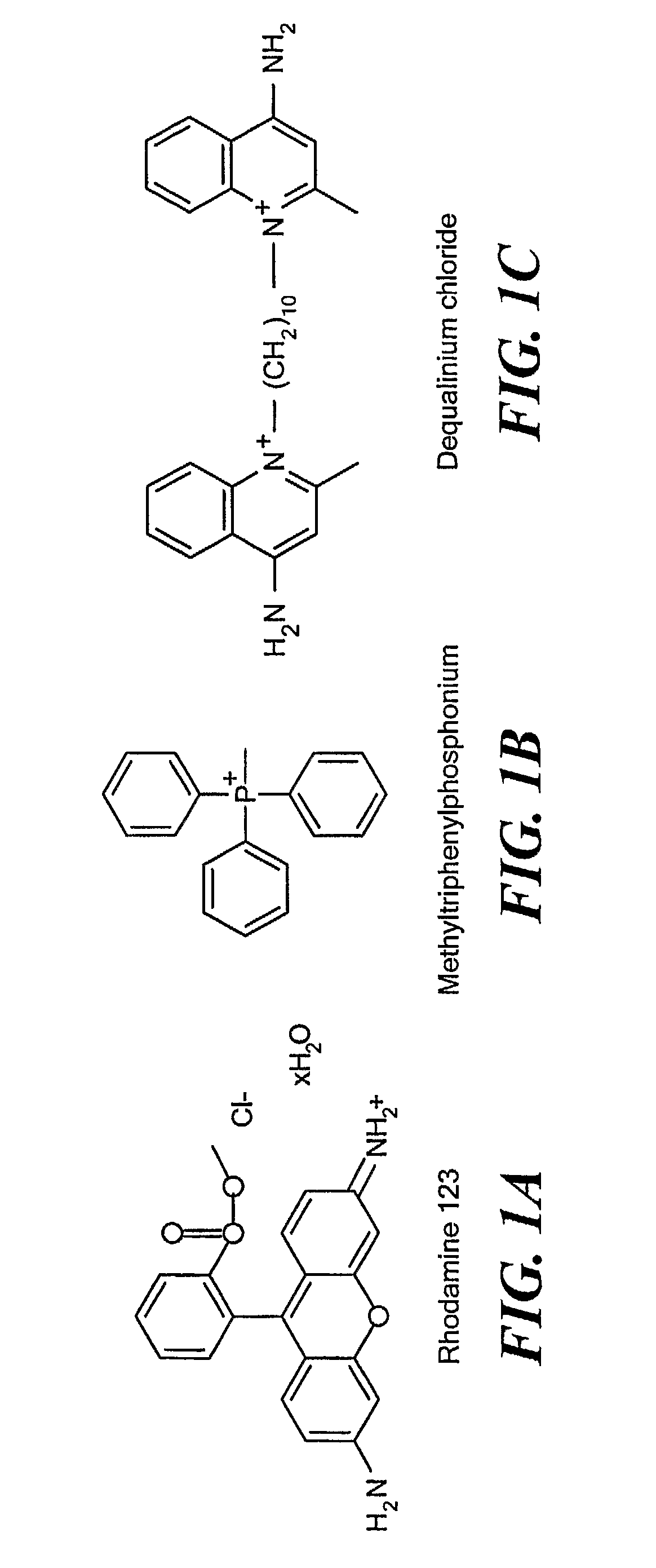
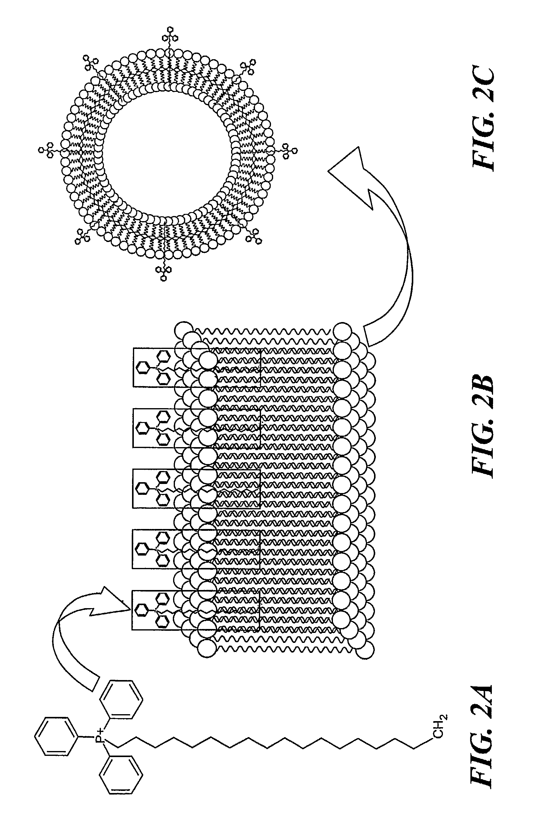
[0035] Stearyl-triphenylphosphonium bromide (according to FIG. 2A) was synthesized by heating stearyl bromide and triphenylphosphine (FIG. 1B) under reflux in xylene according to a protocol for the synthesis of analogous tertiary phosphonium salts (see, Materials and Methods). Isolation by column chromatography on silica gel and recrystallization from diethylether yielded a chromatographically pure product, which was identified by 1H-NMR as described in Materials and Methods. FIG. 3 shows the 31P-NMR spectrum of STPP. The observed 31P chemical shift of STPP is at 25.34 ppm, well within the range of 20.9-26.2 ppm as described for a series of alkyl- and aryl triphenylphosphonium salts (Kiddle, 2000). In comparison, the 31P chemical shift of triphenylphosphine was found to be −4.48 ppm, which is in perfect agreeme...

example ii

Preparation and Characterization of Liposomes According to the Invention with Surface-Linked Triphenylphosphonium Cations

[0036] The preparation of liposomes in the presence of hydrophilic molecules that have been hydrophobized via linkage to fatty acid or phospholipid derivatives results in the covalent “anchoring” of the hydrophilic moiety to the liposomal surface. Liposomes according to the invention were prepared in the presence of STPP according to standard protocols (Lasch et al., 2003). FIG. 2B shows schematically the alkyl (i.e., stearyl) residue mediated “anchoring” of the triphenylphosphonium cation in the liposomal phospholipid bilayer membrane. STPP liposomes, i.e., liposomes with surface-linked triphenylphosphonium cations, were isolated using a Sephadex G-15 column and characterized by 31P-NMR (FIG. 4), size distribution analysis (FIG. 5) and zeta potential measurements (FIG. 6).

[0037] As expected (see, FIG. 4), the 31P-NMR spectrum of STPP-liposomes shows two chemica...

example iii

Intracellular Distribution of STPP Liposomes

[0038] To study the cellular uptake and intracellular distribution of STPP liposomes, cells of the breast cancer cell line BT 20 were incubated with fluorescence-labeled STPP liposomes for 1 h in serum-free medium. To remove non-internalized liposomes, cells were thoroughly washed and allowed to grow for another hour in complete medium. Typically obtained epifluorescence microscopic images are shown in FIGS. 7A and 7B. FIG. 7A displays cells incubated with STPP liposomes that have been labeled by incorporation of 0.5 mol % Rhodamine-PE, while FIG. 7B shows cells, the mitochondria of which have been specifically stained with Mitotracker red. On comparing FIG. 7A with FIG. 7B, it can be seen that cells incubated with STPP liposomes display the same distinct fluorescence pattern as cells stained with the mitochondria-specific dye. Such a comparison of staining patterns has been used by Filipovska et al. (2004) to reveal the localization of l...


PUM

PropertyMeasurementUnit
Amphiphilicaaaaaaaaaa
Solubility (mass)aaaaaaaaaa
Hydrophilicityaaaaaaaaaa

Abstract

Mitochondriotropic phospholipid vesicles, i.e., mitochondriotropic liposomes, that comprise a hydrophobized amphiphilic delocalized cation, such as those comprising, e.g., a triphenylphosphonium or a quinolinium moiety, incorporated into the phospholipid membrane of the vesicles, or liposomes, are disclosed. The hydrophobized portion of the amphiphilic delocalized cation, e.g., a fatty acid or other phospholipid derivative, is embedded in the phospholipid membrane of the liposome, and the amphiphilic portion of the cation is exposed on the surface of the liposome. Mitochondriotropic liposomes constitute a mitochondria-targeted drug delivery system, permitting the transport of a high payload of therapeutic water-soluble molecules in their native (i.e., active) state specifically and exclusively to mitochondria in living mammalian cells.

Description

CROSS REFERENCE TO RELATED APPLICATIONS [0001] This application claims the priority of U.S. Provisional Application No. 60 / 657,802 filed Mar. 2, 2005 entitled, MITOCHONDRIOTROPIC PHOSPHOLIPID VESICLES, the whole of which is hereby incorporated by reference herein.STATEMENT REGARDING FEDERALLY SPONSORED RESEARCH OR DEVELOPMENT [0002] N / A BACKGROUND OF THE INVENTION [0003] Since the first demonstration in 1988 that mitochondrial DNA (mtDNA) base substitution and deletion mutations are linked to human disease, a variety of degenerative diseases have been associated with mtDNA mutations (Wallace, 1994). Mitochondria are vital for the cell's energy metabolism and for the regulation of programmed cell death. In addition, mitochondria are critically involved in the modulation of intracellular calcium concentration and the mitochondrial respiratory chain. Consequently mitochondrial dysfunction either causes or at least contributes to a large number of human diseases. [0004] Mitochondria are...

Claims


Application Information

Patent Timeline
IPC IPC(8): A61K9/127A61K31/70A61P43/00A61K38/00
CPCA61K9/1272A61P43/00
Inventor WEISSIG, VOLKMARBODDAPATI, SARATHI V.HANSON, ROBERT N.TORCHILIN, VLADIMIR P.
Owner NORTHEASTERN UNIV
Why Eureka
Try Eureka