
Method for Determining Responsiveness to Chk1 Inhibitors

a technology of chk1 inhibitor and responsiveness, applied in the field of determining responsiveness to chk1 inhibitor, can solve the problem of severely limited use of both approaches

Inactive Publication Date: 2008-06-19
ASTRAZENECA AB
View PDF2 Cites 10 Cited by
  • Summary
  • Abstract
  • Description
  • Claims
  • Application Information

AI Technical Summary

Benefits of technology

[0074]Phospho-H2AX was also examined by IHC as described above in Example 1 except with the following changes. HeLa or A549 cells were plated into 96 well plates and treated with or without 100 nM gemcitabine and increasing concentrations of CHK inhibitor (0 to 1 μM) for 5 hours prior to fixation and staining. A 1:400 dilution of a mouse monoclonal anti-phospho H2AX Serine 139 (Upstate, N.Y.) followed by a 1:300 dilution of Alexa Fluor 594 anti-mouse IgG (Molecular Probes, OR) was incubated on fixed cells. Quantitation was performed using Cellomics Cell Health algorithms and reported as %phospho-Hp2AX positive cells. FIG. 7 shows the quantitation demonstrating a robust dose dependent increase in %phospho-H2AX positive cells when cells are treated with the combination of gemcitabine and CHK inhibitor. It was observed using immunostaining that phosphorylated H2AX is present throughout the nucleus, not limited to foci.

Problems solved by technology

Chemotherapy and radiation exposure are currently the major options for the treatment of cancer, but the utility of both of these approaches is severely limited by drastic adverse effects on normal tissue, and the frequent development of tumor cell resistance.

Method used


Image

  • Method for Determining Responsiveness to Chk1 Inhibitors
  • Method for Determining Responsiveness to Chk1 Inhibitors
  • Method for Determining Responsiveness to Chk1 Inhibitors

Examples


example 1

Demonstration of Activation of CHK1 following DNA Damage in Tumor Cell LineS, and Xenograft Samples

[0055]A. In vitro Studies:

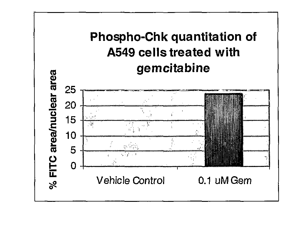
[0056]In brief, 3×10⁶ SW620 and or HT29 cells were plated into 10 cm tissue culture dishes and allowed to adhere for 24 hours at 37° C. Cell were then treated with a titration of DNA damaging agents (SN-38 (CQ International, China), doxorubicin (Sigma-Aldrich, MO) or gemcitabine (CQ International, China)). Cells were then harvested after an 8 hour incubation with DNA damaging agents in Onyx lysis buffer with protease and phosphatase inhibitors (20 mM Tris, 137 mM Na Cl, 1 nm EGTA, 1% Triton X, 10% glycerol, 1.5 mM MgCl2, 1:100 dilution of Calbiochem Phophatase Inhibitor Cocktail set II, 10 mM β-glycerophosphate, 1 mM DTT, 7 μg / ml PMSF, 20KIU / ml aprotinin, 1 mM Pefabloc, 0.1 mg / ml leupeptin). Protein lysates were then generated, protein concentration determined and equal protein loaded and analyzed by Western blot analysis using a 1:100 dilution of phospho-CHK ...

example 2

Demonstration of Increased Phospho-CHK Following Combination Treatment of Gemcitabine and CHK1 inhtibitor in Tumor Cell Lines, Xenograft Samples and Surrogate Tissues

[0060]A. In vitro Studies

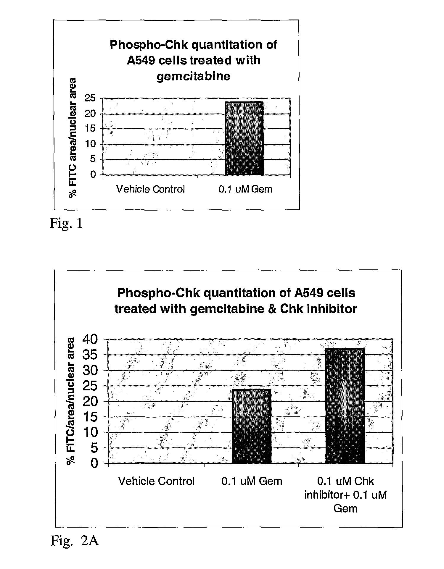
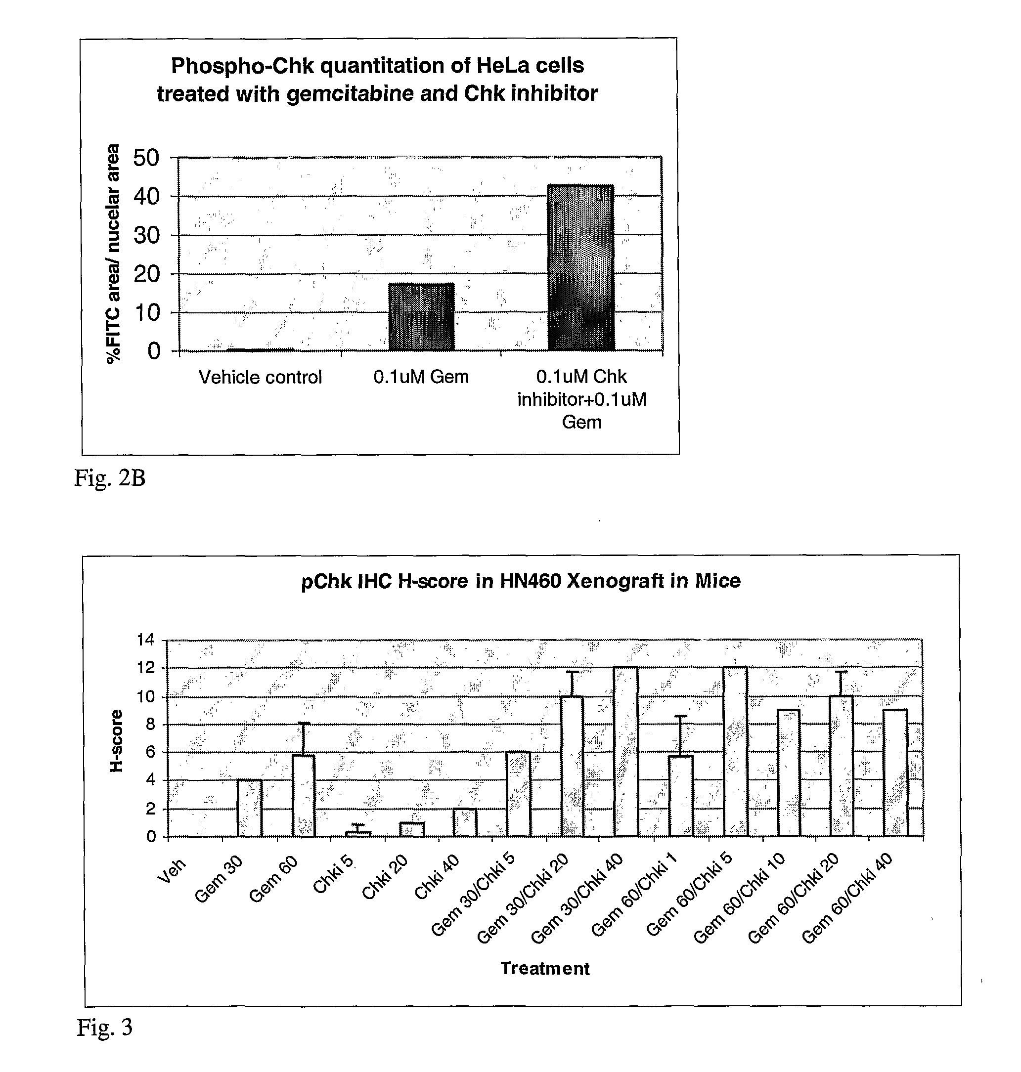
[0061]In brief, 3×10⁶ SW620 were plated into 10 cm tissue culture dishes and allowed to adhere for 24 hours at 37° C. Cell were then treated with 100 nM gemcitabine or 30 nM, 100 nM or 500 nM CHK inhibitor or a combination of 100 nM gemcitabine and 30 nM, 100 nM or 500 nM for 8 hours. One set of cells were then harvested for Western blot analysis. Media containing compounds were removed from the remaining cells and fresh tissue culture media added back. Cells were then harvested 22 hours later, protein lysates generated as described above and analyzed by Western blot analysis using the phosphor-CHK antibody. Western blot analysis demonstrated an increase in phospho-CHK following gemcitabine treatment alone and a dose dependent increase over gemcitabine alone in phospho-CHK following the combinat...

example 3

Demonstration of Decreased DNA Damage Foci Following Inhibition of CHK1 Kinase

[0068]A. In vitro Studies:

[0069]HeLa cells were plated on cover slips and allowed to adhere for 24 hours at 37° C. Cells were then treated for 5 hours with 100 nM Adriamycin™ and 500 nM CHK inhibitor. Cells were then treated as described in Example 1. Primary antibody used for the studies was a 1:120 dilution of rabbit polyclonal anti-53BP1 (Novus, CO) and secondary antibody was a 1:300 dilution of Alexa Fluor 488 anti-rabbit IgG (Molecular Probes, OR). Foci were quantiatated by measuring fluorescent intensity per number of nuclei (determined by Hoescht staining) using Metaphorph analysis. FIG. 5 demonstrates a decrease in the fluorescent intensity when CHK inhibitor is combined with Doxorubicin hydrochloride (Adriamycin™).


PUM

PropertyMeasurementUnit
timeaaaaaaaaaa
pHaaaaaaaaaa
tumor cell resistanceaaaaaaaaaa

Abstract

The invention is directed at a method for predicting a patient's responsiveness to a CHK1 inhibitor The method includes providing a sample from a test patient who is being treated with a DNA damaging agent; and determining for the presence of an activated CHK1 signal transduction pathway molecule, wherein the presence of an activated CHK1 signal transduction pathway molecule is indicative that a CHK1 inhibitor should be administered to the patient.

Description

FIELD OF THE INVENTION[0001]The present invention relates to the use of a biomarker as an indicator of a therapeutic agent's activity within a test cancer patient. The biomarker of the present invention can be used, for example, to determine when to administer a therapeutic agent or to select a therapeutically effective dose of a therapeutic agent to a patient.BACKGROUND OF THE INVENTION[0002]Chemotherapy and radiation exposure are currently the major options for the treatment of cancer, but the utility of both of these approaches is severely limited by drastic adverse effects on normal tissue, and the frequent development of tumor cell resistance. It is therefore highly desirable to improve the efficacy of such treatments in a way that does not increase the toxicity associated with them. One way to achieve this is by the use of specific potentiating agents. Typically, these agents are designed to be used in combination with existing or novel agents.[0003]An individual cell replicat...

Claims


Application Information

Patent Timeline
Patent Type & Authority Applications(United States)
IPC IPC(8): A61K39/395C12Q1/48A61K38/02A61P35/02G01N33/53A61K31/7052
CPCC12Q1/485G01N33/6872G01N33/57496A61P29/00A61P35/00A61P35/02C12Q1/00G01N33/53G01N33/68G01N33/574
Inventor ZABLUDOFF, SONYABERGERON, LOUISE
Owner ASTRAZENECA AB
Learn More