Opto-acoustic imaging devices and methods

a technology of optical acoustic imaging and optical probe, which is applied in the field of optical imaging, can solve the problems of increased risk of ami and insufficient co-registration level of combined catheters, and achieve the effects of saving valuable space within the catheter body, facilitating proper co-registration, and high precision

Active Publication Date: 2008-07-03
LIGHTLAB IMAGING
View PDF68 Cites 172 Cited by
  • Summary
  • Abstract
  • Description
  • Claims
  • Application Information

AI Technical Summary

Benefits of technology

[0007]One advantage of the invention is the aligned nature of the OCT and ultrasound sensors such that co-registration of the cross-sectional images obtained by the two sensors can be obtained with high precision. Previous descriptions of such combined catheters did not provide the co-registration levels needed. Co-registration is important because coronary morphology changes rapidly, often in less than a millimeter of longitudinal distance.
[0008]It is another object of the invention to describe a sensor structure wherein two probe beams are orientated at substantially the same angle with respect to the longitudinal axis of the catheter. Again, this is to facilitate proper co-registration of the images. Differing launch angles of the probe beams implies that the two images diverge each other with depth. Computational correction of this divergence is complex and can lead to errors in image presentation.
[0009]It is another object of this invention to describe efficient methods of providing both optical and electrical energy to the rotating sensor assembly at the tip of the catheter. Using various torque wire and coated fibers to acts as co-axial signal lines saves valuable space within a catheter body.
[0010]It is a further object of the invention to describe mechanisms and configurations of the probe that will simultaneously reduce unwanted parasitic acoustical and optical back-reflections while still providing an aligned and otherwise functional probe assembly.
[0011]It is yet another object of the invention to describe efficient rotary mechanisms for coupling both electrical and optical energy simultaneously into the catheter.
[0012]It is another object of the invention to describe a combined probe utilizing capacitive micro-machined ultrasonic transducers (CMUT) to create a dual element probe such that both the ultrasound and optical beams focus on substantially the same tissue spot simultaneously.

Problems solved by technology

Conversely, deep lipid pools beneath a thin cap greatly increases the risk of an AMI.
Previous descriptions of such combined catheters did not provide the co-registration levels needed.

Method used


Image

  • Opto-acoustic imaging devices and methods
  • Opto-acoustic imaging devices and methods
  • Opto-acoustic imaging devices and methods

Examples


Embodiment Construction

[0034]The following description refers to the accompanying drawings that illustrate certain embodiments of the present invention. Other embodiments are possible and modifications may be made to the embodiments without departing from the spirit and scope of the invention. Therefore, the following detailed description is not meant to limit the present invention. Rather, the scope of the present invention is defined by the appended claims.

[0035]It should be understood that the order of the steps of the methods of the invention is immaterial so long as the invention remains operable. Moreover, two or more steps may be conducted simultaneously or in a different order than recited herein unless otherwise specified.

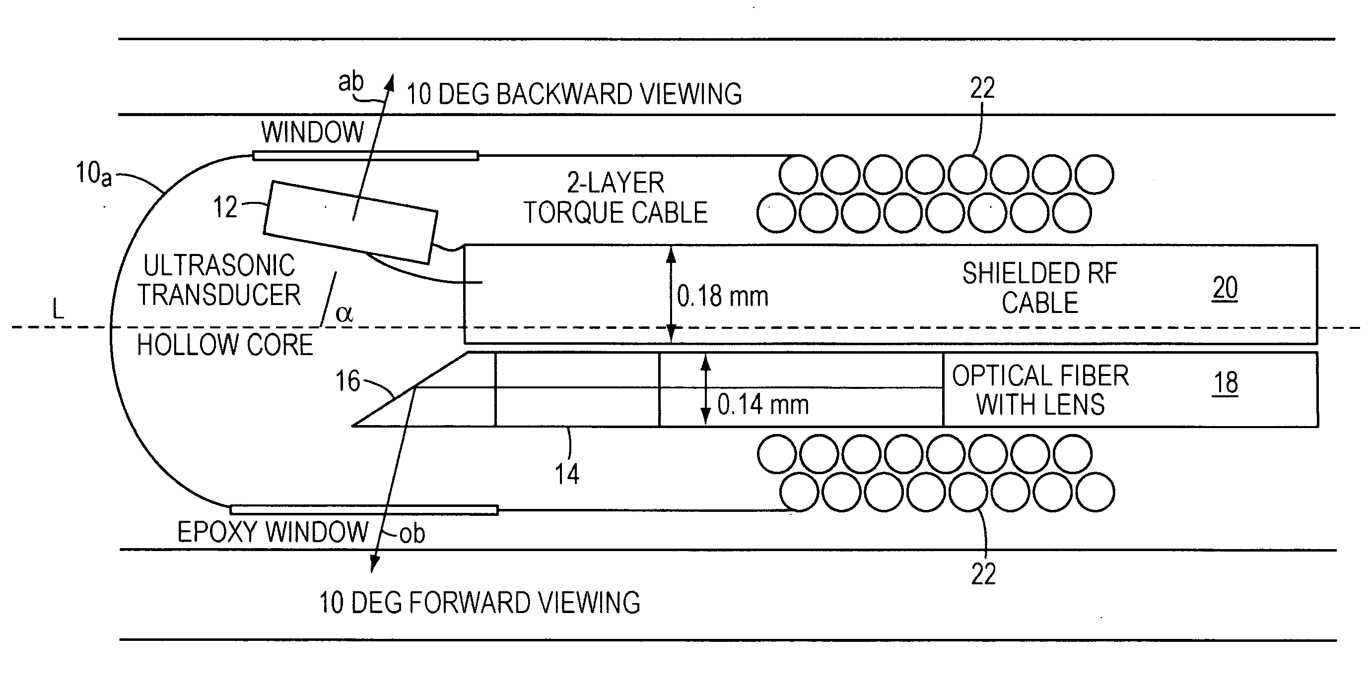
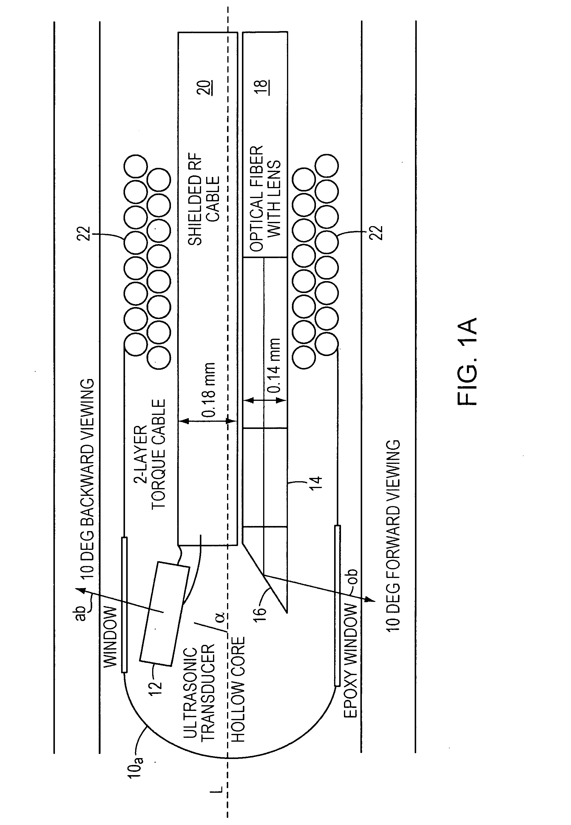
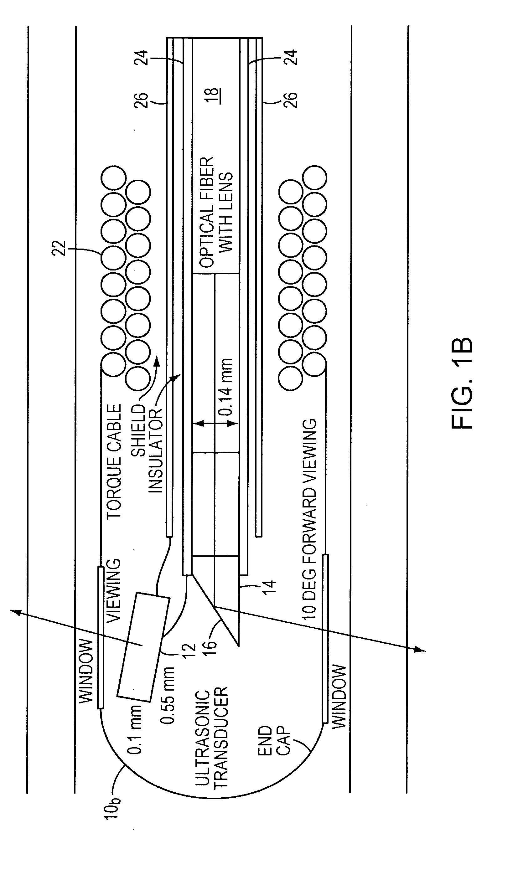
[0036]FIG. 1A illustrates a portion of an imaging probe 10a, using a conventional IVUS ultrasonic transducer 12, an optical transducer 14 which includes an angled-tip optical lens assembly 16 attached to a single mode fiber 18, a standard miniature RF cable 20 delivering power t...


PUM


Abstract

In one aspect, the invention relates to a probe. The probe includes a sheath, a flexible, bi-directionally rotatable, optical subsystem positioned within the sheath, the optical subsystem comprising a transmission fiber, the optical subsystem capable of transmitting and collecting light of a predetermined range of wavelengths along a first beam having a predetermined beam size. The probe also includes an ultrasound subsystem, the ultrasound subsystem positioned within the sheath and adapted to propagate energy of a predetermined range of frequencies along a second beam having a second predetermined beam size, wherein a portion of the first and second beams overlap a region during a scan.

Description

RELATED APPLICATIONS[0001]This application claims priority to U.S. Provisional Patent Application 60 / 857,573 filed on Nov. 8, 2006, the disclosure of which is herein incorporated by reference in its entirety.FIELD OF INVENTION[0002]This invention relates to the field of optical imaging and more specifically to the design of fiber-optic probes for optical coherence tomography (OCT) and other optical imaging technologies, such as ultrasound.BACKGROUND[0003]In recent years, the underlying cause of sudden heart attacks (acute myocardial infarctions or AMI) has been the subject of much attention. The older prevailing theory of gradual occlusion of the coronary artery has been superseded by a new theory based on extensive histopathologic evidence that AMI is the result of a rupture in the coronary artery wall, specifically a rupture of a “vulnerable plaque.” A vulnerable plaque, also known as Thin-Capped Fibro-Artheroma (TCFA), is characterized by a thin fibrous cap covering a lipid pool ...

Claims


Application Information

Patent Timeline
Patent Type & Authority Applications(United States)
IPC IPC(8): A61B8/00
CPCA61B5/0066A61B5/0095A61B8/445A61B8/12A61B8/4461A61B5/6852
Inventor SCHMITT, JOSEPH M.PETERSEN, CHRISTOPHEROHASHI, TORUNAKAMATSU, TETSUYA
Owner LIGHTLAB IMAGING
Why Eureka
Try Eureka