Use of Endogenous Fluorescence to Identify Invading Metastatic Breast Tumor Cells

a metastatic breast tumor and endogenous fluorescence technology, applied in the field of endogenous fluorescence to identify invading metastatic breast tumor cells, can solve the problems of radioactive decay, low fluorescence emission, and relatively low two-photon absorption, and achieve the effect of accurate characterization

Active Publication Date: 2008-09-04
WISCONSIN ALUMNI RES FOUND
View PDF17 Cites 35 Cited by
  • Summary
  • Abstract
  • Description
  • Claims
  • Application Information

AI Technical Summary

Benefits of technology

[0009]The present invention broadly provides methods and systems for detecting, identifying, and characterizing conditions in humans and in other animal subjects, including diseases and other disorders, by analyzing fluorescence from endogenous flavin adenine dinucleotide (FAD) fluorophors present in biological materials and samples. In particular embodiments, the invention relates to conditions of the human breast including cancers such as carcinoma. Methods and systems are provided for detecting, locating, and characterizing tumors, particularly epithelial tumors, via nonlinear optical imaging techniques capable of accurately characterizing fluorescence intensities and fluorescent lifetime parameters from endogenous FAD fluorophors present in a test tissue sample. Nonlinear optical imaging techniques beneficial in the present invention include multiphoton microscopy (MPM), multiphoton fluorescent lifetime imaging microscopy (FLIM), harmonic generation microscopy, and spectral lifetime imaging microscopy (SLIM) techniques providing high resolution multi dimensional imaging (including two-dimensional and three-dimensional) of test tissue samples. The methods and systems support versatile implementation including providing in vivo analysis (including application to a patient sample in situ over real time), and in vitro analysis (including ex vivo as analogous to classical histopathology) of test tissue samples. Methods of the present invention have applications for clinical pathology, optical biopsy and endoscopy. In some embodiments, for example, the present methods analyze the fluorescent properties of endogenous FAD biomarkers in biological materials and samples to identify and characterize invading metastatic breast tumor cells.
[0010]The present invention further and preferably for some applications provides noninvasive and nondestructive methods and systems that are capable of identifying and characterizing cancer in non-fixed, non-sectioned, and / or non-stained tissue, including excised tissue samples and tissues in whole organisms. The present invention provides highly sensitive, selective and specific methods that are capable of directly evaluating subsurface tumor development and progression over the spectrum from a normal state to advanced cancer states, including invasive and metastatic states. The present methods and systems are capable of detecting and characterizing (e.g., classifying, staging etc.) cancer in a range of tissue types, including breast tissue and epithelial tissue. In some specific applications, for example, the present methods are utilized to support clinical diagnosis and treatment in connection with epithelial tumor development, progression, local invasion and metastasis. Systems and methods of the present invention provide diagnostic information that is different from, and can be complementary to, information provided by other diagnostic platforms, such as mammography, ultrasound and conventional histological staining techniques. Methods of the present invention can be carried out rapidly, in real time and, therefore, are useful tools to support biopsy, surgical resection and other clinical diagnostic and treatment procedures for cancer.
[0011]In one aspect, the invention provides a method of evaluating a test tissue sample for the diagnosis of cancer. A method of this aspect comprises the steps of: (i) providing the test tissue sample from a test subject, wherein the test tissue sample comprises a mammary tissue component; (ii) generating a test image or test imaging data from the test tissue sample using a nonlinear optical imaging technique; and (iii) analyzing the test image or test imaging data of the test tissue sample by measuring fluorescence intensities, fluorescence lifetime values or both of fluorescence from FAD in the test tissue sample, thereby evaluating the test tissue sample for the diagnosis of cancer. In an embodiment, the test tissue sample is a breast tissue sample and / or the test tissue sample is intact and non-fixed. Evaluation of fluorescence information from FAD in the present techniques is beneficial because it is an endogenous fluorophor in a range of important tissues from humans and other species and is present at concentrations allowing for effective multiphoton fluorescence characterization. Further, use of an endogenous fluorophor in the present methods eliminates the need for providing imaging agents to the sample, such as optical contrast agents or fluorescent probes, thereby enabling sensitive optical evaluation without requiring a change in the composition of the test tissue sample.
[0016]A variety of reference images, reference data and reference tissues are useful in methods of the present invention. In an embodiment, FAD fluorescence intensity and / or fluorescence lifetime reference values can each independently relate to a reference tissue having a normal condition (i.e., noncancerous) or a disease condition at a particular disease stage or historical time point. For example, FAD fluorescence intensity and / or FAD fluorescence lifetime reference values can be a previously assessed value from the same or different test sample from the same patient or a different patient. When reference values are derived from the same patient, there can be a particular diagnostic advantage in such values serving as an internal control, whether or not temporally synchronized with the test tissue sample. In the specific example where historical reference values reflect a disease state, the reference values can serve to evaluate the present test tissue sample while also providing other information, e.g., regarding the level or rate of change. When the reference values reflect a disease state, there can be a diagnostic advantage in facilitating a comparison of the reference image with the test image to accurately assess the test sample. In a preferred embodiment, greater diagnostic information is achieved by accruing and evaluating multiple reference values corresponding to a plurality of FAD fluorescence references images (e.g., FAD fluorescence intensity or lifetime images) and / or reference image data.
[0023]Test images and test image data is generated in the present invention by exciting endogenous FAD fluorophors and measuring the resulting fluorescence from FAD fluorophors in a test tissue sample. Imaging techniques employing multiphoton excitation allow the use of electromagnetic radiation in the visible or near infrared radiation regions of the spectrum for excitation that effectively penetrates many test tissue samples without resulting in photoinduced degradation or other unwanted changes of composition. In an embodiment, for example, selective imaging of endogenous FAD is achieved by multiphoton excitation using electromagnetic radiation having wavelengths selected over the range of 860 nm to 940 nm, preferably for some applications 890 nanometers, or the 2-photon cross section surrounding the excitation maximum of 450 nm for FAD, and detection of fluorescence having wavelengths selected over the range of 510 nm to 550 nm, preferably for some applications 530 nanometers. Multiphoton excitation also provides the benefit of accessing superior image resolution in fluorescence intensity and fluorescence lifetime images relative to conventional fluorescence excitation techniques.
[0025]In another aspect, the invention provides a method of diagnosing breast cancer that is complementary to conventional breast cancer screening and / or diagnostic techniques. In an embodiment, methods of the present invention allow improved identification and characterization of invasive tumor cells and cells exhibiting metastatic potential. In an embodiment, methods of the present invention allow improved characterization of metastatic potential in cancerous tissue, including epithelial tumors.

Problems solved by technology

In two photon fluorescence excitation techniques, for example, absorption of two lower energy photons simultaneously excites an electronic transition in a fluorophor, thereby causing radiative decay resulting in fluorescence emission of a single higher energy photon.
As the probability of two photon absorption is relatively low (for example, as compared to single photon absorption), excitation in this technique is limited to a spatially confined focused region of the excitation beam having a sufficiently high intensity of photons.
Although the potential for such applications, including endoscopy and optical biopsy applications, is clear, these techniques have not yet matured to the point so as to provide a robust clinical tool.

Method used


Image

  • Use of Endogenous Fluorescence to Identify Invading Metastatic Breast Tumor Cells
  • Use of Endogenous Fluorescence to Identify Invading Metastatic Breast Tumor Cells
  • Use of Endogenous Fluorescence to Identify Invading Metastatic Breast Tumor Cells

Examples


example 1

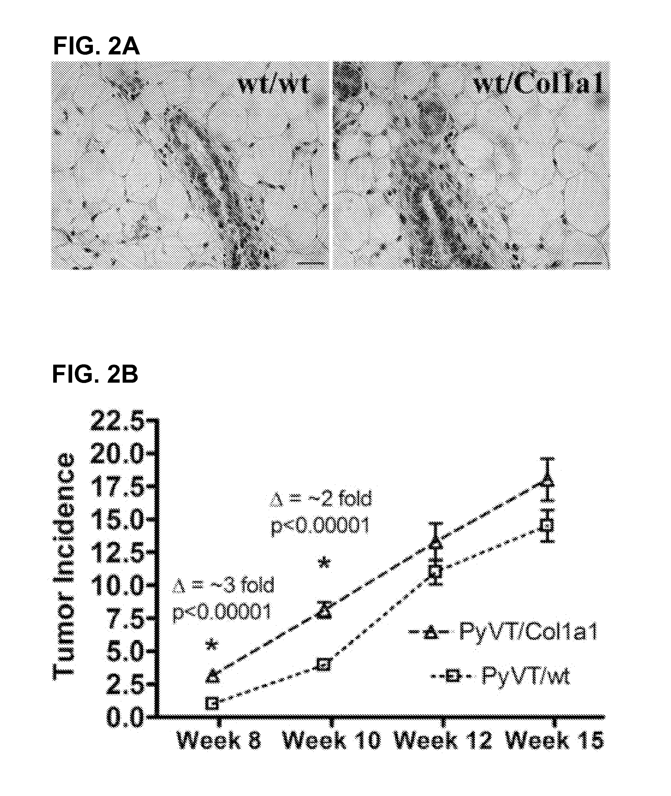
Collagen Density Promotes Mammary Tumor Initiation and Progression

Abstract

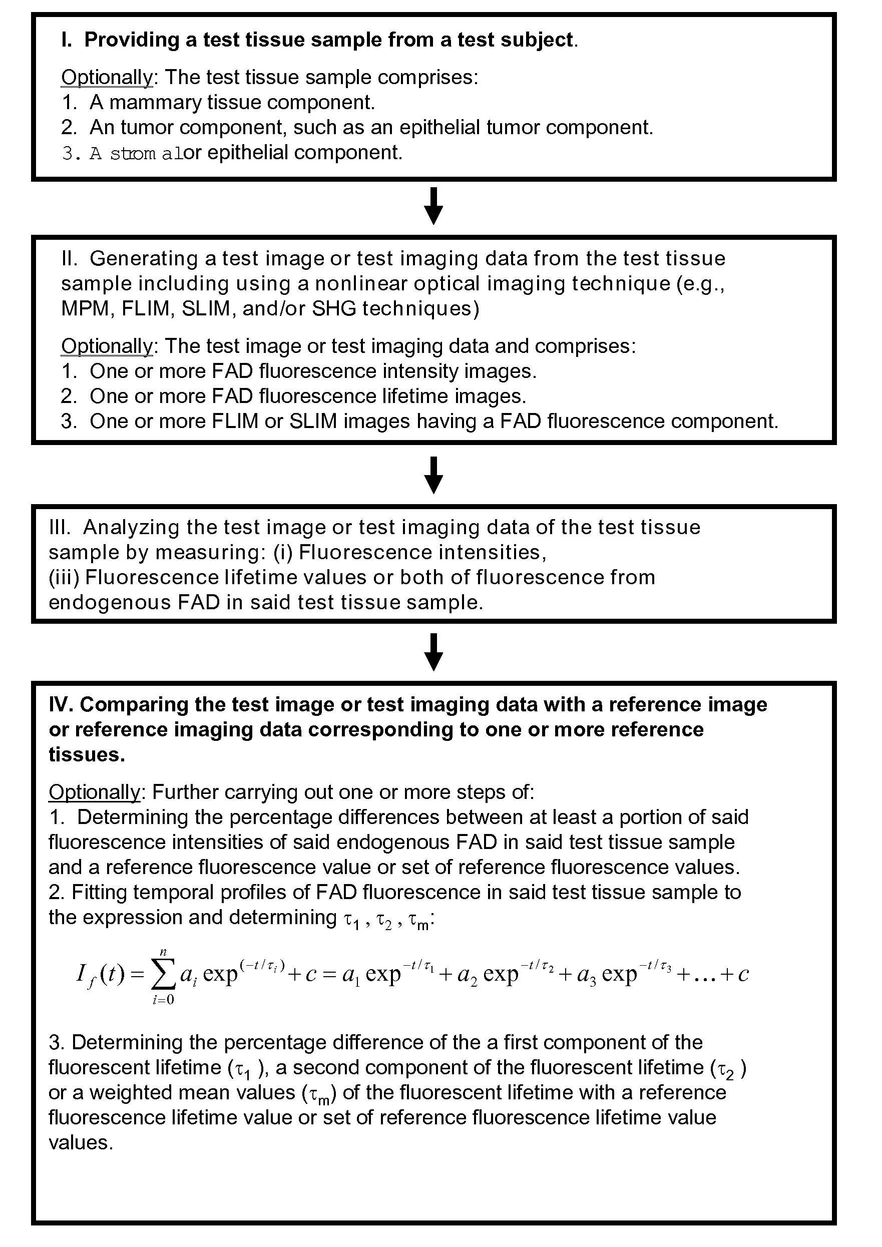
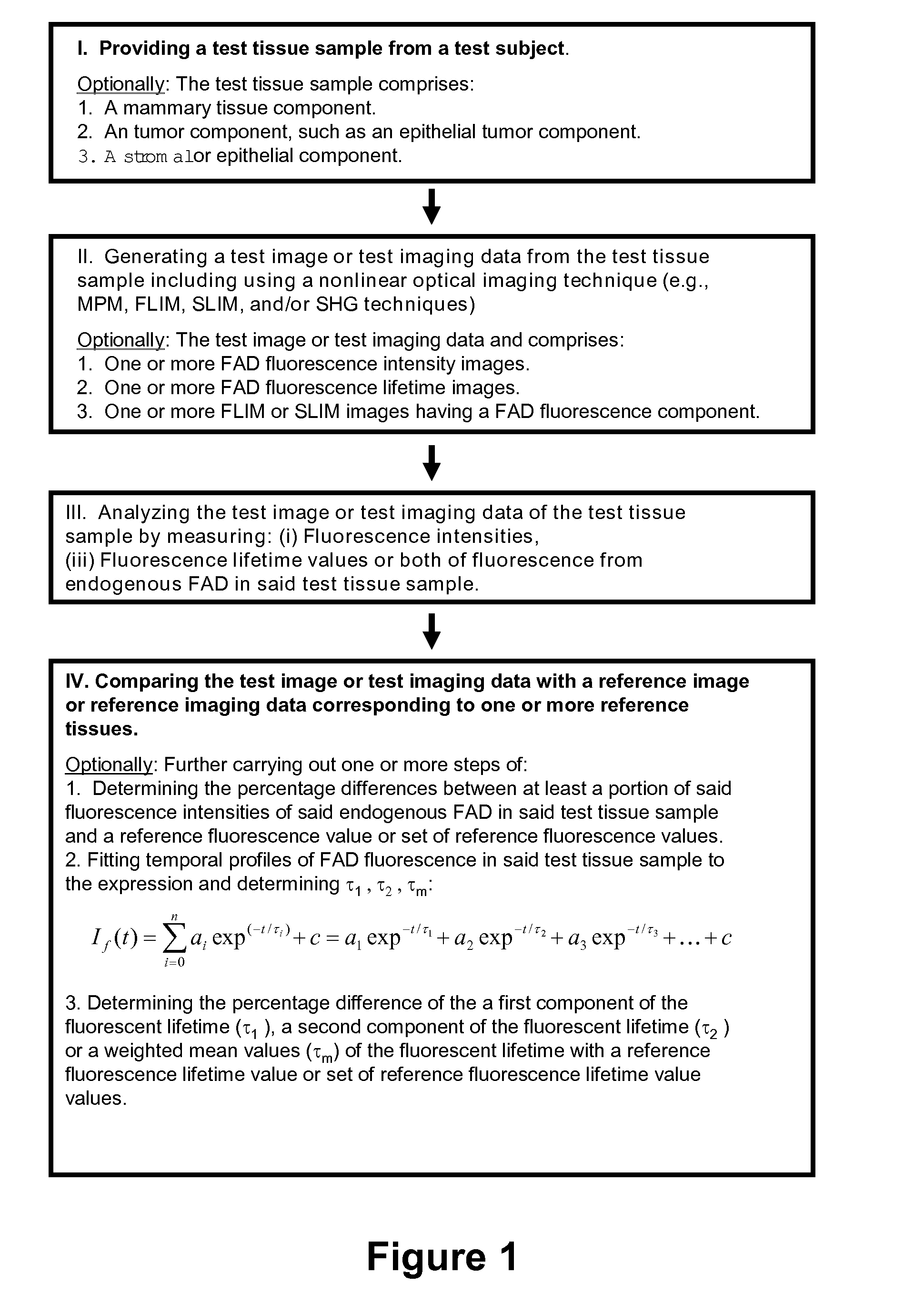
[0054]Mammographically dense breast tissue is one of the greatest risk factors for developing breast carcinoma. Despite the strong clinical correlation, breast density has not been causally linked to tumorigenesis, largely because no animal system has existed for studying breast tissue density. Thus, the influence of the extracellular-matrix on breast carcinoma development and the underlying molecular mechanisms are not understood. Importantly, areas of high breast density are associated with increased stromal collagen. In this Example we demonstrate that increased stromal collagen in mouse mammary tissue increases tumor formation ˜3-fold and results in a more invasive phenotype. Using nonlinear optical imaging approaches we demonstrate that local invasion is facilitated by stromal collagen re-organization and that this behavior is increased in collagen dense tissues. Additionally, we identify a metabolic sign...


PUM


Abstract

The present invention broadly provides methods and systems for detecting, identifying, and characterizing conditions, including diseases and other disorders in human or other animal subjects, by analyzing fluorescence from endogenous flavin adenine dinucleotide (FAD) fluorophors present in biological materials and samples. In particular embodiments, the invention relates to conditions of the human breast including cancers such as carcinoma. Methods and systems are provided for detecting, locating, and characterizing tumors and precancerous tissue via nonlinear optical imaging techniques capable of accurately characterizing fluorescence intensities and fluorescent lifetime parameters from endogenous FAD fluorophors present in a test tissue sample.

Description

CROSS-REFERENCE TO RELATED APPLICATIONS[0001]This application claims priority under 35 U.S.C. 119(e) to U.S. Provisional Patent Application 60 / 892,687 filed Mar. 2, 2007, which is hereby incorporated by reference in its entirety.STATEMENT REGARDING FEDERALLY SPONSORED RESEARCH OR DEVELOPMENT[0002]This invention was made with government support awarded by the following agencies: NIH grants CA076537 and EB000184 and Department of Defense Grant Number W81XWH-04-1-0428. The United States government has certain rights in the invention.BACKGROUND OF INVENTION[0003]Biomedical imaging plays a central role in a large number of diagnostic and therapeutic procedures including visualizing external and internal anatomical and physiological structures, features, and systems; evaluating complex biological events in the body at the organ, tissue, cellular, and molecular levels, and facilitating image guided surgery techniques. Imaging allows physicians and other health care professionals to detect ...

Claims


Application Information

Patent Timeline
Patent Type & Authority Applications(United States)
IPC IPC(8): G06K9/00
CPCG01N33/57415A61B5/0059
Inventor PROVENZANO, PAOLO P.KEELY, PATRICIA JOELICEIRI, KEVIN W.WHITE, JOHN G.
Owner WISCONSIN ALUMNI RES FOUND
Why Eureka
Try Eureka